|
Medium Spiny Neurons
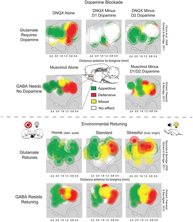
Medium spiny neurons (MSNs), also known as spiny projection neurons (SPNs), are a special type of GABAergic inhibitory cell representing 95% of neurons within the human striatum, a basal ganglia structure. Medium spiny neurons have two primary phenotypes (characteristic types): D1-type MSNs of the direct pathway and D2-type MSNs of the indirect pathway. Most striatal MSNs contain only D1-type or D2-type dopamine receptors, but a subpopulation of MSNs exhibit both phenotypes. Direct pathway MSNs excite their ultimate basal ganglia output structure (such as the thalamus) and promote associated behaviors; these neurons express D1-type dopamine receptors, adenosine A1 receptors, dynorphin peptides, and substance P peptides. Indirect pathway MSNs inhibit their output structure and in turn inhibit associated behaviors; these neurons express D2-type dopamine receptors, adenosine A2A receptors (A2A), heterotetramers, and enkephalin. Both types express glutamate receptors (NMDAR and A ... [...More Info...] [...Related Items...] OR: [Wikipedia] [Google] [Baidu] |
Nucleus Accumbens
The nucleus accumbens (NAc or NAcc; also known as the accumbens nucleus, or formerly as the ''nucleus accumbens septi'', Latin for "nucleus adjacent to the septum") is a region in the basal forebrain rostral to the preoptic area of the hypothalamus. The nucleus accumbens and the olfactory tubercle collectively form the ventral striatum. The ventral striatum and dorsal striatum collectively form the striatum, which is the main component of the basal ganglia. The dopaminergic neurons of the mesolimbic pathway project onto the GABAergic medium spiny neurons of the nucleus accumbens and olfactory tubercle. Each cerebral hemisphere has its own nucleus accumbens, which can be divided into two structures: the nucleus accumbens core and the nucleus accumbens shell. These substructures have different morphology and functions. Different NAcc subregions (core vs shell) and neuron subpopulations within each region ( D1-type vs D2-type medium spiny neurons) are responsible for dif ... [...More Info...] [...Related Items...] OR: [Wikipedia] [Google] [Baidu] |
Substance P
Substance P (SP) is an undecapeptide (a peptide composed of a chain of 11 amino acid residues) and a member of the tachykinin neuropeptide family. It is a neuropeptide, acting as a neurotransmitter and as a neuromodulator. Substance P and its closely related neurokinin A (NKA) are produced from a polyprotein precursor after differential splicing of the preprotachykinin A gene. The deduced amino acid sequence of substance P is as follows: * Arg Pro Lys Pro Gln Gln Phe Phe Gly Leu Met (RPKPQQFFGLM) with an amidation at the C-terminus. Substance P is released from the terminals of specific sensory nerves. It is found in the brain and spinal cord and is associated with inflammatory processes and pain. Discovery The original discovery of Substance P (SP) was in 1931 by Ulf von Euler and John H. Gaddum as a tissue extract that caused intestinal contraction ''in vitro''. Its tissue distribution and biologic actions were further investigated over the following decades. Th ... [...More Info...] [...Related Items...] OR: [Wikipedia] [Google] [Baidu] |
NAcc Shell
The nucleus accumbens (NAc or NAcc; also known as the accumbens nucleus, or formerly as the ''nucleus accumbens septi'', Latin for "nucleus adjacent to the septum") is a region in the basal forebrain rostral to the preoptic area of the hypothalamus. The nucleus accumbens and the olfactory tubercle collectively form the ventral striatum. The ventral striatum and dorsal striatum collectively form the striatum, which is the main component of the basal ganglia. The dopaminergic neurons of the mesolimbic pathway project onto the GABAergic medium spiny neurons of the nucleus accumbens and olfactory tubercle. Each cerebral hemisphere has its own nucleus accumbens, which can be divided into two structures: the nucleus accumbens core and the nucleus accumbens shell. These substructures have different morphology and functions. Different NAcc subregions (core vs shell) and neuron subpopulations within each region (D1-type vs D2-type medium spiny neurons) are responsible for different cog ... [...More Info...] [...Related Items...] OR: [Wikipedia] [Google] [Baidu] |
DRD2
Dopamine receptor D2, also known as D2R, is a protein that, in humans, is encoded by the ''DRD2'' gene. After work from Paul Greengard's lab had suggested that dopamine receptors were the site of action of antipsychotic drugs, several groups, including those of Solomon Snyder and Philip Seeman used a radiolabeled antipsychotic drug to identify what is now known as the dopamine D2 receptor. The dopamine D2 receptor is the main receptor for most antipsychotic drugs. The structure of DRD2 in complex with the atypical antipsychotic risperidone has been determined. Function D2 receptors are coupled to Gi subtype of G protein. This G protein-coupled receptor inhibits adenylyl cyclase activity. In mice, regulation of D2R surface expression by the neuronal calcium sensor-1 (NCS-1) in the dentate gyrus is involved in exploration, synaptic plasticity and memory formation. Studies have shown potential roles for D2R in retrieval of fear memories in the prelimbic cortex and in discrimina ... [...More Info...] [...Related Items...] OR: [Wikipedia] [Google] [Baidu] |
DRD1
Dopamine receptor D1, also known as DRD1. It is one of the two types of D1-like receptor family - receptors D1 and D5. It is a protein that in humans is encoded by the DRD1 gene. Tissue distribution D1 receptors are the most abundant kind of dopamine receptor in the central nervous system. Northern blot and in situ hybridization show that the mRNA expression of DRD1 is highest in the dorsal striatum ( caudate and putamen) and ventral striatum (nucleus accumbens and olfactory tubercle). Lower levels occur in the basolateral amygdala, cerebral cortex, septum, thalamus, and hypothalamus. Function D1 receptors regulate the memory, learning, and the growth of neurons, also is used in the reward system and locomotor activity, mediating some behaviors and modulating dopamine receptor D2-mediated events. They play a role in addiction by facilitating the gene expression changes that occur in the nucleus accumbens during addiction. They are Gs/a coupled and can stimulate neurons ... [...More Info...] [...Related Items...] OR: [Wikipedia] [Google] [Baidu] |
CB1 Receptor
Cannabinoid receptor type 1 (CB1), also known as cannabinoid receptor 1, is a G protein-coupled cannabinoid receptor that in humans is encoded by the ''CNR1'' gene. The human CB1 receptor is expressed in the peripheral nervous system and central nervous system. It is activated by: endocannabinoids, a group of retrograde neurotransmitters that include anandamide and 2-arachidonoylglycerol (2-AG); plant phytocannabinoids, such as the compound THC which is an active ingredient of the psychoactive drug cannabis; and, synthetic analogs of THC. CB1 is antagonized by the phytocannabinoid tetrahydrocannabivarin (THCV). The primary endogenous agonist of the human CB1 receptor is anandamide. Structure The CB1 receptor shares the structure characteristic of all G-protein-coupled receptors, possessing seven transmembrane domains connected by three extracellular and three intracellular loops, an extracellular N-terminal tail, and an intracellular C-terminal tail. The receptor may exist ... [...More Info...] [...Related Items...] OR: [Wikipedia] [Google] [Baidu] |
Muscarinic Acetylcholine Receptor M4
The muscarinic acetylcholine receptor M4, also known as the cholinergic receptor, muscarinic 4 (CHRM4), is a protein that, in humans, is encoded by the ''CHRM4'' gene. Function M4 muscarinic receptors are coupled to Gi/o heterotrimeric proteins. They function as inhibitory autoreceptors for acetylcholine. Activation of M4 receptors inhibits acetylcholine release in the striatum. The M2 subtype of acetylcholine receptor functions similarly as an inhibitory autoreceptor to acetylcholine release, albeit functioning actively primarily in the hippocampus and cerebral cortex. Muscarinic acetylcholine receptors possess a regulatory effect on dopaminergic neurotransmission. Activation of M4 receptors in the striatum inhibit D1-induced locomotor stimulation in mice. M4 receptor-deficient mice exhibit increased locomotor simulation in response to D1 agonists, amphetamine and cocaine. Neurotransmission in the striatum influences extrapyramidal motor control, thus alterations in M4 ac ... [...More Info...] [...Related Items...] OR: [Wikipedia] [Google] [Baidu] |
Muscarinic Acetylcholine Receptor M1
The muscarinic acetylcholine receptor M1, also known as the cholinergic receptor, muscarinic 1, is a muscarinic receptor that in humans is encoded by the ''CHRM1'' gene. It is localized to 11q13. This receptor is found mediating slow EPSP at the ganglion in the postganglionic nerve, is common in exocrine glands and in the CNS. It is predominantly found bound to G proteins of class Gq that use upregulation of phospholipase C and, therefore, inositol trisphosphate and intracellular calcium as a signalling pathway. A receptor so bound would not be susceptible to CTX or PTX. However, Gi (causing a downstream decrease in cAMP) and Gs (causing an increase in cAMP) have also been shown to be involved in interactions in certain tissues, and so would be susceptible to PTX and CTX respectively. Effects * EPSP in autonomic ganglia * Secretion from salivary glands * Gastric acid secretion from stomach * In CNS (memory?) * Vagally-induced bronchoconstriction * Mediating olfac ... [...More Info...] [...Related Items...] OR: [Wikipedia] [Google] [Baidu] |
Acetylcholine Receptor
An acetylcholine receptor (abbreviated AChR) is an integral membrane protein that responds to the binding of acetylcholine, a neurotransmitter. Classification Like other transmembrane receptors, acetylcholine receptors are classified according to their "pharmacology," or according to their relative affinities and sensitivities to different molecules. Although all acetylcholine receptors, by definition, respond to acetylcholine, they respond to other molecules as well. *Nicotinic acetylcholine receptors (''nAChR'', also known as " ionotropic" acetylcholine receptors) are particularly responsive to nicotine. The nicotine ACh receptor is also a Na+, K+ and Ca2+ ion channel. *Muscarinic acetylcholine receptors (''mAChR'', also known as " metabotropic" acetylcholine receptors) are particularly responsive to muscarine. Nicotinic and muscarinic are two main kinds of "cholinergic" receptors. Receptor types Molecular biology has shown that the nicotinic and muscarinic receptors belo ... [...More Info...] [...Related Items...] OR: [Wikipedia] [Google] [Baidu] |
AMPAR
The α-amino-3-hydroxy-5-methyl-4-isoxazolepropionic acid receptor (also known as AMPA receptor, AMPAR, or quisqualate receptor) is an ionotropic transmembrane receptor for glutamate ( iGluR) that mediates fast synaptic transmission in the central nervous system (CNS). It has been traditionally classified as a non-NMDA-type receptor, along with the kainate receptor. Its name is derived from its ability to be activated by the artificial glutamate analog AMPA. The receptor was first named the "quisqualate receptor" by Watkins and colleagues after a naturally occurring agonist quisqualate and was only later given the label "AMPA receptor" after the selective agonist developed by Tage Honore and colleagues at the Royal Danish School of Pharmacy in Copenhagen. The ''GRIA2''-encoded AMPA receptor ligand binding core (GluA2 LBD) was the first glutamate receptor ion channel domain to be crystallized. Structure and function Subunit composition AMPARs are composed of four types of subun ... [...More Info...] [...Related Items...] OR: [Wikipedia] [Google] [Baidu] |
NMDAR
The ''N''-methyl-D-aspartate receptor (also known as the NMDA receptor or NMDAR), is a glutamate receptor and ion channel found in neurons. The NMDA receptor is one of three types of ionotropic glutamate receptors, the other two being AMPA and kainate receptors. Depending on its subunit composition, its ligands are glutamate and glycine (or D-serine). However, the binding of the ligands is typically not sufficient to open the channel as it may be blocked by Mg2+ ions which are only removed when the neuron is sufficiently depolarized. Thus, the channel acts as a “coincidence detector” and only once both of these conditions are met, the channel opens and it allows positively charged ions (cations) to flow through the cell membrane. The NMDA receptor is thought to be very important for controlling synaptic plasticity and mediating learning and memory functions. The NMDA receptor is ionotropic, meaning it is a protein which allows the passage of ions through the cell memb ... [...More Info...] [...Related Items...] OR: [Wikipedia] [Google] [Baidu] |
Glutamate Receptor
Glutamate receptors are synaptic and non synaptic receptors located primarily on the membranes of neuronal and glial cells. Glutamate (the conjugate base of glutamic acid) is abundant in the human body, but particularly in the nervous system and especially prominent in the human brain where it is the body's most prominent neurotransmitter, the brain's main excitatory neurotransmitter, and also the precursor for GABA, the brain's main inhibitory neurotransmitter. Glutamate receptors are responsible for the glutamate-mediated postsynaptic excitation of neural cells, and are important for neural communication, memory formation, learning, and regulation. Glutamate receptors are implicated in a number of neurological conditions. Their central role in excitotoxicity and prevalence in the central nervous system has been linked or speculated to be linked to many neurodegenerative diseases, and several other conditions have been further linked to glutamate receptor gene ... [...More Info...] [...Related Items...] OR: [Wikipedia] [Google] [Baidu] |


