|
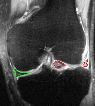
Medial Meniscus
The medial meniscus is a fibrocartilage semicircular band that spans the knee joint medially, located between the medial condyle of the femur and the medial condyle of the tibia.Platzer (2004), p 208 It is also referred to as the internal semilunar fibrocartilage. The medial meniscus has more of a crescent shape while the lateral meniscus is more circular. The anterior aspects of both menisci are connected by the transverse ligament. It is a common site of injury, especially if the knee is twisted. Structure The meniscus attaches to the tibia via coronary ligaments. Its anterior end, thin and pointed, is attached to the anterior intercondyloid fossa of the tibia, in front of the anterior cruciate ligament; Its posterior end is fixed to the posterior intercondyloid fossa of the tibia, between the attachments of the lateral meniscus and the posterior cruciate ligament. It is fused with the tibial collateral ligament which makes it far less mobile than the lateral meniscus. The ... [...More Info...] [...Related Items...] OR: [Wikipedia] [Google] [Baidu] |
Coronary Ligament Of The Knee
The coronary ligaments of the knee (also known as meniscotibial ligaments) are portions of the joint capsule which connect the inferior edges of the fibrocartilaginous menisci to the periphery of the tibial plateaus. Structure The coronary ligaments of the knee are continuous with the joint capsule and the menisci. Function The coronary ligaments function to connect parts of the outside, inferior edges of the medial and lateral menisci to the joint capsule of the knee. The medial meniscus also has firm attachments laterally to the ''intercondylar area of the tibia'' and medially to the tibial collateral ligament. The lateral meniscus has firm attachments medially to the intercondylar area via the ends of the meniscus, and posteromedially via the posterior meniscofemoral ligament, which attaches the posterior limb of the meniscus to the posterior cruciate ligament The posterior cruciate ligament (PCL) is a ligament in each knee of humans and various other animals. It works as a ... [...More Info...] [...Related Items...] OR: [Wikipedia] [Google] [Baidu] |
Tibia
The tibia (; ), also known as the shinbone or shankbone, is the larger, stronger, and anterior (frontal) of the two bones in the leg below the knee in vertebrates (the other being the fibula, behind and to the outside of the tibia); it connects the knee with the ankle. The tibia is found on the medial side of the leg next to the fibula and closer to the median plane. The tibia is connected to the fibula by the interosseous membrane of leg, forming a type of fibrous joint called a syndesmosis with very little movement. The tibia is named for the flute ''tibia''. It is the second largest bone in the human body, after the femur. The leg bones are the strongest long bones as they support the rest of the body. Structure In human anatomy, the tibia is the second largest bone next to the femur. As in other vertebrates the tibia is one of two bones in the lower leg, the other being the fibula, and is a component of the knee and ankle joints. The ossification or formation of the bone ... [...More Info...] [...Related Items...] OR: [Wikipedia] [Google] [Baidu] |
Tibial Collateral Ligament
The medial collateral ligament (MCL), or tibial collateral ligament (TCL), is one of the four major ligaments of the knee. It is on the medial (inner) side of the knee joint in humans and other primates. Its primary function is to resist outward turning forces on the knee. Structure It is a broad, flat, membranous band, situated slightly posterior on the medial side of the knee joint. It is attached proximally to the medial epicondyle of the femur immediately below the adductor tubercle; below to the medial condyle of the tibia and medial surface of its body. It resists forces that would push the knee medially, which would otherwise produce valgus deformity. The fibers of the posterior part of the ligament are short and incline backward as they descend; they are inserted into the tibia above the groove for the semimembranosus muscle. The anterior part of the ligament is a flattened band, about 10 centimeters long, which inclines forward as it descends. It is inserted into th ... [...More Info...] [...Related Items...] OR: [Wikipedia] [Google] [Baidu] |
Meniscal Cartilage Replacement Therapy
Left knee-joint from behind, showing interior ligaments Meniscal cartilage replacement therapy is surgical replacement of the meniscus of the knee as a treatment for where the meniscus is so damaged that it would otherwise need to be removed. Anatomy The meniscus is a C-shaped piece of fibrocartilage located at the peripheral aspect of the knee joint that offers lubrication and nutrition to the joint. Each knee has two menisci, medial and lateral, whose purpose is to provide space between the tibia and the femur, preventing friction and allowing for the diffusion of articular cartilage. The majority of the meniscus has no blood supply. As a result, if the meniscus is damaged, from trauma or with age, it is unable to undergo the body’s normal healing process. Therefore, a torn piece can begin to move inside the joint, get caught between the bones, and cause pain, swelling, and decreased mobility. However, recent research has called into question whether many meniscus tears ... [...More Info...] [...Related Items...] OR: [Wikipedia] [Google] [Baidu] |
Unhappy Triad
The unhappy triad, also known as a blown knee among other names, is an injury to the anterior cruciate ligament, medial collateral ligament, and meniscus. Analysis during the 1990s indicated that this 'classic' O'Donoghue triad is actually an unusual clinical entity among athletes with knee injuries. Some authors mistakenly believe that in this type of injury, "combined anterior cruciate and medial collateral ligament (ACL- MCL) disruptions that were incurred during athletic endeavors" always present with concomitant medial meniscus injury. However, the 1990 analysis showed that lateral meniscus tears are more common than medial meniscus tears in conjunction with sprains of the ACL. Symptoms *Pain in affected knee *Stiffness and swelling in affected knee *Catching or locking of the knee in affected knee *Instability of the knee with twisting or side-to-side movements (The sensation of the knee "giving out"). *Inability to move the knee through its full range of motion Cause The ... [...More Info...] [...Related Items...] OR: [Wikipedia] [Google] [Baidu] |
Cartilage
Cartilage is a resilient and smooth type of connective tissue. In tetrapods, it covers and protects the ends of long bones at the joints as articular cartilage, and is a structural component of many body parts including the rib cage, the neck and the bronchial tubes, and the intervertebral discs. In other taxa, such as chondrichthyans, but also in cyclostomes, it may constitute a much greater proportion of the skeleton. It is not as hard and rigid as bone, but it is much stiffer and much less flexible than muscle. The matrix of cartilage is made up of glycosaminoglycans, proteoglycans, collagen fibers and, sometimes, elastin. Because of its rigidity, cartilage often serves the purpose of holding tubes open in the body. Examples include the rings of the trachea, such as the cricoid cartilage and carina. Cartilage is composed of specialized cells called chondrocytes that produce a large amount of collagenous extracellular matrix, abundant ground substance that is rich in pro ... [...More Info...] [...Related Items...] OR: [Wikipedia] [Google] [Baidu] |
Tear Of Meniscus
A tear of a meniscus is a rupturing of one or more of the fibrocartilage strips in the knee called menisci. When doctors and patients refer to "torn cartilage" in the knee, they actually may be referring to an injury to a meniscus at the top of one of the tibiae. Menisci can be torn during innocuous activities such as walking or squatting. They can also be torn by traumatic force encountered in sports or other forms of physical exertion. The traumatic action is most often a twisting movement at the knee while the leg is bent. In older adults, the meniscus can be damaged following prolonged 'wear and tear'. Especially acute injuries (typically in younger, more active patients) can lead to displaced tears which can cause mechanical symptoms such as clicking, catching, or locking during motion of the joint. The joint will be in pain when in use, but when there is no load, the pain goes away. A tear of the medial meniscus can occur as part of the unhappy triad, together with a tear ... [...More Info...] [...Related Items...] OR: [Wikipedia] [Google] [Baidu] |
Obesity
Obesity is a medical condition, sometimes considered a disease, in which excess body fat has accumulated to such an extent that it may negatively affect health. People are classified as obese when their body mass index (BMI)—a person's weight divided by the square of the person's height—is over ; the range is defined as overweight. Some East Asian countries use lower values to calculate obesity. Obesity is a major cause of disability and is correlated with various diseases and conditions, particularly cardiovascular diseases, type 2 diabetes, obstructive sleep apnea, certain types of cancer, and osteoarthritis. Obesity has individual, socioeconomic, and environmental causes. Some known causes are diet, physical activity, automation, urbanization, genetic susceptibility, medications, mental disorders, economic policies, endocrine disorders, and exposure to endocrine-disrupting chemicals. While a majority of obese individuals at any given time are attempting to ... [...More Info...] [...Related Items...] OR: [Wikipedia] [Google] [Baidu] |
Osteoarthritis
Osteoarthritis (OA) is a type of degenerative joint disease that results from breakdown of joint cartilage and underlying bone which affects 1 in 7 adults in the United States. It is believed to be the fourth leading cause of disability in the world. The most common symptoms are joint pain and stiffness. Usually the symptoms progress slowly over years. Initially they may occur only after exercise but can become constant over time. Other symptoms may include joint swelling, decreased range of motion, and, when the back is affected, weakness or numbness of the arms and legs. The most commonly involved joints are the two near the ends of the fingers and the joint at the base of the thumbs; the knee and hip joints; and the joints of the neck and lower back. Joints on one side of the body are often more affected than those on the other. The symptoms can interfere with work and normal daily activities. Unlike some other types of arthritis, only the joints, not internal organs, are af ... [...More Info...] [...Related Items...] OR: [Wikipedia] [Google] [Baidu] |
Synovial Fluid
Synovial fluid, also called synovia, elp 1/sup> is a viscous, non-Newtonian fluid found in the cavities of synovial joints. With its egg white–like consistency, the principal role of synovial fluid is to reduce friction between the articular cartilage of synovial joints during movement. Synovial fluid is a small component of the transcellular fluid component of extracellular fluid. Structure The inner membrane of synovial joints is called the synovial membrane and secretes synovial fluid into the joint cavity. Synovial fluid is an ultrafiltrate from plasma, and contains proteins derived from the blood plasma and proteins that are produced by cells within the joint tissues. The fluid contains hyaluronan secreted by fibroblast-like cells in the synovial membrane, lubricin (proteoglycan 4; PRG4) secreted by the surface chondrocytes of the articular cartilage and interstitial fluid filtered from the blood plasma. This fluid forms a thin layer (roughly 50 μm) at the surface of c ... [...More Info...] [...Related Items...] OR: [Wikipedia] [Google] [Baidu] |
Vascularized
Angiogenesis is the physiological process through which new blood vessels form from pre-existing vessels, formed in the earlier stage of vasculogenesis. Angiogenesis continues the growth of the vasculature by processes of sprouting and splitting. Vasculogenesis is the embryonic formation of endothelial cells from mesoderm cell precursors, and from neovascularization, although discussions are not always precise (especially in older texts). The first vessels in the developing embryo form through vasculogenesis, after which angiogenesis is responsible for most, if not all, blood vessel growth during development and in disease. Angiogenesis is a normal and vital process in growth and development, as well as in wound healing and in the formation of granulation tissue. However, it is also a fundamental step in the transition of tumors from a benign state to a malignant one, leading to the use of angiogenesis inhibitors in the treatment of cancer. The essential role of angiogenesis in t ... [...More Info...] [...Related Items...] OR: [Wikipedia] [Google] [Baidu] |
Medial Collateral Ligament
The medial collateral ligament (MCL), or tibial collateral ligament (TCL), is one of the four major ligaments of the knee. It is on the medial (inner) side of the knee joint in humans and other primates. Its primary function is to resist outward turning forces on the knee. Structure It is a broad, flat, membranous band, situated slightly posterior on the medial side of the knee joint. It is attached proximally to the medial epicondyle of the femur immediately below the adductor tubercle; below to the medial condyle of the tibia and medial surface of its body. It resists forces that would push the knee medially, which would otherwise produce valgus deformity. The fibers of the posterior part of the ligament are short and incline backward as they descend; they are inserted into the tibia above the groove for the semimembranosus muscle. The anterior part of the ligament is a flattened band, about 10 centimeters long, which inclines forward as it descends. It is inserted into ... [...More Info...] [...Related Items...] OR: [Wikipedia] [Google] [Baidu] |


