|
Mandibular Division Of The Trigeminal Nerve
In neuroanatomy, the mandibular nerve (V) is the largest of the three divisions of the trigeminal nerve, the fifth cranial nerve (CN V). Unlike the other divisions of the trigeminal nerve ( ophthalmic nerve, maxillary nerve) which contain only afferent fibers, the mandibular nerve contains both afferent and efferent fibers. These nerve fibers innervate structures of the lower jaw and face, such as the tongue, lower lip, and chin. The mandibular nerve also innervates the muscles of mastication. Structure Course The large sensory root of mandibular nerve emerges from the lateral part of the trigeminal ganglion and exits the cranial cavity through the foramen ovale. The motor root (Latin: ''radix motoria'' s. ''portio minor''), the small motor root of the trigeminal nerve, passes under the trigeminal ganglion and through the foramen ovale to unite with the sensory root just outside the skull. The mandibular nerve immediately passes between tensor veli palatini, which is med ... [...More Info...] [...Related Items...] OR: [Wikipedia] [Google] [Baidu] [Amazon] |
Mandibular Division
In neuroanatomy, the mandibular nerve (V) is the largest of the three divisions of the trigeminal nerve, the fifth Cranial nerves, cranial nerve (CN V). Unlike the other divisions of the trigeminal nerve (ophthalmic nerve, maxillary nerve) which contain only Afferent nerve fiber, afferent fibers, the mandibular nerve contains both afferent and Efferent nerve fiber, efferent fibers. These nerve fibers innervate structures of the lower jaw and face, such as the tongue, lower lip, and chin. The mandibular nerve also innervates the muscles of mastication. Structure Course The large sensory root of mandibular nerve emerges from the lateral part of the trigeminal ganglion and exits the cranial cavity through the Foramen ovale (skull), foramen ovale. The motor root (Latin: ''radix motoria'' s. ''portio minor''), the small motor root of the trigeminal nerve, passes under the trigeminal ganglion and through the Foramen ovale (skull), foramen ovale to unite with the sensory root just out ... [...More Info...] [...Related Items...] OR: [Wikipedia] [Google] [Baidu] [Amazon] |
Foramen Ovale (skull)
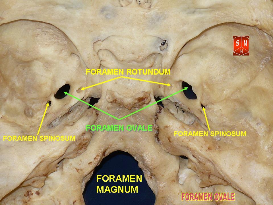
The foramen ovale (En: oval window) is a hole in the posterior part of the sphenoid bone, posterolateral to the foramen rotundum. It is one of the larger of the several holes (the foramina) in the skull. It transmits the mandibular nerve, a branch of the trigeminal nerve. Structure The foramen ovale is an opening in the greater wing of the sphenoid bone. The foramen ovale is one of two cranial foramina in the greater wing, the other being the foramen spinosum. The foramen ovale is posterolateral to the foramen rotundum and anteromedial to the foramen spinosum. Posterior and medial to the foramen is the opening for the carotid canal. Contents The following structures pass through foramen ovale: * mandibular nerve (CN V) (a branch of the trigeminal nerve (CN V)) *accessory meningeal artery * lesser petrosal nerve (a branch of the glossopharyngeal nerve) * an emissary vein connecting the cavernous sinus with the pterygoid plexus * (occasionally) meningeal branch o ... [...More Info...] [...Related Items...] OR: [Wikipedia] [Google] [Baidu] [Amazon] |
Lingual Nerve
The lingual nerve carries sensory innervation from the anterior two-thirds of the tongue. It contains fibres from both the mandibular division of the trigeminal nerve (CN V) and from the facial nerve (CN VII). The fibres from the trigeminal nerve are for touch, pain and temperature (general sensation), and the ones from the facial nerve are for taste (special sensation). Structure Origin The lingual nerve arises from the posterior trunk of mandibular nerve (CN V) within the infratemporal fossa. Course The lingual nerve first courses deep to the lateral pterygoid muscle and superior to the tensor veli palatini muscle; while passing between these two muscle, it is joined by the chorda tympani, and often by a communicating branch from the inferior alveolar nerve. The nerve then comes to pass inferoanteriorly upon the medial pterygoid muscle towards the medial aspect of the ramus of mandible, eventually meeting the mandible at the junction of the ramus and body of mandibl ... [...More Info...] [...Related Items...] OR: [Wikipedia] [Google] [Baidu] [Amazon] |
Auriculotemporal Nerve
The auriculotemporal nerve is a sensory branch of the mandibular nerve (CN V3) that runs with the superficial temporal artery and vein, and provides sensory innervation to parts of the external ear, scalp, and temporomandibular joint. The nerve also conveys post-ganglionic parasympathetic fibres from the otic ganglion to the parotid gland. Structure Origin The auriculotemporal nerve arises from the posterior division of the mandibular nerve (CN V3) (which is itself a branch of the trigeminal nerve (CN V)). It arises by two roots that circle around either side of the middle meningeal artery before uniting to form a single nerve. Course Roots of the auriculotemporal nerve circle around both sides of the middle meningeal artery before uniting to form a single nerve. The nerve passes deep to the neck of the mandible - between it and the sphenomandibular ligament - and then courses deep to the lateral pterygoid muscle. It issues parotid branches and then turns superiorly, ... [...More Info...] [...Related Items...] OR: [Wikipedia] [Google] [Baidu] [Amazon] |
Lateral Pterygoid Nerve
The lateral pterygoid nerve (or external pterygoid nerve) is a branch of the anterior division of the mandibular nerve. It usually originates as two separate branches that travel near the buccal nerve, and enter the deep surfaces of the superior and inferior heads of the lateral pterygoid muscle. Nerve pathway * trigeminal nerve (CN V) * mandibular nerve In neuroanatomy, the mandibular nerve (V) is the largest of the three divisions of the trigeminal nerve, the fifth Cranial nerves, cranial nerve (CN V). Unlike the other divisions of the trigeminal nerve (ophthalmic nerve, maxillary nerve) which ... (V3) * anterior division of mandibular nerve Variation Some authors describe the lateral pterygoid nerve as a single branch of the anterior division of the mandibular nerve which then bifurcates to enter the two heads of the lateral pterygoid muscle. References Mandibular nerve {{Neuroanatomy-stub ... [...More Info...] [...Related Items...] OR: [Wikipedia] [Google] [Baidu] [Amazon] |
Buccal Nerve
The buccal nerve (long buccal nerve) is a sensory nerve of the face arising from the mandibular nerve (CN V3) (which is itself a branch of the trigeminal nerve). It conveys sensory information from the skin of the cheek, and parts of the oral mucosa, periodontium, and gingiva. Structure Origin The buccal nerve is a branch of the anterior division of the mandibular nerve (CN V3). It is the only sensory branch of the anterior division. Course and relations After branching from the anterior trunk of the mandibular nerve (CN V3), the buccal nerve passes between the two heads of the lateral pterygoid muscle, underneath the tendon of the temporalis muscle. It then passes anterior to the ramus of the mandible to first course deep to the masseter muscle, and finally anteroinferiorly upon surface of the buccinator muscle before piercing this muscle. Communications It connects with the buccal branches of the facial nerve on the surface of the buccinator muscle. It gives off m ... [...More Info...] [...Related Items...] OR: [Wikipedia] [Google] [Baidu] [Amazon] |
Deep Temporal Nerves
The deep temporal nerves are typically two nerves (one anterior and one posterior) which arise from the mandibular nerve (CN V3) and provide motor innervation to the temporalis muscle. Structure Origin They usually arise from (the anterior division of) the mandibular nerve (CN V3). Course They pass superior to the superior border of the lateral pterygoid muscle. They ascend to the temporal fossa and enter the deep surface of the temporalis muscle. Distribution The deep temporal nerves provide motor innervation to the temporalis muscle. The deep temporal nerves also have articular branches which provide a minor contribution to the innervation of the temporomandibular joint In anatomy, the temporomandibular joints (TMJ) are the two joints connecting the jawbone to the skull. It is a bilateral Synovial joint, synovial articulation between the temporal bone of the skull above and the condylar process of mandible be .... Variation Number There are usually two deep te ... [...More Info...] [...Related Items...] OR: [Wikipedia] [Google] [Baidu] [Amazon] |
Masseteric Nerve
The masseteric nerve is a nerve of the face. It is a branch of the mandibular nerve (CN V3). It passes through the mandibular notch to reach masseter muscle. It provides motor innervation the masseter muscle, and sensory innervation to the temporomandibular joint. Structure Origin The masseteric nerve is a branch of (the anterior division of) the mandibular nerve (CN V3) (itself a branch of the trigeminal nerve (CN V)). Course It passes laterally superior to the lateral pterygoid muscle, anterior to the temporomandibular joint, and posterior to the tendon of the temporalis muscle. It crosses (the posterior portion of) the mandibular notch alongside the masseteric artery before branching out upon the surface of the masseter muscle, then entering the muscle. Distribution The masseteric nerve provides motor innervation the masseter muscle. It additionally sends articular (sensory) branches to the temporomandibular joint. Clinical significance The masseteric nerve ma ... [...More Info...] [...Related Items...] OR: [Wikipedia] [Google] [Baidu] [Amazon] |
Medial Pterygoid Nerve
The medial pterygoid nerve (nerve to medial pterygoid, or internal pterygoid nerve) is a nerve of the head. It is a branch of the mandibular nerve (CN V3). It supplies the medial pterygoid muscle, the tensor veli palatini muscle, and the tensor tympani muscle. Structure Origin The medial pterygoid nerve is a slender branch of the mandibular nerve (CN V3) (itself a branch of the trigeminal nerve (CN V)). Course It passes through the otic ganglion (without synapsing). It penetrates the deep surface of the medial pterygoid muscle. It issues 1-2 twigs which traverse the otic ganglion (without synapsing) to reach and innervate the tensor tympani muscle, and tensor veli palatini muscle. Distribution The medial pterygoid nerve supplies the medial pterygoid muscle, tensor tympani muscle The tensor tympani is a muscle within the middle ear, located in the bony canal above the bony part of the auditory tube, and connects to the malleus bone. Its role is to dampen loud sound ... [...More Info...] [...Related Items...] OR: [Wikipedia] [Google] [Baidu] [Amazon] |
Meningeal Branch Of The Mandibular Nerve
The meningeal branch of the mandibular nerve (also known as the nervus spinosus) is a sensory branch of the mandibular nerve (CN V3) that enters the middle cranial fossa through either the foramen spinosum or foramen ovale to innervate the meninges of this fossa as well as the mastoid air cells. Anatomy Branches It divides into two branches - anterior and posterior - which accompany the main divisions of the middle meningeal artery and supply the dura mater: * The ''anterior branch'' communicates with the meningeal branch of the maxillary nerve. * The ''posterior branch'' also supplies the mucous lining of the mastoid cells The mastoid cells (also called air cells of Lenoir or mastoid cells of Lenoir) are air-filled cavities within the mastoid process of the temporal bone of the cranium. The mastoid cells are a form of skeletal pneumaticity. Infection in these cells .... References External links Overview at tufts.edu* Mandibular nerve Meninges {{Neur ... [...More Info...] [...Related Items...] OR: [Wikipedia] [Google] [Baidu] [Amazon] |
Lateral Pterygoid Muscle
The lateral pterygoid muscle (or external pterygoid muscle) is a muscle of mastication. It has two heads. It lies superior to the medial pterygoid muscle. It is supplied by pterygoid branches of the maxillary artery, and the lateral pterygoid nerve (from the mandibular nerve, CN V3). It depresses and protrudes the mandible. When each muscle works independently, they can move the mandible side to side. Structure The lateral pterygoid muscle has an upper head and a lower head. * The upper head originates on the infratemporal surface and infratemporal crest of the greater wing of the sphenoid bone. It inserts onto the articular disc and fibrous capsule of the temporomandibular joint. * The lower head originates on the lateral surface of the lateral pterygoid plate. It inserts onto the pterygoid fovea at the neck of the condyloid process of the mandible. It lies superior to the medial pterygoid muscle. Blood supply The lateral pterygoid muscle is supplied by pterygoid b ... [...More Info...] [...Related Items...] OR: [Wikipedia] [Google] [Baidu] [Amazon] |
