|
Major Urinary Protein
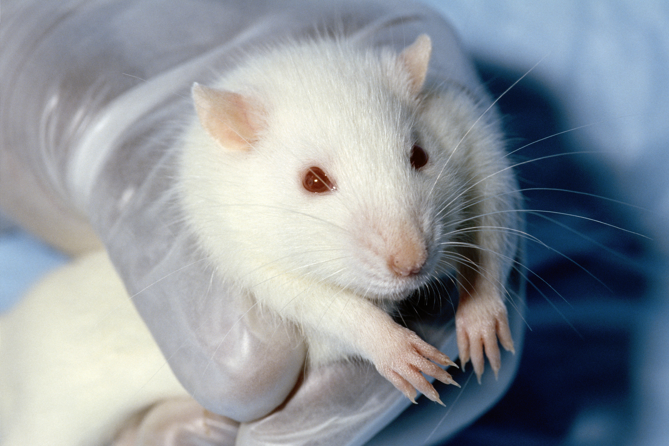
Major urinary proteins (Mups), also known as α2u-globulins, are a subfamily of proteins found in abundance in the urine and other secretions of many animals. Mups provide a small range of identifying information about the donor animal, when detected by the vomeronasal organ of the receiving animal. They belong to a larger family of proteins known as lipocalins. Mups are encoded by a cluster of genes, located adjacent to each other on a single stretch of DNA, that varies greatly in number between species: from at least 21 functional genes in mice to none in humans. Mup proteins form a characteristic glove shape, encompassing a ligand-binding pocket that accommodates specific small organic chemicals. Urinary proteins were first reported in rodents in 1932, during studies by Thomas Addis into the cause of proteinuria. They are potent human allergens and are largely responsible for a number of animal allergies, including to cats, horses and rodents. Their endogenous function with ... [...More Info...] [...Related Items...] OR: [Wikipedia] [Google] [Baidu] |
Kairomone
A kairomone (a coinage using the Greek καιρός ''opportune moment'', paralleling pheromone"kairomone, n.". OED Online. September 2012. Oxford University Press. http://www.oed.com/view/Entry/241005?redirectedFrom=kairomone (accessed 3 October 2012).) is a semiochemical, emitted by an organism, which mediates interspecific interactions in a way that benefits an individual of another species which receives it and harms the emitter. This "eavesdropping" is often disadvantageous to the producer (though other benefits of producing the substance may outweigh this cost, hence its persistence over evolutionary time). The kairomone improves the fitness of the recipient and in this respect differs from an allomone (which is the opposite: it benefits the producer and harms the receiver) and a synomone (which benefits both parties). The term is mostly used in the field of entomology (the study of insects). Two main ecological cues are provided by kairomones; they generally either indicat ... [...More Info...] [...Related Items...] OR: [Wikipedia] [Google] [Baidu] |
Submaxillary
The paired submandibular glands (historically known as submaxillary glands) are major salivary glands located beneath the floor of the mouth. They each weigh about 15 grams and contribute some 60–67% of unstimulated saliva secretion; on stimulation their contribution decreases in proportion as the parotid secretion rises to 50%. The average length of the normal human submandibular salivary gland is approximately 27mm, while the average width is approximately 14.3mm. Structure Lying superior to the digastric muscles, each submandibular gland is divided into superficial and deep lobes, which are separated by the mylohyoid muscle: * The superficial lobe comprises most of the gland, with the mylohyoid muscle runs under it * The deep lobe is the smaller part Secretions are delivered into the submandibular duct on the deep portion after which they hook around the posterior edge of the mylohyoid muscle and proceed on the superior surface laterally. The excretory ducts are then cross ... [...More Info...] [...Related Items...] OR: [Wikipedia] [Google] [Baidu] |
Parotid
The parotid gland is a major salivary gland in many animals. In humans, the two parotid glands are present on either side of the mouth and in front of both ears. They are the largest of the salivary glands. Each parotid is wrapped around the mandibular ramus, and secretes serous saliva through the parotid duct into the mouth, to facilitate mastication and swallowing and to begin the digestion of starches. There are also two other types of salivary glands; they are submandibular and sublingual glands. Sometimes accessory parotid glands are found close to the main parotid glands. Etymology The word ''parotid'' literally means "beside the ear". From Greek παρωτίς (stem παρωτιδ-) : (gland) behind the ear < παρά - pará : in front, and οὖς - ous (stem ὠτ-, ōt-) : ear. Structure The parotid glands are a pair of mainly |
Lacrimal Gland
The lacrimal glands are paired exocrine glands, one for each eye, found in most terrestrial vertebrates and some marine mammals, that secrete the aqueous layer of the tear film. In humans, they are situated in the upper lateral region of each orbit, in the lacrimal fossa of the orbit formed by the frontal bone. Inflammation of the lacrimal glands is called dacryoadenitis. The lacrimal gland produces tears which are secreted by the lacrimal ducts, and flow over the ocular surface, and then into canals that connect to the lacrimal sac. From that sac, the tears drain through the lacrimal duct into the nose. Anatomists divide the gland into two sections, a palpebral lobe, or portion, and an orbital lobe or portion. The smaller ''palpebral lobe'' lies close to the eye, along the inner surface of the eyelid; if the upper eyelid is everted, the palpebral portion can be seen. The orbital lobe of the gland, contains fine interlobular ducts that connect the orbital lobe and the palpebra ... [...More Info...] [...Related Items...] OR: [Wikipedia] [Google] [Baidu] |
Exocrine Gland
Exocrine glands are glands that secrete substances on to an epithelial surface by way of a duct. Examples of exocrine glands include sweat, salivary, mammary, ceruminous, lacrimal, sebaceous, prostate and mucous. Exocrine glands are one of two types of glands in the human body, the other being endocrine glands, which secrete their products directly into the bloodstream. The liver and pancreas are both exocrine and endocrine glands; they are exocrine glands because they secrete products—bile and pancreatic juice—into the gastrointestinal tract through a series of ducts, and endocrine because they secrete other substances directly into the bloodstream. Exocrine sweat glands are part of the integumentary system; they have eccrine and apocrine types. Classification Structure Exocrine glands contain a glandular portion and a duct portion, the structures of which can be used to classify the gland. * The duct portion may be branched (called compound) or unbranched (called simple ... [...More Info...] [...Related Items...] OR: [Wikipedia] [Google] [Baidu] |
Kidney
The kidneys are two reddish-brown bean-shaped organs found in vertebrates. They are located on the left and right in the retroperitoneal space, and in adult humans are about in length. They receive blood from the paired renal arteries; blood exits into the paired renal veins. Each kidney is attached to a ureter, a tube that carries excreted urine to the bladder. The kidney participates in the control of the volume of various body fluids, fluid osmolality, acid–base balance, various electrolyte concentrations, and removal of toxins. Filtration occurs in the glomerulus: one-fifth of the blood volume that enters the kidneys is filtered. Examples of substances reabsorbed are solute-free water, sodium, bicarbonate, glucose, and amino acids. Examples of substances secreted are hydrogen, ammonium, potassium and uric acid. The nephron is the structural and functional unit of the kidney. Each adult human kidney contains around 1 million nephrons, while a mouse kidney contains on ... [...More Info...] [...Related Items...] OR: [Wikipedia] [Google] [Baidu] |
Liver
The liver is a major Organ (anatomy), organ only found in vertebrates which performs many essential biological functions such as detoxification of the organism, and the Protein biosynthesis, synthesis of proteins and biochemicals necessary for digestion and growth. In humans, it is located in the quadrant (anatomy), right upper quadrant of the abdomen, below the thoracic diaphragm, diaphragm. Its other roles in metabolism include the regulation of Glycogen, glycogen storage, decomposition of red blood cells, and the production of hormones. The liver is an accessory digestive organ that produces bile, an alkaline fluid containing cholesterol and bile acids, which helps the fatty acid degradation, breakdown of fat. The gallbladder, a small pouch that sits just under the liver, stores bile produced by the liver which is later moved to the small intestine to complete digestion. The liver's highly specialized biological tissue, tissue, consisting mostly of hepatocytes, regulates a w ... [...More Info...] [...Related Items...] OR: [Wikipedia] [Google] [Baidu] |
Animal Testing
Animal testing, also known as animal experimentation, animal research, and ''in vivo'' testing, is the use of non-human animals in experiments that seek to control the variables that affect the behavior or biological system under study. This approach can be contrasted with field studies in which animals are observed in their natural environments or habitats. Experimental research with animals is usually conducted in universities, medical schools, pharmaceutical companies, defense establishments, and commercial facilities that provide animal-testing services to the industry. The focus of animal testing varies on a continuum from pure research, focusing on developing fundamental knowledge of an organism, to applied research, which may focus on answering some questions of great practical importance, such as finding a cure for a disease. Examples of applied research include testing disease treatments, breeding, defense research, and toxicology, including cosmetics testing. In edu ... [...More Info...] [...Related Items...] OR: [Wikipedia] [Google] [Baidu] |
Etiology
Etiology (pronounced ; alternatively: aetiology or ætiology) is the study of causation or origination. The word is derived from the Greek (''aitiología'') "giving a reason for" (, ''aitía'', "cause"); and ('' -logía''). More completely, etiology is the study of the causes, origins, or reasons behind the way that things are, or the way they function, or it can refer to the causes themselves. The word is commonly used in medicine (pertaining to causes of disease) and in philosophy, but also in physics, psychology, government, geography, spatial analysis, theology, and biology, in reference to the causes or origins of various phenomena. In the past, when many physical phenomena were not well understood or when histories were not recorded, myths often arose to provide etiologies. Thus, an etiological myth, or origin myth, is a myth that has arisen, been told over time or written to explain the origins of various social or natural phenomena. For example, Virgil's ''Aeneid'' is ... [...More Info...] [...Related Items...] OR: [Wikipedia] [Google] [Baidu] |
Albumin
Albumin is a family of globular proteins, the most common of which are the serum albumins. All the proteins of the albumin family are water-soluble, moderately soluble in concentrated salt solutions, and experience heat denaturation. Albumins are commonly found in blood plasma and differ from other blood proteins in that they are not glycosylated. Substances containing albumins are called ''albuminoids''. A number of blood transport proteins are evolutionarily related in the albumin family, including serum albumin, alpha-fetoprotein, vitamin D-binding protein and afamin. This family is only found in vertebrates. ''Albumins'' in a less strict sense can mean other proteins that coagulate under certain conditions. See for lactalbumin, ovalbumin and plant "2S albumin". Function Albumins in general are transport proteins that bind to various ligands and carry them around. Human types include: * Human serum albumin is the main protein of human blood plasma. It makes up around 50 ... [...More Info...] [...Related Items...] OR: [Wikipedia] [Google] [Baidu] |
Bright's Disease
Bright's disease is a historical classification of kidney diseases that are described in modern medicine as acute or chronic nephritis. It was characterized by swelling and the presence of albumin in the urine, and was frequently accompanied by high blood pressure and heart disease. Signs and symptoms The symptoms and signs of Bright's disease were first described in 1827 by the English physician Richard Bright, after whom the disease was named. In his ''Reports of Medical Cases'', he described 25 cases of dropsy ( edema) which he attributed to kidney disease. Symptoms and signs included: inflammation of serous membranes, hemorrhages, apoplexy, convulsions, blindness and coma. Many of these cases were found to have albumin in their urine (detected by the spoon and candle-heat coagulation), and showed striking morbid changes of the kidneys at autopsy. The triad of dropsy, albumin in the urine, and kidney disease came to be regarded as characteristic of Bright's disease. Sub ... [...More Info...] [...Related Items...] OR: [Wikipedia] [Google] [Baidu] |


