|
Macula Utriculi
The utricle and saccule are the two otolith organs in the vertebrate inner ear. They are part of the balancing system (membranous labyrinth) in the vestibule of the bony labyrinth (small oval chamber). They use small stones and a viscous fluid to stimulate hair cells to detect motion and orientation. The utricle detects linear accelerations and head-tilts in the horizontal plane. The word utricle comes . Structure The utricle is larger than the saccule and is of an oblong form, compressed transversely, and occupies the upper and back part of the vestibule, lying in contact with the recessus ellipticus and the part below it. Macula The macula of utricle (macula acustica utriculi) is a small (2 by 3 mm) thickening lying horizontally on the floor of the utricle where the epithelium contains vestibular hair cells that allow a person to perceive changes in latitudinal acceleration as well as the effects of gravity; it receives the utricular filaments of the acoustic nerve. T ... [...More Info...] [...Related Items...] OR: [Wikipedia] [Google] [Baidu] |
Equilibrioception
The sense of balance or equilibrioception is the perception of balance and spatial orientation. It helps prevent humans and nonhuman animals from falling over when standing or moving. Equilibrioception is the result of a number of sensory systems working together; the eyes (visual system), the inner ears (vestibular system), and the body's sense of where it is in space (proprioception) ideally need to be intact. The vestibular system, the region of the inner ear where three semicircular canals converge, works with the visual system to keep objects in focus when the head is moving. This is called the vestibulo-ocular reflex (VOR). The balance system works with the visual and skeletal systems (the muscles and joints and their sensors) to maintain orientation or balance. Visual signals sent to the brain about the body's position in relation to its surroundings are processed by the brain and compared to information from the vestibular and skeletal systems. Vestibular system In the ... [...More Info...] [...Related Items...] OR: [Wikipedia] [Google] [Baidu] |
Hair Cell
Hair cells are the sensory receptors of both the auditory system and the vestibular system in the ears of all vertebrates, and in the lateral line organ of fishes. Through mechanotransduction, hair cells detect movement in their environment. In mammals, the auditory hair cells are located within the spiral organ of Corti on the thin basilar membrane in the cochlea of the inner ear. They derive their name from the tufts of stereocilia called ''hair bundles'' that protrude from the apical surface of the cell into the fluid-filled cochlear duct. The stereocilia number from 50-100 in each cell while being tightly packed together and decrease in size the further away they are located from the kinocilium. The hair bundles are arranged as stiff columns that move at their base in response to stimuli applied to the tips. Mammalian cochlear hair cells are of two anatomically and functionally distinct types, known as outer, and inner hair cells. Damage to these hair cells results in ... [...More Info...] [...Related Items...] OR: [Wikipedia] [Google] [Baidu] |
Stereocilia
Stereocilia (or stereovilli or villi) are non-motile apical cell modifications. They are distinct from cilia and microvilli, but are closely related to microvilli. They form single "finger-like" projections that may be branched, with normal cell membrane characteristics. They contain actin. Stereocilia are found in the vas deferens, the epididymis, and the sensory cells of the inner ear. Structure Stereocilia are cylindrical and non-motile. They are much longer and thicker than microvilli, form single "finger-like" projections that may be branched, and have more of the characteristics of the cellular membrane proper. Like microvilli, they contain actin and lack an axoneme. This distinguishes them from cilia. They do not have a Basal body at their base since they do not contain microtubules. They may or may not be covered by a glycocalyx coating. They have no fixed arrangement, different to the structure present in kinocilium. Function Stereocilia are found in: *the vas ... [...More Info...] [...Related Items...] OR: [Wikipedia] [Google] [Baidu] |
Ductus Endolymphaticus
From the posterior wall of the saccule a canal, the endolymphatic duct, is given off; this duct is joined by the ductus utriculosaccularis, and then passes along the aquaeductus vestibuli and ends in a blind pouch (endolymphatic sac) on the posterior surface of the petrous portion of the temporal bone, where it is in contact with the dura mater. Disorders of the endolymphatic duct include Meniere's Disease and Enlarged Vestibular Aqueduct Large vestibular aqueduct is a structural deformity of the inner ear. Enlargement of this duct is one of the most common inner ear deformities and is commonly associated with hearing loss during childhood. The term was first discovered in 1791 by .... Additional images File:Gray902.png, Transverse section through head of fetal sheep, in the region of the labyrinth. X 30. File:Gray927.png, Transverse section of a human semicircular canal and duct References External links *The Endolymphatic Duct and Sac Vestibular system { ... [...More Info...] [...Related Items...] OR: [Wikipedia] [Google] [Baidu] |
Semicircular Ducts
In mathematics (and more specifically geometry), a semicircle is a one-dimensional locus of points that forms half of a circle. The full arc of a semicircle always measures 180° (equivalently, radians, or a half-turn). It has only one line of symmetry (reflection symmetry). In non-technical usage, the term "semicircle" is sometimes used to refer to a half- disk, which is a two-dimensional geometric shape that also includes the diameter segment from one end of the arc to the other as well as all the interior points. By Thales' theorem, any triangle inscribed in a semicircle with a vertex at each of the endpoints of the semicircle and the third vertex elsewhere on the semicircle is a right triangle, with a right angle at the third vertex. All lines intersecting the semicircle perpendicularly are concurrent at the center of the circle containing the given semicircle. Uses A semicircle can be used to construct the arithmetic and geometric means of two lengths using strai ... [...More Info...] [...Related Items...] OR: [Wikipedia] [Google] [Baidu] |
Nystagmus
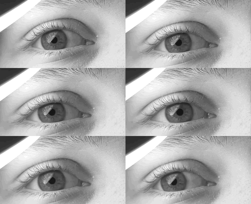
Nystagmus is a condition of involuntary (or voluntary, in some cases) eye movement. Infants can be born with it but more commonly acquire it in infancy or later in life. In many cases it may result in reduced or limited vision. Due to the involuntary movement of the eye, it has been called "dancing eyes". In normal eyesight, while the head rotates about an axis, distant visual images are sustained by rotating eyes in the opposite direction of the respective axis. The semicircular canals in the vestibule of the ear sense angular acceleration, and send signals to the nuclei for eye movement in the brain. From here, a signal is relayed to the extraocular muscles to allow one's gaze to fix on an object as the head moves. Nystagmus occurs when the semicircular canals are stimulated (e.g., by means of the caloric test, or by disease) while the head is stationary. The direction of ocular movement is related to the semicircular canal that is being stimulated. There are two key form ... [...More Info...] [...Related Items...] OR: [Wikipedia] [Google] [Baidu] |
Statoconia
An otolith ( grc-gre, ὠτο-, ' ear + , ', a stone), also called statoconium or otoconium or statolith, is a calcium carbonate structure in the saccule or utricle of the inner ear, specifically in the vestibular system of vertebrates. The saccule and utricle, in turn, together make the ''otolith organs''. These organs are what allows an organism, including humans, to perceive linear acceleration, both horizontally and vertically (gravity). They have been identified in both extinct and extant vertebrates. Counting the annual growth rings on the otoliths is a common technique in estimating the age of fish. Description Endolymphatic infillings such as otoliths are structures in the saccule and utricle of the inner ear, specifically in the vestibular labyrinth of all vertebrates (fish, amphibians, reptiles, mammals and birds). In vertebrates, the saccule and utricle together make the ''otolith organs''. Both statoconia and otoliths are used as gravity, balance, movement, and d ... [...More Info...] [...Related Items...] OR: [Wikipedia] [Google] [Baidu] |
Kinocilium
A kinocilium is a special type of cilium on the apex of hair cells located in the sensory epithelium of the vertebrate inner ear. Anatomy in humans Kinocilia are found on the apical surface of hair cells and are involved in both the morphogenesis of the hair bundle and mechanotransduction. Vibrations (either by movement or sound waves) cause displacement of the hair bundle, resulting in depolarization or hyperpolarization of the hair cell. The depolarization of the hair cells in both instances causes signal transduction via neurotransmitter release. Role in hair bundle morphogenesis Each hair cell has a single, microtubular kinocilium. Before morphogenesis of the hair bundle, the kinocilium is found in the center of the apical surface of the hair cell surrounded by 20-300 microvilli. During hair bundle morphogenesis, the kinocilium moves to the cell periphery dictating hair bundle orientation. As the kinocilium does not move, microvilli surrounding it begin to elongate and form ac ... [...More Info...] [...Related Items...] OR: [Wikipedia] [Google] [Baidu] |
Acoustic Nerve
The cochlear nerve (also auditory nerve or acoustic nerve) is one of two parts of the vestibulocochlear nerve, a cranial nerve present in amniotes, the other part being the vestibular nerve. The cochlear nerve carries auditory sensory information from the cochlea of the inner ear directly to the brain. The other portion of the vestibulocochlear nerve is the vestibular nerve, which carries spatial orientation information to the brain from the semicircular canals, also known as semicircular ducts. Anatomy and connections In terms of anatomy, an auditory nerve fiber is either bipolar or unipolar, with its distal projection being called the peripheral process, and its proximal projection being called the axon; these two projections are also known as the "peripheral axon" and the "central axon", respectively. The peripheral process is sometimes referred to as a dendrite, although that term is somewhat inaccurate. Unlike the typical dendrite, the peripheral process generates and con ... [...More Info...] [...Related Items...] OR: [Wikipedia] [Google] [Baidu] |
Saccular Nerve
The saccular nerve is a nerve which supplies the macula of the saccule The saccule is a bed of sensory cells in the inner ear. It translates head movements into neural impulses for the brain to interpret. The saccule detects linear accelerations and head tilts in the vertical plane. When the head moves verticall .... References External links Ear {{Neuroanatomy-stub ... [...More Info...] [...Related Items...] OR: [Wikipedia] [Google] [Baidu] |
Macula
The macula (/ˈmakjʊlə/) or macula lutea is an oval-shaped pigmented area in the center of the retina of the human eye and in other animals. The macula in humans has a diameter of around and is subdivided into the umbo, foveola, foveal avascular zone, fovea, parafovea, and perifovea areas. The anatomical macula at a size of is much larger than the clinical macula which, at a size of , corresponds to the anatomical fovea. The macula is responsible for the central, high-resolution, color vision that is possible in good light; and this kind of vision is impaired if the macula is damaged, for example in macular degeneration. The clinical macula is seen when viewed from the pupil, as in ophthalmoscopy or retinal photography. The term macula lutea comes from Latin ''macula'', "spot", and ''lutea'', "yellow". Structure The macula is an oval-shaped pigmented area in the center of the retina of the human eye and other animal eyes. Its center is shifted slightly away from the ... [...More Info...] [...Related Items...] OR: [Wikipedia] [Google] [Baidu] |


