|
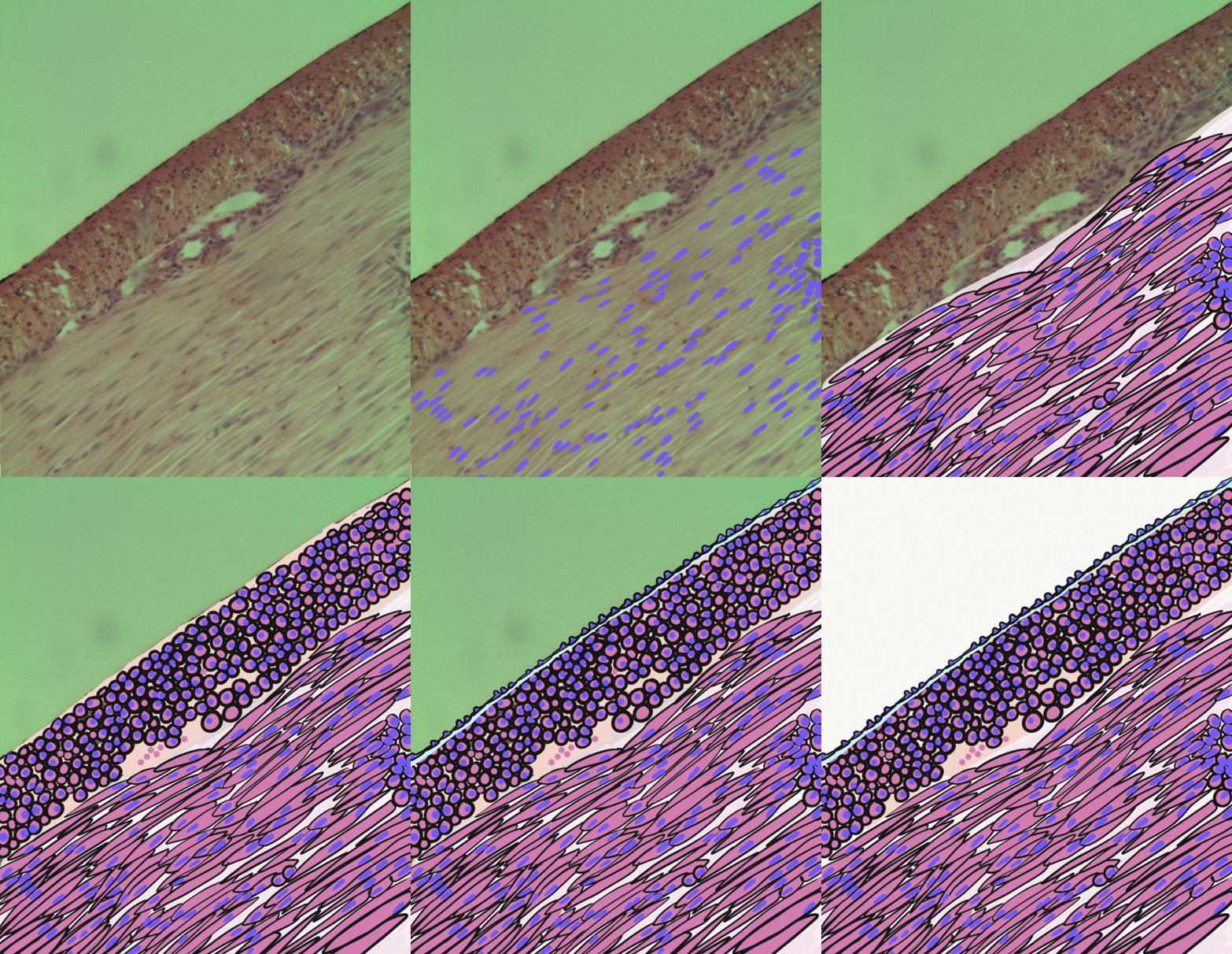
Muscularis Mucosae
The lamina muscularis mucosae (or muscularis mucosae) is a thin layer (lamina) of muscle of the gastrointestinal tract, located outside the lamina propria, and separating it from the submucosa. It is present in a continuous fashion from the esophagus to the upper rectum (the exact nomenclature of the rectum's muscle layers is still being debated). A discontinuous muscularis mucosae–like muscle layer is present in the urinary tract, from the renal pelvis to the bladder; as it is discontinuous, it should not be regarded as a true muscularis mucosae. In the gastrointestinal tract, the term ''mucosa'' or ''mucous membrane'' refers to the combination of epithelium, lamina propria, and (where it occurs) muscularis mucosae.H.G. Burkitt et al., ''Wheater's Functional Histology, 3rd ed.'' The etymology suggests this, since the Latin names translate to "the mucosa's own special layer" (''lamina propria mucosae'') and "muscular layer of the mucosa" (''lamina muscularis mucosae''). The m ... [...More Info...] [...Related Items...] OR: [Wikipedia] [Google] [Baidu] |
Duodenum
The duodenum is the first section of the small intestine in most higher vertebrates, including mammals, reptiles, and birds. In fish, the divisions of the small intestine are not as clear, and the terms anterior intestine or proximal intestine may be used instead of duodenum. In mammals the duodenum may be the principal site for iron absorption. The duodenum precedes the jejunum and ileum and is the shortest part of the small intestine. In humans, the duodenum is a hollow jointed tube about 25–38 cm (10–15 inches) long connecting the stomach to the middle part of the small intestine. It begins with the duodenal bulb and ends at the suspensory muscle of duodenum. Duodenum can be divided into four parts: the first (superior), the second (descending), the third (horizontal) and the fourth (ascending) parts. Structure The duodenum is a C-shaped structure lying adjacent to the stomach. It is divided anatomically into four sections. The first part of the duodenu ... [...More Info...] [...Related Items...] OR: [Wikipedia] [Google] [Baidu] |
Renal Pelvis
The renal pelvis or pelvis of the kidney is the funnel-like dilated part of the ureter in the kidney. It is formed by the covnvergence of the major calyces, acting as a funnel for urine flowing from the major calyces to the ureter. It has a mucous membrane and is covered with transitional epithelium and an underlying lamina propria of loose-to-dense connective tissue. The renal pelvis is situated within the renal sinus alongside the other structures of the renal sinus. The renal pelvis is the location of several kinds of kidney cancer and is affected by infection in pyelonephritis. Clinical significance The renal pelvis is the location of several kinds of kidney cancer and is affected by infection in pyelonephritis. A large "staghorn" kidney stone may block all or part of the renal pelvis. The size of the renal pelvis plays a major role in the grading of hydronephrosis. Normally, the anteroposterior diameter of the renal pelvis is less than 4 mm in fetuses up to 32 wee ... [...More Info...] [...Related Items...] OR: [Wikipedia] [Google] [Baidu] |
Crypt (anatomy)
Crypts are anatomical structures that are narrow but deep invaginations into a larger structure. One common type of anatomical crypt is the Crypts of Lieberkühn. However, it is not the only type: some types of tonsils The tonsils are a set of lymphoid organs facing into the aerodigestive tract, which is known as Waldeyer's tonsillar ring and consists of the adenoid tonsil, two tubal tonsils, two palatine tonsils, and the lingual tonsils. These organs pla ... also have crypts. Because these crypts allow external access to the deep portions of the tonsils, these tonsils are more vulnerable to infection. References External links * - "Lymphoid Tissues and Organs: tonsil" Histology of crypt of tonsil at siumed.edu Anatomy {{Anatomy-stub ... [...More Info...] [...Related Items...] OR: [Wikipedia] [Google] [Baidu] |
Gland
In animals, a gland is a group of cells in an animal's body that synthesizes substances (such as hormones) for release into the bloodstream ( endocrine gland) or into cavities inside the body or its outer surface ( exocrine gland). Structure Development Every gland is formed by an ingrowth from an epithelial surface. This ingrowth may in the beginning possess a tubular structure, but in other instances glands may start as a solid column of cells which subsequently becomes tubulated. As growth proceeds, the column of cells may split or give off offshoots, in which case a compound gland is formed. In many glands, the number of branches is limited, in others (salivary, pancreas) a very large structure is finally formed by repeated growth and sub-division. As a rule, the branches do not unite with one another, but in one instance, the liver, this does occur when a reticulated compound gland is produced. In compound glands the more typical or secretory epithelium is found formi ... [...More Info...] [...Related Items...] OR: [Wikipedia] [Google] [Baidu] |
Smooth Muscle Tissue
Smooth muscle is an involuntary non- striated muscle, so-called because it has no sarcomeres and therefore no striations (''bands'' or ''stripes''). It is divided into two subgroups, single-unit and multiunit smooth muscle. Within single-unit muscle, the whole bundle or sheet of smooth muscle cells contracts as a syncytium. Smooth muscle is found in the walls of hollow organs, including the stomach, intestines, bladder and uterus; in the walls of passageways, such as blood, and lymph vessels, and in the tracts of the respiratory, urinary, and reproductive systems. In the eyes, the ciliary muscles, a type of smooth muscle, dilate and contract the iris and alter the shape of the lens. In the skin, smooth muscle cells such as those of the arrector pili cause hair to stand erect in response to cold temperature or fear. Structure Gross anatomy Smooth muscle is grouped into two types: single-unit smooth muscle, also known as visceral smooth muscle, and multiunit smooth mus ... [...More Info...] [...Related Items...] OR: [Wikipedia] [Google] [Baidu] |
Epithelium
Epithelium or epithelial tissue is one of the four basic types of animal tissue, along with connective tissue, muscle tissue and nervous tissue. It is a thin, continuous, protective layer of compactly packed cells with a little intercellular matrix. Epithelial tissues line the outer surfaces of organs and blood vessels throughout the body, as well as the inner surfaces of cavities in many internal organs. An example is the epidermis, the outermost layer of the skin. There are three principal shapes of epithelial cell: squamous (scaly), columnar, and cuboidal. These can be arranged in a singular layer of cells as simple epithelium, either squamous, columnar, or cuboidal, or in layers of two or more cells deep as stratified (layered), or ''compound'', either squamous, columnar or cuboidal. In some tissues, a layer of columnar cells may appear to be stratified due to the placement of the nuclei. This sort of tissue is called pseudostratified. All glands are made up of epi ... [...More Info...] [...Related Items...] OR: [Wikipedia] [Google] [Baidu] |
Mucous Membrane
A mucous membrane or mucosa is a membrane that lines various cavities in the body of an organism and covers the surface of internal organs. It consists of one or more layers of epithelial cells overlying a layer of loose connective tissue. It is mostly of endodermal origin and is continuous with the skin at body openings such as the eyes, eyelids, ears, inside the nose, inside the mouth, lips, the genital areas, the urethral opening and the anus. Some mucous membranes secrete mucus, a thick protective fluid. The function of the membrane is to stop pathogens and dirt from entering the body and to prevent bodily tissues from becoming dehydrated. Structure The mucosa is composed of one or more layers of epithelial cells that secrete mucus, and an underlying lamina propria of loose connective tissue. The type of cells and type of mucus secreted vary from organ to organ and each can differ along a given tract. Mucous membranes line the digestive, respiratory and reproduct ... [...More Info...] [...Related Items...] OR: [Wikipedia] [Google] [Baidu] |
Urinary Bladder
The urinary bladder, or simply bladder, is a hollow organ in humans and other vertebrates that stores urine from the kidneys before disposal by urination. In humans the bladder is a distensible organ that sits on the pelvic floor. Urine enters the bladder via the ureters and exits via the urethra. The typical adult human bladder will hold between 300 and (10.14 and ) before the urge to empty occurs, but can hold considerably more. The Latin phrase for "urinary bladder" is ''vesica urinaria'', and the term ''vesical'' or prefix ''vesico -'' appear in connection with associated structures such as vesical veins. The modern Latin word for "bladder" – ''cystis'' – appears in associated terms such as cystitis (inflammation of the bladder). Structure In humans, the bladder is a hollow muscular organ situated at the base of the pelvis. In gross anatomy, the bladder can be divided into a broad , a body, an apex, and a neck. The apex (also called the vertex) is directed fo ... [...More Info...] [...Related Items...] OR: [Wikipedia] [Google] [Baidu] |
Urinary System
The urinary system, also known as the urinary tract or renal system, consists of the kidneys, ureters, bladder, and the urethra. The purpose of the urinary system is to eliminate waste from the body, regulate blood volume and blood pressure, control levels of electrolytes and metabolites, and regulate blood pH. The urinary tract is the body's drainage system for the eventual removal of urine. The kidneys have an extensive blood supply via the renal arteries which leave the kidneys via the renal vein. Each kidney consists of functional units called nephrons. Following filtration of blood and further processing, wastes (in the form of urine) exit the kidney via the ureters, tubes made of smooth muscle fibres that propel urine towards the urinary bladder, where it is stored and subsequently expelled from the body by urination ( voiding). The female and male urinary system are very similar, differing only in the length of the urethra. Urine is formed in the kidneys through ... [...More Info...] [...Related Items...] OR: [Wikipedia] [Google] [Baidu] |
Lamina
Lamina may refer to: Science and technology * Planar lamina, a two-dimensional planar closed surface with mass and density, in mathematics * Laminar flow, (or streamline flow) occurs when a fluid flows in parallel layers, with no disruption between the layers * Lamina (algae), a structure in seaweeds * Lamina (anatomy), with several meanings * Lamina (leaf), the flat part of a leaf, an organ of a plant * Lamina, the largest petal of a floret in an aster family flowerhead: see * ''Lamina'' (spider), a genus in the family Toxopidae * Lamina (neuropil), the most peripheral neuropil of the insect visual system *Nuclear lamina, another structure of a living cell *Basal lamina, a structure of a living cell * Lamina propria, the connective part of the mucous * Lamina of the vertebral arch *Lamination (geology), a layering structure in sedimentary rocks usually less than 1 cm in thickness * Laminae, a part of the horse hoof * Laminae, another name for the core lamiids, a clade in b ... [...More Info...] [...Related Items...] OR: [Wikipedia] [Google] [Baidu] |
Rectum
The rectum is the final straight portion of the large intestine in humans and some other mammals, and the gut in others. The adult human rectum is about long, and begins at the rectosigmoid junction (the end of the sigmoid colon) at the level of the third sacral vertebra or the sacral promontory depending upon what definition is used. Its diameter is similar to that of the sigmoid colon at its commencement, but it is dilated near its termination, forming the rectal ampulla. It terminates at the level of the anorectal ring (the level of the puborectalis sling) or the dentate line, again depending upon which definition is used. In humans, the rectum is followed by the anal canal which is about long, before the gastrointestinal tract terminates at the anal verge. The word rectum comes from the Latin '' rectum intestinum'', meaning ''straight intestine''. Structure The rectum is a part of the lower gastrointestinal tract. The rectum is a continuation of the sigmoi ... [...More Info...] [...Related Items...] OR: [Wikipedia] [Google] [Baidu] |
Esophagus
The esophagus (American English) or oesophagus (British English; both ), non-technically known also as the food pipe or gullet, is an organ in vertebrates through which food passes, aided by peristaltic contractions, from the pharynx to the stomach. The esophagus is a fibromuscular tube, about long in adults, that travels behind the trachea and heart, passes through the diaphragm, and empties into the uppermost region of the stomach. During swallowing, the epiglottis tilts backwards to prevent food from going down the larynx and lungs. The word ''oesophagus'' is from Ancient Greek οἰσοφάγος (oisophágos), from οἴσω (oísō), future form of φέρω (phérō, “I carry”) + ἔφαγον (éphagon, “I ate”). The wall of the esophagus from the lumen outwards consists of mucosa, submucosa (connective tissue), layers of muscle fibers between layers of fibrous tissue, and an outer layer of connective tissue. The mucosa is a stratified squamous epith ... [...More Info...] [...Related Items...] OR: [Wikipedia] [Google] [Baidu] |


