|
Muscles Of Respiration
The muscles of respiration are the muscles that contribute to inhalation and exhalation, by aiding in the expansion and contraction of the thoracic cavity. The diaphragm and, to a lesser extent, the intercostal muscles drive respiration during quiet breathing. The elasticity of these muscles is crucial to the health of the respiratory system and to maximize its functional capabilities. Diaphragm The diaphragm is the major muscle responsible for breathing. It is a thin, dome-shaped muscle that separates the abdominal cavity from the thoracic cavity. During inhalation, the diaphragm contracts, so that its center moves caudally (downward) and its edges move cranially (upward). This compresses the abdominal cavity, raises the ribs upward and outward and thus expands the thoracic cavity. This expansion draws air into the lungs. When the diaphragm relaxes, elastic recoil of the lungs causes the thoracic cavity to contract, forcing air out of the lungs, and returning to its dome-s ... [...More Info...] [...Related Items...] OR: [Wikipedia] [Google] [Baidu] |
Muscle
Skeletal muscles (commonly referred to as muscles) are organs of the vertebrate muscular system and typically are attached by tendons to bones of a skeleton. The muscle cells of skeletal muscles are much longer than in the other types of muscle tissue, and are often known as muscle fibers. The muscle tissue of a skeletal muscle is striated – having a striped appearance due to the arrangement of the sarcomeres. Skeletal muscles are voluntary muscles under the control of the somatic nervous system. The other types of muscle are cardiac muscle which is also striated and smooth muscle which is non-striated; both of these types of muscle tissue are classified as involuntary, or, under the control of the autonomic nervous system. A skeletal muscle contains multiple fascicles – bundles of muscle fibers. Each individual fiber, and each muscle is surrounded by a type of connective tissue layer of fascia. Muscle fibers are formed from the fusion of developmental myoblasts ... [...More Info...] [...Related Items...] OR: [Wikipedia] [Google] [Baidu] |
Respiratory Distress
Shortness of breath (SOB), also medically known as dyspnea (in AmE) or dyspnoea (in BrE), is an uncomfortable feeling of not being able to breathe well enough. The American Thoracic Society defines it as "a subjective experience of breathing discomfort that consists of qualitatively distinct sensations that vary in intensity", and recommends evaluating dyspnea by assessing the intensity of its distinct sensations, the degree of distress and discomfort involved, and its burden or impact on the patient's activities of daily living. Distinct sensations include effort/work to breathe, chest tightness or pain, and "air hunger" (the feeling of not enough oxygen). The tripod position is often assumed to be a sign. Dyspnea is a normal symptom of heavy physical exertion but becomes pathological if it occurs in unexpected situations, when resting or during light exertion. In 85% of cases it is due to asthma, pneumonia, cardiac ischemia, interstitial lung disease, congestive heart failu ... [...More Info...] [...Related Items...] OR: [Wikipedia] [Google] [Baidu] |
Levatores Costarum Muscles
The ''Levatores costarum'' (), twelve in number on either side, are small tendinous and fleshy bundles, which arise from the ends of the transverse processes of the seventh cervical and upper eleven thoracic vertebrae They pass obliquely downward and laterally, like the fibers of the Intercostales externi, and each is inserted into the outer surface of the rib immediately below the vertebra from which it takes origin, between the tubercle and the angle (''Levatores costarum breves''). Each of the four lower muscles divides into two fasciculi, one of which is inserted as above described; the other passes down to the second rib below its origin (''Levatores costarum longi''). They have a role in forceful inspiration. See also * Iliocostalis * Interspinales muscles * Intertransversarii muscle * Longissimus * Spinalis The spinalis is a portion of the erector spinae, a bundle of muscles and tendons, located nearest to the spine. It is divided into three parts: Spinalis dorsi ... [...More Info...] [...Related Items...] OR: [Wikipedia] [Google] [Baidu] |
Serratus Posterior Inferior Muscle
The serratus posterior inferior muscle, also known as the posterior serratus muscle, is a muscle of the human body. Structure The muscle is situated at the junction of the thoracic and lumbar regions. It has an irregularly quadrilateral form, broader than the serratus posterior superior muscle, and separated from it by a wide interval. It arises by a thin aponeurosis from the spinous processes of the lower two thoracic and upper two or three lumbar vertebrae. Passing obliquely upward and lateralward, it becomes fleshy, and divides into four flat digitations. These are inserted into the inferior borders of the lower four ribs, a little beyond their angles. The thin aponeurosis of origin is intimately blended with the thoracolumbar fascia, and aponeurosis of the latissimus dorsi muscle. Function The serratus posterior inferior draws the lower ribs backward and downward to assist in rotation and extension of the trunk. This movement of the ribs may also contribute to inha ... [...More Info...] [...Related Items...] OR: [Wikipedia] [Google] [Baidu] |
Serratus Posterior Superior Muscle
The serratus posterior superior muscle is a thin, quadrilateral muscle. It is situated at the upper back part of the thorax, deep to the rhomboid muscles. Structure The serratus posterior superior muscle arises by an aponeurosis from the lower part of the nuchal ligament, from the spinous processes of C7, T1, T2, and sometimes T3, and from the supraspinal ligament. It is inserted, by four fleshy digitations into the upper borders of the second, third, fourth, and fifth ribs past the angle of the rib. Function The serratus posterior superior muscle elevates the second to fifth ribs. This aids deep respiration. Additional images File:Serratus posterior superior muscle animation small.gif, Position of serratus posterior superior muscle (shown in red). File:Serratus posterior superior.jpg, Serratus posterior superior muscles are labeled at center left and center right. See also * Serratus anterior muscle * Serratus posterior inferior muscle The serratus poster ... [...More Info...] [...Related Items...] OR: [Wikipedia] [Google] [Baidu] |
Quadratus Lumborum Muscle
The quadratus lumborum muscle, informally called the ''QL'', is a paired muscle of the left and right posterior abdominal wall. It is the deepest abdominal muscle, and commonly referred to as a back muscle. Each is irregular and quadrilateral in shape. The quadratus lumborum muscles originate from the wings of the ilium; their insertions are on the transverse processes of the upper four lumbar vertebrae plus the lower posterior border of the twelfth rib. Contraction of one of the pair of muscles causes '' lateral flexion'' of the lumbar spine, ''elevation'' of the pelvis, or both. Contraction of both causes ''extension'' of the lumbar spine. A disorder of the quadratus lumborum muscles is pain due to muscle fatigue from constant contraction due to prolonged sitting, such as at a computer or in a car.Core Topics in Pain, p. 131, Anita Holdcraft and Sian Jaggar, 2005. Kyphosis and weak gluteal muscles can also contribute to the likelihood of quadratus lumborum pain. Structure Th ... [...More Info...] [...Related Items...] OR: [Wikipedia] [Google] [Baidu] |
Iliocostalis
Iliocostalis muscle is the muscle immediately lateral to the longissimus that is the nearest to the furrow that separates the epaxial muscles from the hypaxial. It lies very deep to the fleshy portion of the serratus posterior muscle. It laterally flexes the vertebral column to the same side. Structure Iliocostalis muscle has a common origin from the iliac crest, the sacrum, the thoracolumbar fascia, and the spinous processes of the vertebrae from T11 to L5. Iliocostalis cervicis (cervicalis ascendens) arises from the angles of the third, fourth, fifth, and sixth ribs, and is inserted into the posterior tubercles of the transverse processes of the fourth, fifth, and sixth cervical vertebrae. Iliocostalis thoracis (musculus accessorius; iliocostalis thoracis) arises by flattened tendons from the upper borders of the angles of the lower six ribs medial to the tendons of insertion of the iliocostalis lumborum; these become muscular, and are inserted into the upper borders o ... [...More Info...] [...Related Items...] OR: [Wikipedia] [Google] [Baidu] |
Erector Spinae Muscles
The erector spinae ( ) or spinal erectors is a set of muscles that straighten and rotate the back. The spinal erectors work together with the glutes ( gluteus maximus, gluteus medius and gluteus minimus) to maintain stable posture standing or sitting. Structure The erector spinae is not just one muscle, but a group of muscles and tendons which run more or less the length of the spine on the left and the right, from the sacrum, or sacral region, and hips to the base of the skull. They are also known as the sacrospinalis group of muscles. These muscles lie on either side of the spinous processes of the vertebrae and extend throughout the lumbar, thoracic, and cervical regions. The erector spinae is covered in the lumbar and thoracic regions by the thoracolumbar fascia, and in the cervical region by the nuchal ligament. This large muscular and tendinous mass varies in size and structure at different parts of the vertebral column. In the sacral region, it is narrow and ... [...More Info...] [...Related Items...] OR: [Wikipedia] [Google] [Baidu] |
Latissimus Dorsi Muscle
The latissimus dorsi () is a large, flat muscle on the back that stretches to the sides, behind the arm, and is partly covered by the trapezius on the back near the midline. The word latissimus dorsi (plural: ''latissimi dorsorum'') comes from Latin and means "broadest uscleof the back", from "latissimus" ( la, broadest)' and "dorsum" ( la, back). The pair of muscles are commonly known as "lats", especially among bodybuilders. The latissimus dorsi is the largest muscle in the upper body. The latissimus dorsi is responsible for extension, adduction, transverse extension also known as horizontal abduction (or horizontal extension), flexion from an extended position, and (medial) internal rotation of the shoulder joint. It also has a synergistic role in extension and lateral flexion of the lumbar spine. Due to bypassing the scapulothoracic joints and attaching directly to the spine, the actions the latissimi dorsi have on moving the arms can also influence the movement of the sc ... [...More Info...] [...Related Items...] OR: [Wikipedia] [Google] [Baidu] |
Trapezius
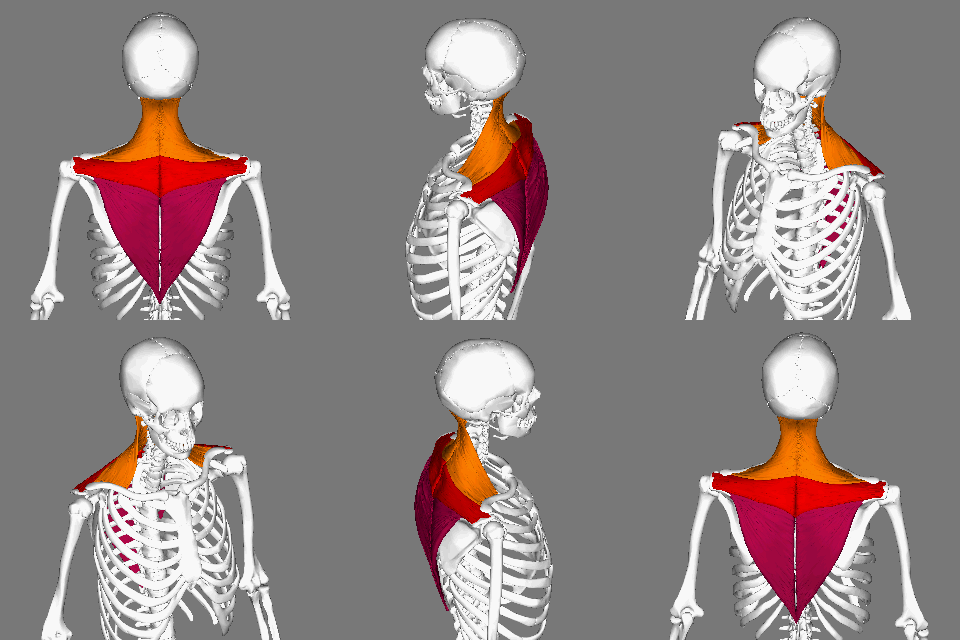
The trapezius is a large paired trapezoid-shaped surface muscle that extends longitudinally from the occipital bone to the lower thoracic vertebrae of the spine and laterally to the spine of the scapula. It moves the scapula and supports the arm. The trapezius has three functional parts: an upper (descending) part which supports the weight of the arm; a middle region (transverse), which retracts the scapula; and a lower (ascending) part which medially rotates and depresses the scapula. Name and history The trapezius muscle resembles a trapezium, also known as a trapezoid, or diamond-shaped quadrilateral. The word "spinotrapezius" refers to the human trapezius, although it is not commonly used in modern texts. In other mammals, it refers to a portion of the analogous muscle. Similarly, the term "tri-axle back plate" was historically used to describe the trapezius muscle. Structure The ''superior'' or ''upper'' (or descending) fibers of the trapezius originate from the ... [...More Info...] [...Related Items...] OR: [Wikipedia] [Google] [Baidu] |
Pectoralis Minor
Pectoralis minor muscle () is a thin, triangular muscle, situated at the upper part of the chest, beneath the pectoralis major in the human body. Structure Attachments Pectoralis minor muscle arises from the upper margins and outer surfaces of the third, fourth, and fifth ribs, near their costal cartilages and from the aponeuroses covering the intercostalis. The fibers pass superior and lateral and converge to form a flat tendon. This tendon inserts onto the medial border and upper surface of the coracoid process of the scapula. Relations Pectoralis minor muscle forms part of the anterior wall of the axilla. It is covered anteriorly (superficially) by the clavipectoral fascia. The medial pectoral nerve pierces the pectoralis minor and the clavipectoral fascia. In attaching to the coracoid process, the pectoralis minor forms a 'bridge' - structures passing into the upper limb from the thorax will pass directly underneath.http://www.teachmeanatomy.com/muscles-of-the-pecto ... [...More Info...] [...Related Items...] OR: [Wikipedia] [Google] [Baidu] |
Pectoralis Major
The pectoralis major () is a thick, fan-shaped or triangular convergent muscle, situated at the chest of the human body. It makes up the bulk of the chest muscles and lies under the breast. Beneath the pectoralis major is the pectoralis minor, a thin, triangular muscle. The pectoralis major's primary functions are flexion, adduction, and internal rotation of the humerus. The pectoral major may colloquially be referred to as "pecs", "pectoral muscle", or "chest muscle", because it is the largest and most superficial muscle in the chest area. Structure It arises from the anterior surface of the sternal half of the clavicle from breadth of the half of the anterior surface of the sternum, as low down as the attachment of the cartilage of the sixth or seventh rib; from the cartilages of all the true ribs, with the exception, frequently, of the first or seventh, and from the aponeurosis of the abdominal external oblique muscle. From this extensive origin the fibers converge towa ... [...More Info...] [...Related Items...] OR: [Wikipedia] [Google] [Baidu] |


