|
Multipolar Migration
The development of the nervous system, or neural development (neurodevelopment), refers to the processes that generate, shape, and reshape the nervous system of animals, from the earliest stages of embryonic development to adulthood. The field of neural development draws on both neuroscience and developmental biology to describe and provide insight into the cellular and molecular mechanisms by which complex nervous systems develop, from nematodes and fruit flies to mammals. Defects in neural development can lead to malformations such as holoprosencephaly, and a wide variety of neurological disorders including limb paresis and paralysis, balance and vision disorders, and seizures, and in humans other disorders such as Rett syndrome, Down syndrome and intellectual disability. Overview of vertebrate brain development The vertebrate central nervous system (CNS) is derived from the ectoderm—the outermost germ layer of the embryo. A part of the dorsal ectoderm becomes specifie ... [...More Info...] [...Related Items...] OR: [Wikipedia] [Google] [Baidu] |
Nervous System
In biology, the nervous system is the highly complex part of an animal that coordinates its actions and sensory information by transmitting signals to and from different parts of its body. The nervous system detects environmental changes that impact the body, then works in tandem with the endocrine system to respond to such events. Nervous tissue first arose in wormlike organisms about 550 to 600 million years ago. In vertebrates it consists of two main parts, the central nervous system (CNS) and the peripheral nervous system (PNS). The CNS consists of the brain and spinal cord. The PNS consists mainly of nerves, which are enclosed bundles of the long fibers or axons, that connect the CNS to every other part of the body. Nerves that transmit signals from the brain are called motor nerves or '' efferent'' nerves, while those nerves that transmit information from the body to the CNS are called sensory nerves or '' afferent''. Spinal nerves are mixed nerves that serve both fu ... [...More Info...] [...Related Items...] OR: [Wikipedia] [Google] [Baidu] |
Vertebrate
Vertebrates () comprise all animal taxa within the subphylum Vertebrata () ( chordates with backbones), including all mammals, birds, reptiles, amphibians, and fish. Vertebrates represent the overwhelming majority of the phylum Chordata, with currently about 69,963 species described. Vertebrates comprise such groups as the following: * jawless fish, which include hagfish and lampreys * jawed vertebrates, which include: ** cartilaginous fish (sharks, rays, and ratfish) ** bony vertebrates, which include: *** ray-fins (the majority of living bony fish) *** lobe-fins, which include: **** coelacanths and lungfish **** tetrapods (limbed vertebrates) Extant vertebrates range in size from the frog species ''Paedophryne amauensis'', at as little as , to the blue whale, at up to . Vertebrates make up less than five percent of all described animal species; the rest are invertebrates, which lack vertebral columns. The vertebrates traditionally include the hagfish, which do no ... [...More Info...] [...Related Items...] OR: [Wikipedia] [Google] [Baidu] |
Hindbrain
The hindbrain or rhombencephalon or lower brain is a developmental categorization of portions of the central nervous system in vertebrates. It includes the medulla, pons, and cerebellum. Together they support vital bodily processes. Metencephalon Rhombomeres Rh3-Rh1 form the metencephalon. The metencephalon is composed of the pons and the cerebellum; it contains: * a portion of the fourth (IV) ventricle, * the trigeminal nerve (CN V), * abducens nerve (CN VI), * facial nerve (CN VII), * and a portion of the vestibulocochlear nerve (CN VIII). Myelencephalon Rhombomeres Rh8-Rh4 form the myelencephalon. The myelencephalon forms the medulla oblongata in the adult brain; it contains: * a portion of the fourth ventricle, * the glossopharyngeal nerve (CN IX), * vagus nerve (CN X), * accessory nerve (CN XI), * hypoglossal nerve (CN XII), * and a portion of the vestibulocochlear nerve (CN VIII). Evolution The hindbrain is homologous to a part of the arthropod brain known as the sub- ... [...More Info...] [...Related Items...] OR: [Wikipedia] [Google] [Baidu] |
Mesencephalon
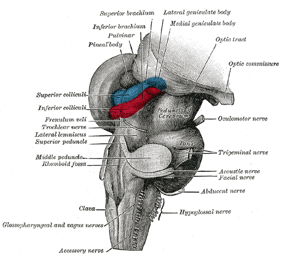
The midbrain or mesencephalon is the forward-most portion of the brainstem and is associated with vision, hearing, motor control, sleep and wakefulness, arousal (alertness), and temperature regulation. The name comes from the Greek ''mesos'', "middle", and ''enkephalos'', "brain". Structure The principal regions of the midbrain are the tectum, the cerebral aqueduct, tegmentum, and the cerebral peduncles. Rostrally the midbrain adjoins the diencephalon (thalamus, hypothalamus, etc.), while caudally it adjoins the hindbrain (pons, medulla and cerebellum). In the rostral direction, the midbrain noticeably splays laterally. Sectioning of the midbrain is usually performed axially, at one of two levels – that of the superior colliculi, or that of the inferior colliculi. One common technique for remembering the structures of the midbrain involves visualizing these cross-sections (especially at the level of the superior colliculi) as the upside-down face of a bea ... [...More Info...] [...Related Items...] OR: [Wikipedia] [Google] [Baidu] |
Midbrain
The midbrain or mesencephalon is the forward-most portion of the brainstem and is associated with vision, hearing, motor control, sleep and wakefulness, arousal (alertness), and temperature regulation. The name comes from the Greek ''mesos'', "middle", and ''enkephalos'', "brain". Structure The principal regions of the midbrain are the tectum, the cerebral aqueduct, tegmentum, and the cerebral peduncles. Rostrally the midbrain adjoins the diencephalon (thalamus, hypothalamus, etc.), while caudally it adjoins the hindbrain (pons, medulla and cerebellum). In the rostral direction, the midbrain noticeably splays laterally. Sectioning of the midbrain is usually performed axially, at one of two levels – that of the superior colliculi, or that of the inferior colliculi. One common technique for remembering the structures of the midbrain involves visualizing these cross-sections (especially at the level of the superior colliculi) as the upside-down face of a be ... [...More Info...] [...Related Items...] OR: [Wikipedia] [Google] [Baidu] |
Prosencephalon
In the anatomy of the brain of vertebrates, the forebrain or prosencephalon is the Anatomical terms of location#Directional terms, rostral (forward-most) portion of the brain. The forebrain (prosencephalon), the midbrain (mesencephalon), and hindbrain (rhombencephalon) are the three Brain vesicle, primary brain vesicles during the early development of the nervous system. The forebrain controls body temperature, reproductive functions, eating, sleeping, and the display of emotions. At the five-vesicle stage, the forebrain separates into the diencephalon (thalamus, hypothalamus, subthalamus, and epithalamus) and the telencephalon which develops into the cerebrum. The cerebrum consists of the cerebral cortex, underlying white matter, and the basal ganglia. In humans, by 5 weeks in utero it is visible as a single portion toward the front of the fetus. At 8 weeks in utero, the forebrain splits into the left and right cerebral hemispheres. When the embryonic forebrain fails to divide ... [...More Info...] [...Related Items...] OR: [Wikipedia] [Google] [Baidu] |
Forebrain
In the anatomy of the brain of vertebrates, the forebrain or prosencephalon is the Anatomical terms of location#Directional terms, rostral (forward-most) portion of the brain. The forebrain (prosencephalon), the midbrain (mesencephalon), and hindbrain (rhombencephalon) are the three Brain vesicle, primary brain vesicles during the early development of the nervous system. The forebrain controls body temperature, reproductive functions, eating, sleeping, and the display of emotions. At the five-vesicle stage, the forebrain separates into the diencephalon (thalamus, hypothalamus, subthalamus, and epithalamus) and the telencephalon which develops into the cerebrum. The cerebrum consists of the cerebral cortex, underlying white matter, and the basal ganglia. In humans, by 5 weeks in utero it is visible as a single portion toward the front of the fetus. At 8 weeks in utero, the forebrain splits into the left and right cerebral hemispheres. When the embryonic forebrain fails to divide ... [...More Info...] [...Related Items...] OR: [Wikipedia] [Google] [Baidu] |
Primary Brain Vesicles
Brain vesicles are the bulge-like features of the early development of the neural tube in vertebrates. Vesicle formation begins shortly after anterior neural tube closure at about embryonic day 9.0 in the mouse and the fourth and fifth gestational week in human development. In zebrafish and chicken embryos, brain vesicles form by about 24 hours and 48 hours post-conception, respectively. Initially there are three primary brain vesicles: prosencephalon, mesencephalon, and rhombencephalon. These develop into five secondary brain vesicles – the prosencephalon is subdivided into the telencephalon and diencephalon, and the rhombencephalon into the metencephalon and myelencephalon. During these early vesicle stages, the walls of the neural tube contain neural stem cells in a region called the neuroepithelium or ventricular zone. These neural stem cells divide rapidly, driving growth of the early brain, but later, these stem cells begin to generate neurons through the process of neur ... [...More Info...] [...Related Items...] OR: [Wikipedia] [Google] [Baidu] |
Neural Tube
In the developing chordate (including vertebrates), the neural tube is the embryonic precursor to the central nervous system, which is made up of the brain and spinal cord. The neural groove gradually deepens as the neural fold become elevated, and ultimately the folds meet and coalesce in the middle line and convert the groove into the closed neural tube. In humans, neural tube closure usually occurs by the fourth week of pregnancy (the 28th day after conception). The ectodermal wall of the tube forms the rudiment of the nervous system. The centre of the tube is the ''neural canal''.It is an important structure for the development of fetus's brain and spine Development The neural tube develops in two ways: primary neurulation and secondary neurulation. Primary neurulation divides the ectoderm into three cell types: * The internally located neural tube * The externally located epidermis * The neural crest cells, which develop in the region between the neural tube and epider ... [...More Info...] [...Related Items...] OR: [Wikipedia] [Google] [Baidu] |
Neural Groove
The neural groove is a shallow median groove of the neural plate between the neural folds of an embryo. The neural plate is a thick sheet of ectoderm surrounded on either side by the neural folds, two longitudinal ridges in front of the primitive streak of the developing embryo.. The groove gradually deepens as the neural folds become elevated, and ultimately the folds meet and coalesce in the middle line and convert the groove into a closed tube, the neural tube or canal, the ectodermal wall of which forms the rudiment of the nervous system. After the coalescence of the neural folds over the anterior end of the primitive streak, the blastopore no longer opens on the surface but into the closed canal of the neural tube, and thus a transitory communication, the neurenteric canal, is established between the neural tube and the primitive digestive tube. The coalescence of the neural folds occurs first in the region of the hind-brain, and from there extends forward and backward; towa ... [...More Info...] [...Related Items...] OR: [Wikipedia] [Google] [Baidu] |
Neural Plate
The neural plate is a key developmental structure that serves as the basis for the nervous system. Cranial to the primitive node of the embryonic primitive streak, ectodermal tissue thickens and flattens to become the neural plate. The region anterior to the primitive node can be generally referred to as the neural plate. Cells take on a columnar appearance in the process as they continue to lengthen and narrow. The ends of the neural plate, known as the neural folds, push the ends of the plate up and together, folding into the neural tube, a structure critical to brain and spinal cord development. This process as a whole is termed primary neurulation. Signaling proteins are also important in neural plate development, and aid in differentiating the tissue destined to become the neural plate. Examples of such proteins include bone morphogenetic proteins and cadherins. Expression of these proteins is essential to neural plate folding and subsequent neural tube formation. Involvem ... [...More Info...] [...Related Items...] OR: [Wikipedia] [Google] [Baidu] |
Neuroectoderm
Neuroectoderm (or neural ectoderm or neural tube epithelium) consists of cells derived from ectoderm. Formation of the neuroectoderm is first step in the development of the nervous system. The neuroectoderm receives bone morphogenetic protein-inhibiting signals from proteins such as noggin, which leads to the development of the nervous system from this tissue. Histologically, these cells are classified as pseudostratified columnar cells. After recruitment from the ectoderm, the neuroectoderm undergoes three stages of development: transformation into the neural plate, transformation into the neural groove (with associated neural folds), and transformation into the neural tube. After formation of the tube, the brain forms into three sections; the hindbrain, the midbrain, and the forebrain. The types of neuroectoderm include: *Neural crest ** pigment cells in the skin **ganglia of the autonomic nervous system **dorsal root ganglia. **facial cartilage ** aorticopulmonary septum of ... [...More Info...] [...Related Items...] OR: [Wikipedia] [Google] [Baidu] |


