|
Mir-133 MicroRNA Precursor Family
mir-133 is a type of non-coding RNA called a microRNA that was first experimentally characterised in mice. Homologues have since been discovered in several other species including invertebrates such as the fruitfly ''Drosophila melanogaster''. Each species often encodes multiple microRNAs with identical or similar mature sequence. For example, in the human genome there are three known miR-133 genes: miR-133a-1, miR-133a-2 and miR-133b found on chromosomes 18, 20 and 6 respectively. The mature sequence is excised from the 3' arm of the hairpin. miR-133 is expressed in muscle tissue and appears to repress the expression of non-muscle genes. Regulation It is proposed that Insulin activates the translocation of SREBP-1c (BHLH) active form from the endoplasmic reticulum (ER) to the nucleus and, concomitantly, induces SREPB-1c expression via PI3K signaling pathway. SREBP-1c mediates MEF2C downregulation through a mechanism that remains to be determined. As a consequence of lowe ... [...More Info...] [...Related Items...] OR: [Wikipedia] [Google] [Baidu] |
Secondary Structure
Protein secondary structure is the three dimensional conformational isomerism, form of ''local segments'' of proteins. The two most common Protein structure#Secondary structure, secondary structural elements are alpha helix, alpha helices and beta sheets, though beta turns and omega loops occur as well. Secondary structure elements typically spontaneously form as an intermediate before the protein protein folding, folds into its three dimensional protein tertiary structure, tertiary structure. Secondary structure is formally defined by the pattern of hydrogen bonds between the Amine, amino hydrogen and carboxyl oxygen atoms in the peptide backbone chain, backbone. Secondary structure may alternatively be defined based on the regular pattern of backbone Dihedral angle#Dihedral angles of proteins, dihedral angles in a particular region of the Ramachandran plot regardless of whether it has the correct hydrogen bonds. The concept of secondary structure was first introduced by Kaj Ulrik ... [...More Info...] [...Related Items...] OR: [Wikipedia] [Google] [Baidu] |
MEF2C
Myocyte-specific enhancer factor 2C also known as MADS box transcription enhancer factor 2, polypeptide C is a protein that in humans is encoded by the ''MEF2C'' gene. MEF2C is a transcription factor in the Mef2 family. Genomics The gene is located at 5q14.3 on the minus (Crick) strand and is 200,723 bases in length. The encoded protein has 473 amino acids with a predicted molecular weight of 51.221 kiloDaltons. Three isoforms have been identified. Several post translational modifications have been identified including phosphorylation on serine-59 and serine-396, sumoylation on lysine-391, acetylation on lysine-4 and proteolytic cleavage. Interactions MEF2C has been shown to interact with: * EP300, * HDAC4, HDAC7, HDAC9, * MAPK7, * SOX18 * SP1, and * TEAD1. * SETD1A Biological significance This gene is involved in cardiac morphogenesis and myogenesis and vascular development. It may also be involved in neurogenesis and in the development of cortical architecture. Mi ... [...More Info...] [...Related Items...] OR: [Wikipedia] [Google] [Baidu] |
Amacrine Cell
Amacrine cells are interneurons in the retina. They are named from the Greek roots ''a–'' ("non"), ''makr–'' ("long") and ''in–'' ("fiber"), because of their short neuronal processes. Amacrine cells are inhibitory neurons, and they project their dendritic arbors onto the inner plexiform layer (IPL), they interact with retinal ganglion cells and/or bipolar cells. Structure Amacrine cells operate at inner plexiform layer (IPL), the second synaptic retinal layer where bipolar cells and retinal ganglion cells form synapses. There are at least 33 different subtypes of amacrine cells based just on their dendrite morphology and stratification. Like horizontal cells, amacrine cells work laterally, but whereas horizontal cells are connected to the output of rod and cone cells, amacrine cells affect the output from bipolar cells, and are often more specialized. Each type of amacrine cell releases one or several neurotransmitters where it connects with other cells. They are often ... [...More Info...] [...Related Items...] OR: [Wikipedia] [Google] [Baidu] |
Runx2
Runt-related transcription factor 2 (RUNX2) also known as core-binding factor subunit alpha-1 (CBF-alpha-1) is a protein that in humans is encoded by the ''RUNX2'' gene. RUNX2 is a key transcription factor associated with osteoblast differentiation. It has also been suggested that Runx2 plays a cell proliferation regulatory role in cell cycle entry and exit in osteoblasts, as well as endothelial cells. Runx2 suppresses pre-osteoblast proliferation by affecting cell cycle progression in the G1 phase. In osteoblasts, the levels of Runx2 is highest in G1 phase and is lowest in S, G2, and M. The comprehensive cell cycle regulatory mechanisms that Runx2 may play are still unknown, although it is generally accepted that the varying activity and levels of Runx2 throughout the cell cycle contribute to cell cycle entry and exit, as well as cell cycle progression. These functions are especially important when discussing bone cancer, particularly osteosarcoma development, that can be a ... [...More Info...] [...Related Items...] OR: [Wikipedia] [Google] [Baidu] |
Bone Morphogenetic Protein 2
Bone morphogenetic protein 2 or BMP-2 belongs to the TGF-β superfamily of proteins. Function BMP-2 like other bone morphogenetic proteins, plays an important role in the development of bone and cartilage. It is involved in the hedgehog pathway, TGF beta signaling pathway, and in cytokine-cytokine receptor interaction. It is also involved in cardiac cell differentiation and epithelial to mesenchymal transition. Like many other proteins from the BMP family, BMP-2 has been demonstrated to potently induce osteoblast differentiation in a variety of cell types. BMP-2 may be involved in white adipogenesis and may have metabolic effects. Interactions Bone morphogenetic protein 2 has been shown to interact with BMPR1A. Clinical use and complications Bone morphogenetic protein 2 is shown to stimulate the production of bone. Recombinant human protein (rhBMP-2) is currently available for orthopaedic usage in the United States. Implantation of BMP-2 is performed using a vari ... [...More Info...] [...Related Items...] OR: [Wikipedia] [Google] [Baidu] |
HCN2
Potassium/sodium hyperpolarization-activated cyclic nucleotide-gated ion channel 2 is a protein that in humans is encoded by the ''HCN2'' gene. Interactions HCN2 has been shown to interact with HCN1 and HCN4. Function The function of the channel is not known although its activation by hyperpolarization alludes to the funny channels in the sinoatrial node of the heart (which form the basis of spontaneous generation of electrical rhythm). These channels have recently been associated with chronic pain and blocking the gene is associated with resolution of neuropathic episodes of pain. See also * Cyclic nucleotide-gated ion channel Cycle, cycles, or cyclic may refer to: Anthropology and social sciences * Cyclic history, a theory of history * Cyclical theory, a theory of American political history associated with Arthur Schlesinger, Sr. * Social cycle, various cycles in soc ... References Further reading * * * * * * * * * * * * * * External link ... [...More Info...] [...Related Items...] OR: [Wikipedia] [Google] [Baidu] |
3' UTR
In molecular genetics, the three prime untranslated region (3′-UTR) is the section of messenger RNA (mRNA) that immediately follows the translation termination codon. The 3′-UTR often contains regulatory regions that post-transcriptionally influence gene expression. During gene expression, an mRNA molecule is transcribed from the DNA sequence and is later translated into a protein. Several regions of the mRNA molecule are not translated into a protein including the 5' cap, 5' untranslated region, 3′ untranslated region and poly(A) tail. Regulatory regions within the 3′-untranslated region can influence polyadenylation, translation efficiency, localization, and stability of the mRNA. The 3′-UTR contains both binding sites for regulatory proteins as well as microRNAs (miRNAs). By binding to specific sites within the 3′-UTR, miRNAs can decrease gene expression of various mRNAs by either inhibiting translation or directly causing degradation of the transcript. The 3� ... [...More Info...] [...Related Items...] OR: [Wikipedia] [Google] [Baidu] |
MiR-1
In cryptography, Mir-1 is a stream cypher algorithm developed by Alexander Maximov. It has been submitted to the eSTREAM project of the eCRYPT network. It has not been selected for focus or for consideration during Phase 2; it has been 'archived'. References Stream ciphers {{crypto-stub ... [...More Info...] [...Related Items...] OR: [Wikipedia] [Google] [Baidu] |
Sequence Conservation
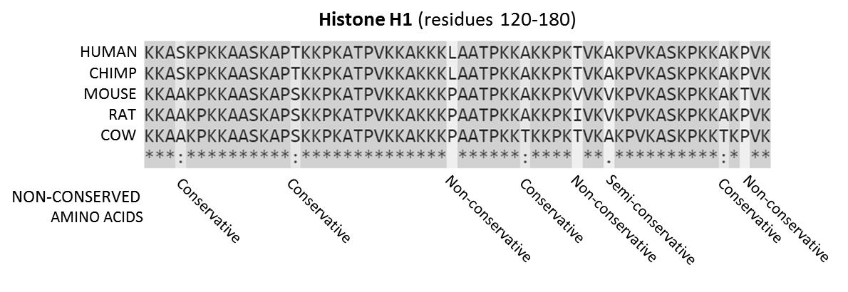
In evolutionary biology, conserved sequences are identical or similar sequences in nucleic acids ( DNA and RNA) or proteins across species ( orthologous sequences), or within a genome ( paralogous sequences), or between donor and receptor taxa ( xenologous sequences). Conservation indicates that a sequence has been maintained by natural selection. A highly conserved sequence is one that has remained relatively unchanged far back up the phylogenetic tree, and hence far back in geological time. Examples of highly conserved sequences include the RNA components of ribosomes present in all domains of life, the homeobox sequences widespread amongst Eukaryotes, and the tmRNA in Bacteria. The study of sequence conservation overlaps with the fields of genomics, proteomics, evolutionary biology, phylogenetics, bioinformatics and mathematics. History The discovery of the role of DNA in heredity, and observations by Frederick Sanger of variation between animal insulins in 1949, promp ... [...More Info...] [...Related Items...] OR: [Wikipedia] [Google] [Baidu] |
Stem-loop
Stem-loop intramolecular base pairing is a pattern that can occur in single-stranded RNA. The structure is also known as a hairpin or hairpin loop. It occurs when two regions of the same strand, usually complementary in nucleotide sequence when read in opposite directions, base-pair to form a double helix that ends in an unpaired loop. The resulting structure is a key building block of many RNA secondary structures. As an important secondary structure of RNA, it can direct RNA folding, protect structural stability for messenger RNA (mRNA), provide recognition sites for RNA binding proteins, and serve as a substrate for enzymatic reactions. Formation and stability The formation of a stem-loop structure is dependent on the stability of the resulting helix and loop regions. The first prerequisite is the presence of a sequence that can fold back on itself to form a paired double helix. The stability of this helix is determined by its length, the number of mismatches or bulges it co ... [...More Info...] [...Related Items...] OR: [Wikipedia] [Google] [Baidu] |


