|
Microscopy, Interference
Interference microscopy involving measurements of differences in the path between two beams of light that have been split. Types include: * Classical interference microscopy * Differential interference contrast microscopy * Fluorescence interference contrast microscopy * Interference reflection microscopy Interference reflection microscopy (IRM), also called Reflection Interference Contrast Microscopy (RICM) or Reflection Contrast Microscopy (RCM) depending on the context, is an optical microscopy technique that leverages interference effects to for ... See also * Phase contrast microscopy References {{science-stub Microscopy ... [...More Info...] [...Related Items...] OR: [Wikipedia] [Google] [Baidu] |
Classical Interference Microscopy
Classical interference microscopy, also called quantitative interference microscopy, uses two separate light beams with much greater lateral separation than that used in phase contrast microscopy or in differential interference microscopy (DIC). In variants of the interference microscope where object and reference beam pass through the same objective, two images are produced of every object (one being the "ghost image"). The two images are separated either laterally within the visual field or at different focal planes, as determined by the optical principles employed. These two images can be a nuisance when they overlap, since they can severely affect the accuracy of mass thickness measurements. Rotation of the preparation may thus be necessary, as in the case of DIC. One of the first usable interference microscopes was designed by Dyson and manufactured by Cooke, Troughton & Simms (later Vickers Instruments), York England. This ingenious optical system achieved interference imagi ... [...More Info...] [...Related Items...] OR: [Wikipedia] [Google] [Baidu] |
Differential Interference Contrast Microscopy
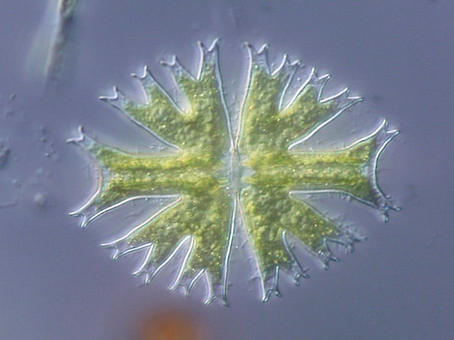
Differential interference contrast (DIC) microscopy, also known as Nomarski interference contrast (NIC) or Nomarski microscopy, is an optical microscopy technique used to enhance the contrast in unstained, transparent samples. DIC works on the principle of interferometry to gain information about the optical path length of the sample, to see otherwise invisible features. A relatively complex optical system produces an image with the object appearing black to white on a grey background. This image is similar to that obtained by phase contrast microscopy but without the bright diffraction halo. The technique was developed by Polish physicist Georges Nomarski in 1952. DIC works by separating a polarized light source into two orthogonally polarized mutually coherent parts which are spatially displaced (sheared) at the sample plane, and recombined before observation. The interference of the two parts at recombination is sensitive to their optical path difference (i.e. the product ... [...More Info...] [...Related Items...] OR: [Wikipedia] [Google] [Baidu] |
Fluorescence Interference Contrast Microscopy
Fluorescence interference contrast (FLIC) microscopy is a microscopic technique developed to achieve z-resolution on the nanometer scale. FLIC occurs whenever fluorescent objects are in the vicinity of a reflecting surface (e.g. Si wafer). The resulting interference between the direct and the reflected light leads to a double sin2 modulation of the intensity, I, of a fluorescent object as a function of distance, h, above the reflecting surface. This allows for the ''nanometer height measurements''. FLIC microscope is well suited to measuring the topography of a membrane that contains fluorescent probes e.g. an artificial lipid bilayer, or a living cell membrane or the structure of fluorescently labeled proteins on a surface. FLIC optical theory General two layer system The optical theory underlying FLIC was developed by Armin Lambacher and Peter Fromherz. They derived a relationship between the observed fluorescence intensity and the distance of the fluorophore from a reflective ... [...More Info...] [...Related Items...] OR: [Wikipedia] [Google] [Baidu] |
Interference Reflection Microscopy
Interference reflection microscopy (IRM), also called Reflection Interference Contrast Microscopy (RICM) or Reflection Contrast Microscopy (RCM) depending on the context, is an optical microscopy technique that leverages interference effects to form an image of an object on a glass surface. The intensity of the signal is a measure of proximity of the object to the glass surface. This technique can be used to study events at the cell membrane without the use of a (fluorescent) label as is the case for TIRF microscopy. History and name In 1964, Adam S. G. Curtis coined the term Interference Reflection Microscopy (IRM), using it in the field of cell biology to study embryonic chick heart fibroblasts. He used IRM to look at adhesion sites and distances of fibroblasts, noting that contact with the glass was mostly limited to the cell periphery and the pseudopodia. In 1975, Johan Sebastiaan Ploem introduced an improvement to IRM (published in a book chapter), which he called Reflection ... [...More Info...] [...Related Items...] OR: [Wikipedia] [Google] [Baidu] |
Phase Contrast Microscopy
__NOTOC__ Phase-contrast microscopy (PCM) is an optical microscopy technique that converts phase shifts in light passing through a transparent specimen to brightness changes in the image. Phase shifts themselves are invisible, but become visible when shown as brightness variations. When light waves travel through a medium other than a vacuum, interaction with the medium causes the wave amplitude and phase to change in a manner dependent on properties of the medium. Changes in amplitude (brightness) arise from the scattering and absorption of light, which is often wavelength-dependent and may give rise to colors. Photographic equipment and the human eye are only sensitive to amplitude variations. Without special arrangements, phase changes are therefore invisible. Yet, phase changes often convey important information. Phase-contrast microscopy is particularly important in biology. It reveals many cellular structures that are invisible with a bright-field microscope, as exemplif ... [...More Info...] [...Related Items...] OR: [Wikipedia] [Google] [Baidu] |

