|
Merkel Cell Cancer
Merkel cell carcinoma (MCC) is a rare and aggressive skin cancer occurring in about 3 people per 1,000,000 members of the population. It is also known as cutaneous APUDoma, primary neuroendocrine carcinoma of the skin, primary small cell carcinoma of the skin, and trabecular carcinoma of the skin. Factors involved in the development of MCC include the Merkel cell polyomavirus (MCPyV or MCV), a weakened immune system, and exposure to ultraviolet radiation. Merkel-cell carcinoma usually arises on the head, neck, and extremities, as well as in the perianal region and on the eyelid. It is more common in people over 60 years old, Caucasian people, and males. MCC is less common in children. Signs and symptoms Merkel cell carcinoma (MCC) usually presents as a firm nodule (up to 2 cm diameter) or mass (>2 cm diameter). These flesh-colored, red, or blue tumors typically vary in size from 0.5 cm (less than one-quarter of an inch) to more than 5 cm (2 inches) in diame ... [...More Info...] [...Related Items...] OR: [Wikipedia] [Google] [Baidu] |
Micrograph
A micrograph or photomicrograph is a photograph or digital image taken through a microscope or similar device to show a magnified image of an object. This is opposed to a macrograph or photomacrograph, an image which is also taken on a microscope but is only slightly magnified, usually less than 10 times. Micrography is the practice or art of using microscopes to make photographs. A micrograph contains extensive details of microstructure. A wealth of information can be obtained from a simple micrograph like behavior of the material under different conditions, the phases found in the system, failure analysis, grain size estimation, elemental analysis and so on. Micrographs are widely used in all fields of microscopy. Types Photomicrograph A light micrograph or photomicrograph is a micrograph prepared using an optical microscope, a process referred to as ''photomicroscopy''. At a basic level, photomicroscopy may be performed simply by connecting a camera to a microscope, th ... [...More Info...] [...Related Items...] OR: [Wikipedia] [Google] [Baidu] |
Lymphoma
Lymphoma is a group of blood and lymph tumors that develop from lymphocytes (a type of white blood cell). In current usage the name usually refers to just the cancerous versions rather than all such tumours. Signs and symptoms may include enlarged lymph nodes, fever, drenching sweats, unintended weight loss, itching, and constantly feeling tired. The enlarged lymph nodes are usually painless. The sweats are most common at night. Many subtypes of lymphomas are known. The two main categories of lymphomas are the non-Hodgkin lymphoma (NHL) (90% of cases) and Hodgkin lymphoma (HL) (10%). The World Health Organization (WHO) includes two other categories as types of lymphoma – multiple myeloma and immunoproliferative diseases. Lymphomas and leukemias are a part of the broader group of tumors of the hematopoietic and lymphoid tissues. Risk factors for Hodgkin lymphoma include infection with Epstein–Barr virus and a history of the disease in the family. Risk factors for common ... [...More Info...] [...Related Items...] OR: [Wikipedia] [Google] [Baidu] |
ATOH1
Protein atonal homolog 1 is a protein that in humans is encoded by the ''ATOH1'' gene. Function This protein belongs to the basic helix-loop-helix (BHLH) family of transcription factors. It activates E-box dependent transcription along with TCF3 (E47). ATOH1 is required for the formation of both neural and non-neural cell types. Using genetic deletion in mice, Atoh1 has been shown to be essential for formation of cerebellar granule neurons, inner ear hair cells, spinal cord interneurons, Merkel cells of the skin, and intestinal secretory cells (goblet, enteroendocrine, and Paneth cells). ATOH1 is a mammalian homolog of the ''Drosophila melanogaster'' gene ''atonal''. ATOH1 is considered part of the Notch signaling pathway. In 2009, ATOH1 was identified as a tumor suppressor gene A tumor suppressor gene (TSG), or anti-oncogene, is a gene that regulates a cell during cell division and replication. If the cell grows uncontrollably, it will result in cancer. When a tumor suppr ... [...More Info...] [...Related Items...] OR: [Wikipedia] [Google] [Baidu] |
PIEZO2
Piezo-type mechanosensitive ion channel component 2 is a protein that in humans is encoded by the PIEZO2 gene. It has a homotrimeric structure, with three blades curving into a nano-dome, with a diameter of 28 nanometers. Function Piezos are large transmembrane proteins conserved among various species, all having between 24 and 36 predicted transmembrane domains. 'Piezo' comes from the Greek 'piesi,' meaning 'pressure.' The PIEZO2 protein has a role in rapidly adapting mechanically activated (MA) currents in somatosensory neurons. Its structure is resolved via a mouse version in 2019, showing the predicted homotrimeric propeller. PIEZO2 is typically found in tissues that respond to physical touch, such as Merkel cells, and is thought to regulate light touch response. Pathology * Gain-of-function mutations in the mechanically activated ion channel PIEZO2 cause a subtype of Distal Arthrogryposis. * Mice without PIEZO2 in their proprioceptive neurons show uncoordinated body mo ... [...More Info...] [...Related Items...] OR: [Wikipedia] [Google] [Baidu] |
Chromogranin A
Chromogranin A or parathyroid secretory protein 1 (gene name CHGA) is a member of the granin family of neuroendocrine secretory proteins. As such, it is located in secretory vesicles of neurons and endocrine cells such as islet beta cell secretory granules in the pancreas. In humans, chromogranin A protein is encoded by the ''CHGA'' gene. Tissue distribution Examples of cells producing chromogranin A (ChgA) are chromaffin cells of the adrenal medulla, paraganglia, enterochromaffin-like cells and beta cells of the pancreas. It is present in islet beta cell secretory granules. chromogranin-A (CgA)+ Pulmonary neuroendocrine cells account for 0.41% of all epithelial cells in the conducting airway, but are absent from the alveoli. Function Chromogranin A is the precursor to several functional peptides including vasostatin-1, vasostatin-2, pancreastatin, catestatin and parastatin. These peptides negatively modulate the neuroendocrine function of the releasing cell (autocrine) or ne ... [...More Info...] [...Related Items...] OR: [Wikipedia] [Google] [Baidu] |
Synaptophysin
Synaptophysin, also known as the major synaptic vesicle protein p38, is a protein that in humans is encoded by the ''SYP'' gene. Genomics The gene is located on the short arm of X chromosome (Xp11.23-p11.22). It is 12,406 bases in length and lies on the minus strand. The encoded protein has 313 amino acids with a predicted molecular weight of 33.845 kDa. Molecular biology The protein is a synaptic vesicle glycoprotein with four transmembrane domains weighing 38kDa. It is present in neuroendocrine cells and in virtually all neurons in the brain and spinal cord that participate in synaptic transmission. It acts as a marker for neuroendocrine tumors, and its ubiquity at the synapse has led to the use of synaptophysin immunostaining for quantification of synapses. The exact function of the protein is unknown: it interacts with the essential synaptic vesicle protein synaptobrevin, but when the synaptophysin gene is experimentally inactivated in animals, they still develop and ... [...More Info...] [...Related Items...] OR: [Wikipedia] [Google] [Baidu] |
Keratin 20
Keratin 20, often abbreviated CK20, is a protein that in humans is encoded by the ''KRT20'' gene. Keratin 20 is a type I cytokeratin. It is a major cellular protein of mature enterocytes and goblet cells and is specifically found in the gastric and intestinal mucosa. In immunohistochemistry, antibodies to CK20 can be used to identify a range of adenocarcinoma arising from epithelia that normally contain the CK20 protein. For example, the protein is commonly found in colorectal cancer, transitional cell carcinomas and in Merkel cell carcinoma, but is absent in lung cancer, prostate cancer Prostate cancer is cancer of the prostate. Prostate cancer is the second most common cancerous tumor worldwide and is the fifth leading cause of cancer-related mortality among men. The prostate is a gland in the male reproductive system that sur ..., and non-mucinous ovarian cancer. It is often used in combination with antibodies to CK7 to distinguish different types of glandular t ... [...More Info...] [...Related Items...] OR: [Wikipedia] [Google] [Baidu] |
Immunohistochemistry
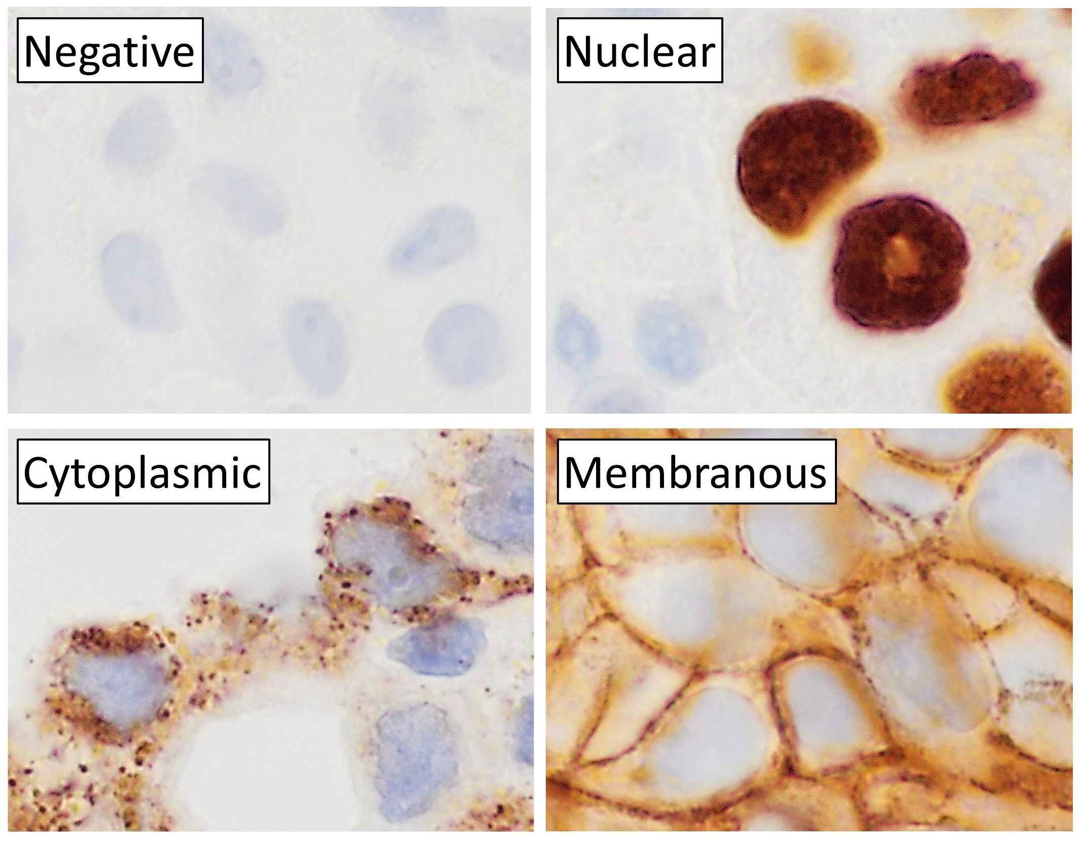
Immunohistochemistry (IHC) is the most common application of immunostaining. It involves the process of selectively identifying antigens (proteins) in cells of a tissue section by exploiting the principle of antibodies binding specifically to antigens in biological tissues. IHC takes its name from the roots "immuno", in reference to antibodies used in the procedure, and "histo", meaning tissue (compare to immunocytochemistry). Albert Coons conceptualized and first implemented the procedure in 1941. Visualising an antibody-antigen interaction can be accomplished in a number of ways, mainly either of the following: * ''Chromogenic immunohistochemistry'' (CIH), wherein an antibody is conjugated to an enzyme, such as peroxidase (the combination being termed immunoperoxidase), that can catalyse a colour-producing reaction. * '' Immunofluorescence'', where the antibody is tagged to a fluorophore, such as fluorescein or rhodamine. Immunohistochemical staining is widely used in the dia ... [...More Info...] [...Related Items...] OR: [Wikipedia] [Google] [Baidu] |
Merkel Cell
Merkel cells, also known as Merkel-Ranvier cells or tactile epithelial cells, are oval-shaped mechanoreceptors essential for light touch sensation and found in the skin of vertebrates. They are abundant in highly sensitive skin like that of the fingertips in humans, and make synaptic contacts with somatosensory afferent nerve fibers. Though it has been reported that Merkel cells are derived from neural crest cells, more recent experiments in mammals have indicated that they are in fact epithelial in origin. Structure Merkel cells are found in the skin and some parts of the mucosa of all vertebrates. In mammalian skin, they are clear cells found in the ''stratum basale'' (at the bottom of sweat duct ridges) of the epidermis approximately 10 μm in diameter. They also occur in epidermal invaginations of the plantar foot surface called rete ridges. Most often, they are associated with sensory nerve endings, when they are known as Merkel nerve endings (also called a Merkel ... [...More Info...] [...Related Items...] OR: [Wikipedia] [Google] [Baidu] |
Metastasize
Metastasis is a pathogenic agent's spread from an initial or primary site to a different or secondary site within the host's body; the term is typically used when referring to metastasis by a cancerous tumor. The newly pathological sites, then, are metastases (mets). It is generally distinguished from cancer invasion, which is the direct extension and penetration by cancer cells into neighboring tissues. Cancer occurs after cells are genetically altered to proliferate rapidly and indefinitely. This uncontrolled proliferation by mitosis produces a primary heterogeneic tumour. The cells which constitute the tumor eventually undergo metaplasia, followed by dysplasia then anaplasia, resulting in a malignant phenotype. This malignancy allows for invasion into the circulation, followed by invasion to a second site for tumorigenesis. Some cancer cells known as circulating tumor cells acquire the ability to penetrate the walls of lymphatic or blood vessels, after which they are able t ... [...More Info...] [...Related Items...] OR: [Wikipedia] [Google] [Baidu] |
Fascia
A fascia (; plural fasciae or fascias; adjective fascial; from Latin: "band") is a band or sheet of connective tissue, primarily collagen, beneath the skin that attaches to, stabilizes, encloses, and separates muscles and other internal organs. Fascia is classified by layer, as superficial fascia, deep fascia, and ''visceral'' or ''parietal'' fascia, or by its function and anatomical location. Like ligaments, aponeuroses, and tendons, fascia is made up of fibrous connective tissue containing closely packed bundles of collagen fibers oriented in a wavy pattern parallel to the direction of pull. Fascia is consequently flexible and able to resist great unidirectional tension forces until the wavy pattern of fibers has been straightened out by the pulling force. These collagen fibers are produced by fibroblasts located within the fascia. Fasciae are similar to ligaments and tendons as they have collagen as their major component. They differ in their location and function: ligament ... [...More Info...] [...Related Items...] OR: [Wikipedia] [Google] [Baidu] |


