|
Median Tongue Bud
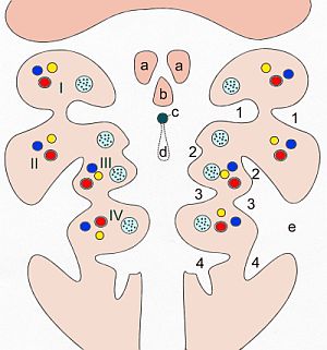
The median tongue bud (also tuberculum impar) marks the beginning of the development of the tongue. It appears as a midline swelling from the first pharyngeal arch late in the fourth week of embryogenesis. In the fifth week, a pair of lateral lingual swelling The tongue is a muscular organ (anatomy), organ in the mouth of a typical tetrapod. It manipulates food for mastication and swallowing as part of the digestive system, digestive process, and is the primary organ of taste. The tongue's upper surfa ...s (or ''distal tongue buds'') develop above and in line with the median tongue bud. These swellings grow downwards towards each other, quickly overgrowing the median tongue bud. The line of the fusion of the distal tongue buds is marked by the median sulcus. References External links * Embryology {{Portal bar, Anatomy ... [...More Info...] [...Related Items...] OR: [Wikipedia] [Google] [Baidu] |
Tongue
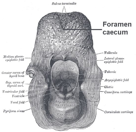
The tongue is a muscular organ (anatomy), organ in the mouth of a typical tetrapod. It manipulates food for mastication and swallowing as part of the digestive system, digestive process, and is the primary organ of taste. The tongue's upper surface (dorsum) is covered by taste buds housed in numerous lingual papillae. It is sensitive and kept moist by saliva and is richly supplied with nerves and blood vessels. The tongue also serves as a natural means of oral hygiene, cleaning the teeth. A major function of the tongue is the enabling of speech in humans and animal communication, vocalization in other animals. The human tongue is divided into two parts, an oral cavity, oral part at the front and a pharynx, pharyngeal part at the back. The left and right sides are also separated along most of its length by a vertical section of connective tissue, fibrous tissue (the lingual septum) that results in a groove, the median sulcus, on the tongue's surface. There are two groups of muscle ... [...More Info...] [...Related Items...] OR: [Wikipedia] [Google] [Baidu] |
Pharyngeal Arch
The pharyngeal arches, also known as visceral arches'','' are structures seen in the embryonic development of vertebrates that are recognisable precursors for many structures. In fish, the arches are known as the branchial arches, or gill arches. In the human embryo, the arches are first seen during the fourth week of development. They appear as a series of outpouchings of mesoderm on both sides of the developing pharynx. The vasculature of the pharyngeal arches is known as the aortic arches. In fish, the branchial arches support the gills. Structure In vertebrates, the pharyngeal arches are derived from all three germ layers (the primary layers of cells that form during embryogenesis). Neural crest cells enter these arches where they contribute to features of the skull and facial skeleton such as bone and cartilage. However, the existence of pharyngeal structures before neural crest cells evolved is indicated by the existence of neural crest-independent mechanisms of pharyng ... [...More Info...] [...Related Items...] OR: [Wikipedia] [Google] [Baidu] |
Human Embryogenesis
Human embryonic development, or human embryogenesis, is the development and formation of the human embryo. It is characterised by the processes of cell division and cellular differentiation of the embryo that occurs during the early stages of development. In biological terms, the development of the human body entails growth from a one-celled zygote to an adult human being. Fertilization occurs when the sperm cell successfully enters and fuses with an egg cell (ovum). The genetic material of the sperm and egg then combine to form the single cell zygote and the germinal stage of development commences. Embryonic development in the human, covers the first eight weeks of development; at the beginning of the ninth week the embryo is termed a fetus. Human embryology is the study of this development during the first eight weeks after fertilization. The normal period of gestation (pregnancy) is about nine months or 40 weeks. The germinal stage refers to the time from fertilization throu ... [...More Info...] [...Related Items...] OR: [Wikipedia] [Google] [Baidu] |
Lateral Lingual Swelling
The tongue is a muscular organ (anatomy), organ in the mouth of a typical tetrapod. It manipulates food for mastication and swallowing as part of the digestive system, digestive process, and is the primary organ of taste. The tongue's upper surface (dorsum) is covered by taste buds housed in numerous lingual papillae. It is sensitive and kept moist by saliva and is richly supplied with nerves and blood vessels. The tongue also serves as a natural means of oral hygiene, cleaning the teeth. A major function of the tongue is the enabling of speech in humans and animal communication, vocalization in other animals. The human tongue is divided into two parts, an oral cavity, oral part at the front and a pharynx, pharyngeal part at the back. The left and right sides are also separated along most of its length by a vertical section of connective tissue, fibrous tissue (the lingual septum) that results in a groove, the median sulcus, on the tongue's surface. There are two groups of muscle ... [...More Info...] [...Related Items...] OR: [Wikipedia] [Google] [Baidu] |
Tongue
The tongue is a muscular organ (anatomy), organ in the mouth of a typical tetrapod. It manipulates food for mastication and swallowing as part of the digestive system, digestive process, and is the primary organ of taste. The tongue's upper surface (dorsum) is covered by taste buds housed in numerous lingual papillae. It is sensitive and kept moist by saliva and is richly supplied with nerves and blood vessels. The tongue also serves as a natural means of oral hygiene, cleaning the teeth. A major function of the tongue is the enabling of speech in humans and animal communication, vocalization in other animals. The human tongue is divided into two parts, an oral cavity, oral part at the front and a pharynx, pharyngeal part at the back. The left and right sides are also separated along most of its length by a vertical section of connective tissue, fibrous tissue (the lingual septum) that results in a groove, the median sulcus, on the tongue's surface. There are two groups of muscle ... [...More Info...] [...Related Items...] OR: [Wikipedia] [Google] [Baidu] |

