|
Medial Geniculate Body
The medial geniculate nucleus (MGN) or medial geniculate body (MGB) is part of the auditory thalamus and represents the thalamic relay between the inferior colliculus (IC) and the auditory cortex (AC). It is made up of a number of sub-nuclei that are distinguished by their neuronal morphology and density, by their afferent and efferent connections, and by the coding properties of their neurons. It is thought that the MGN influences the direction and maintenance of attention. Divisions The MGN has three major divisions; ventral (VMGN), dorsal (DMGN) and medial (MMGN). Whilst the VMGN is specific to auditory information processing, the DMGN and MMGN also receive information from non-auditory pathways. Ventral subnucleus Cell types There are two main cell types in the ventral subnucleus of the medial geniculate body (VMGN): * Thalamocortical relay cells (or principal neurons): The dendritic input to these cells comes from two sets of dendritic trees oriented on opposite poles of th ... [...More Info...] [...Related Items...] OR: [Wikipedia] [Google] [Baidu] |
Midline Nuclear Group
The midline nuclear group (or midline thalamic nuclei) is a region of the thalamus consisting of the following nuclei: * paraventricular nucleus of thalamus (''nucleus paraventricularis thalami'') - not to be confused with paraventricular nucleus of hypothalamus * paratenial nucleus The paratenial nucleus, or parataenial nucleus ( la, nucleus parataenialis), is a component of the midline nuclear group in the thalamus. It is sometimes subdivided into the nucleus parataenialis interstitialis and nucleus parataenialis parvocellu ... (''nucleus parataenialis'') * nucleus reuniens * rhomboid nucleus (''nucleus commissuralis rhomboidalis'') * subfascicular nucleus (''nucleus subfascicularis'') The midline nuclei are often called "nonspecific" in that they project widely to the cortex and elsewhere. This has led to the assumption that they may be involved in general functions such as alerting. However, anatomical connections might suggest more specific functions, with the paraventr ... [...More Info...] [...Related Items...] OR: [Wikipedia] [Google] [Baidu] |
Thalamus
The thalamus (from Greek θάλαμος, "chamber") is a large mass of gray matter located in the dorsal part of the diencephalon (a division of the forebrain). Nerve fibers project out of the thalamus to the cerebral cortex in all directions, allowing hub-like exchanges of information. It has several functions, such as the relaying of sensory signals, including motor signals to the cerebral cortex and the regulation of consciousness, sleep, and alertness. Anatomically, it is a paramedian symmetrical structure of two halves (left and right), within the vertebrate brain, situated between the cerebral cortex and the midbrain. It forms during embryonic development as the main product of the diencephalon, as first recognized by the Swiss embryologist and anatomist Wilhelm His Sr. in 1893. Anatomy The thalamus is a paired structure of gray matter located in the forebrain which is superior to the midbrain, near the center of the brain, with nerve fibers projecting out to the ... [...More Info...] [...Related Items...] OR: [Wikipedia] [Google] [Baidu] |
Tonotopy
In physiology, tonotopy (from Greek tono = frequency and topos = place) is the spatial arrangement of where sounds of different frequency are processed in the brain. Tones close to each other in terms of frequency are represented in topologically neighbouring regions in the brain. Tonotopic maps are a particular case of topographic organization, similar to retinotopy in the visual system. Tonotopy in the auditory system begins at the cochlea, the small snail-like structure in the inner ear that sends information about sound to the brain. Different regions of the basilar membrane in the organ of Corti, the sound-sensitive portion of the cochlea, vibrate at different sinusoidal frequencies due to variations in thickness and width along the length of the membrane. Nerves that transmit information from different regions of the basilar membrane therefore encode frequency tonotopically. This tonotopy then projects through the vestibulocochlear nerve and associated midbrain structures ... [...More Info...] [...Related Items...] OR: [Wikipedia] [Google] [Baidu] |
Interneuron
Interneurons (also called internuncial neurons, relay neurons, association neurons, connector neurons, intermediate neurons or local circuit neurons) are neurons that connect two brain regions, i.e. not direct motor neurons or sensory neurons. Interneurons are the central nodes of neural circuits, enabling communication between sensory or motor neurons and the central nervous system (CNS). They play vital roles in reflexes, neuronal oscillations, and neurogenesis in the adult mammalian brain. Interneurons can be further broken down into two groups: local interneurons and relay interneurons. Local interneurons have short axons and form circuits with nearby neurons to analyze small pieces of information. Relay interneurons have long axons and connect circuits of neurons in one region of the brain with those in other regions. However, interneurons are generally considered to operate mainly within local brain areas. The interaction between interneurons allow the brain to perform c ... [...More Info...] [...Related Items...] OR: [Wikipedia] [Google] [Baidu] |
Dendrite
Dendrites (from Greek δένδρον ''déndron'', "tree"), also dendrons, are branched protoplasmic extensions of a nerve cell that propagate the electrochemical stimulation received from other neural cells to the cell body, or soma, of the neuron from which the dendrites project. Electrical stimulation is transmitted onto dendrites by upstream neurons (usually via their axons) via synapses which are located at various points throughout the dendritic tree. Dendrites play a critical role in integrating these synaptic inputs and in determining the extent to which action potentials are produced by the neuron. Dendritic arborization, also known as dendritic branching, is a multi-step biological process by which neurons form new dendritic trees and branches to create new synapses. The morphology of dendrites such as branch density and grouping patterns are highly correlated to the function of the neuron. Malformation of dendrites is also tightly correlated to impaired nervous syste ... [...More Info...] [...Related Items...] OR: [Wikipedia] [Google] [Baidu] |
Superior Olivary Nucleus
The superior olivary complex (SOC) or superior olive is a collection of brainstem nuclei that functions in multiple aspects of hearing and is an important component of the ascending and descending auditory pathways of the auditory system. The SOC is intimately related to the trapezoid body: most of the cell groups of the SOC are dorsal (posterior in primates) to this axon bundle while a number of cell groups are embedded in the trapezoid body. Overall, the SOC displays a significant interspecies variation, being largest in bats and rodents and smaller in primates. Physiology The superior olivary nucleus plays a number of roles in hearing. The medial superior olive (MSO) is a specialized nucleus that is believed to measure the time difference of arrival of sounds between the ears (the interaural time difference or ITD). The ITD is a major cue for determining the azimuth of sounds, i.e., localising them on the azimuthal plane – their degree to the left or the right. The lateral ... [...More Info...] [...Related Items...] OR: [Wikipedia] [Google] [Baidu] |
Superior Colliculus
In neuroanatomy, the superior colliculus () is a structure lying on the roof of the mammalian midbrain. In non-mammalian vertebrates, the homologous structure is known as the optic tectum, or optic lobe. The adjective form ''tectal'' is commonly used for both structures. In mammals, the superior colliculus forms a major component of the midbrain. It is a paired structure and together with the paired inferior colliculi forms the corpora quadrigemina. The superior colliculus is a layered structure, with a pattern that is similar to all mammals. The layers can be grouped into the superficial layers ( stratum opticum and above) and the deeper remaining layers. Neurons in the superficial layers receive direct input from the retina and respond almost exclusively to visual stimuli. Many neurons in the deeper layers also respond to other modalities, and some respond to stimuli in multiple modalities. The deeper layers also contain a population of motor-related neurons, capable of activat ... [...More Info...] [...Related Items...] OR: [Wikipedia] [Google] [Baidu] |
Auditory Cortex
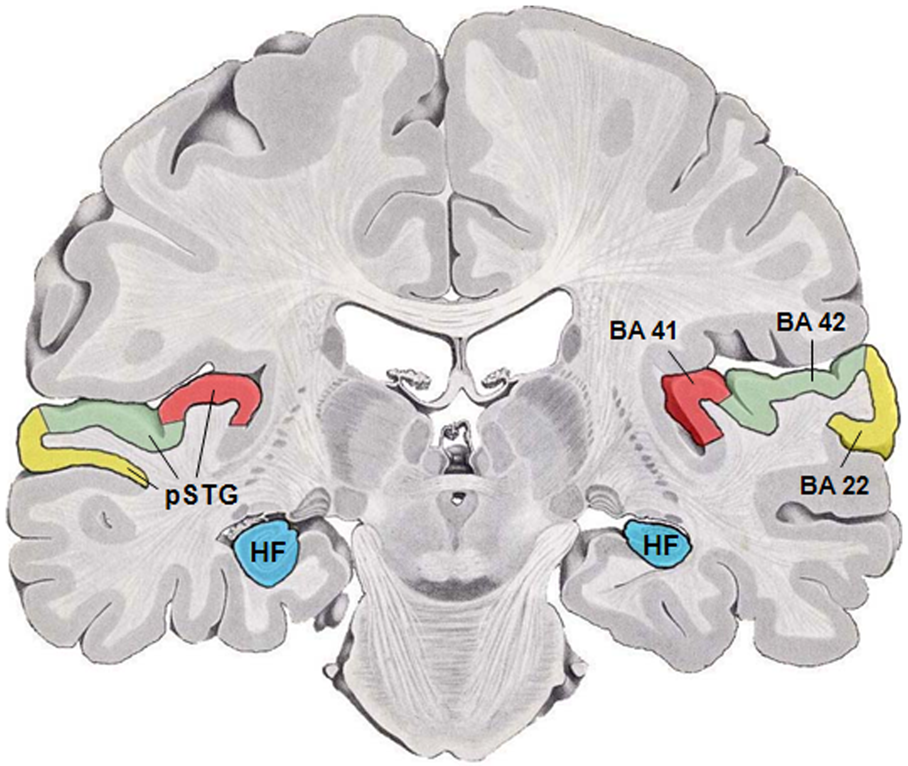
The auditory cortex is the part of the temporal lobe that processes auditory information in humans and many other vertebrates. It is a part of the auditory system, performing basic and higher functions in hearing, such as possible relations to language switching.Cf. Pickles, James O. (2012). ''An Introduction to the Physiology of Hearing'' (4th ed.). Bingley, UK: Emerald Group Publishing Limited, p. 238. It is located bilaterally, roughly at the upper sides of the temporal lobes – in humans, curving down and onto the medial surface, on the superior temporal plane, within the lateral sulcus and comprising parts of the transverse temporal gyri, and the superior temporal gyrus, including the planum polare and planum temporale (roughly Brodmann areas 41 and 42, and partially 22). The auditory cortex takes part in the spectrotemporal, meaning involving time and frequency, analysis of the inputs passed on from the ear. The cortex then filters and passes on the information to th ... [...More Info...] [...Related Items...] OR: [Wikipedia] [Google] [Baidu] |
Reticular Nucleus Of The Thalamus
The thalamic reticular nucleus is part of the ventral thalamus that forms a capsule around the thalamus laterally. However, recent evidence from mice and fish question this statement and define it as a dorsal thalamic structure. It is separated from the thalamus by the external medullary lamina. Reticular cells are GABAergic, and have discoid dendritic arbors in the plane of the nucleus. Thalamic Reticular Nucleus is variously abbreviated TRN, RTN, NRT, and RT. Input and output The thalamic reticular nucleus receives input from the cerebral cortex The cerebral cortex, also known as the cerebral mantle, is the outer layer of neural tissue of the cerebrum of the brain in humans and other mammals. The cerebral cortex mostly consists of the six-layered neocortex, with just 10% consistin ... and dorsal thalamic nuclei. Most input comes from collaterals of fibers passing through the thalamic reticular nucleus. Primary thalamic reticular nucleus efferent fibers project to d ... [...More Info...] [...Related Items...] OR: [Wikipedia] [Google] [Baidu] |
Inferior Colliculus
The inferior colliculus (IC) (Latin for ''lower hill'') is the principal midbrain nucleus of the auditory pathway and receives input from several peripheral brainstem nuclei in the auditory pathway, as well as inputs from the auditory cortex. The inferior colliculus has three subdivisions: the central nucleus, a dorsal cortex by which it is surrounded, and an external cortex which is located laterally. Its bimodal neurons are implicated in auditory-somatosensory interaction, receiving projections from somatosensory nuclei. This multisensory integration may underlie a filtering of self-effected sounds from vocalization, chewing, or respiration activities. The inferior colliculi together with the superior colliculi form the eminences of the corpora quadrigemina, and also part of the tectal region of the midbrain. The inferior colliculus lies caudal to its counterpart – the superior colliculus – above the trochlear nerve, and at the base of the projection of the medial genicu ... [...More Info...] [...Related Items...] OR: [Wikipedia] [Google] [Baidu] |
Auditory Cortex
The auditory cortex is the part of the temporal lobe that processes auditory information in humans and many other vertebrates. It is a part of the auditory system, performing basic and higher functions in hearing, such as possible relations to language switching.Cf. Pickles, James O. (2012). ''An Introduction to the Physiology of Hearing'' (4th ed.). Bingley, UK: Emerald Group Publishing Limited, p. 238. It is located bilaterally, roughly at the upper sides of the temporal lobes – in humans, curving down and onto the medial surface, on the superior temporal plane, within the lateral sulcus and comprising parts of the transverse temporal gyri, and the superior temporal gyrus, including the planum polare and planum temporale (roughly Brodmann areas 41 and 42, and partially 22). The auditory cortex takes part in the spectrotemporal, meaning involving time and frequency, analysis of the inputs passed on from the ear. The cortex then filters and passes on the information to th ... [...More Info...] [...Related Items...] OR: [Wikipedia] [Google] [Baidu] |


