|
Longissimus Muscle
The longissimus ( la, the longest one) is the muscle lateral to the semispinalis muscles. It is the longest subdivision of the erector spinae muscles that extends forward into the transverse processes of the posterior cervical vertebrae. Structure Longissimus thoracis et lumborum The longissimus thoracis et lumborum is the intermediate and largest of the continuations of the erector spinae. In the lumbar region (longissimus lumborum), where it is as yet blended with the iliocostalis, some of its fibers are attached to the whole length of the posterior surfaces of the transverse processes and the accessory processes of the lumbar vertebrae, and to the anterior layer of the lumbodorsal fascia. In the thoracic region (longissimus thoracis), it is inserted, by rounded tendons, into the tips of the transverse processes of all the thoracic vertebrae, and by fleshy processes into the lower nine or ten ribs between their tubercles and angles. Longissimus cervicis The longissimus cervic ... [...More Info...] [...Related Items...] OR: [Wikipedia] [Google] [Baidu] |
Transverse Process
The spinal column, a defining synapomorphy shared by nearly all vertebrates, Hagfish are believed to have secondarily lost their spinal column is a moderately flexible series of vertebrae (singular vertebra), each constituting a characteristic irregular bone whose complex structure is composed primarily of bone, and secondarily of hyaline cartilage. They show variation in the proportion contributed by these two tissue types; such variations correlate on one hand with the cerebral/caudal rank (i.e., location within the backbone), and on the other with phylogenetic differences among the vertebrate taxa. The basic configuration of a vertebra varies, but the bone is its ''body'', with the central part of the body constituting the ''centrum''. The upper (closer to) and lower (further from), respectively, the cranium and its central nervous system surfaces of the vertebra body support attachment to the intervertebral discs. The posterior part of a vertebra forms a vertebral a ... [...More Info...] [...Related Items...] OR: [Wikipedia] [Google] [Baidu] |
Lumbar Vertebrae
The lumbar vertebrae are, in human anatomy, the five vertebrae between the rib cage and the pelvis. They are the largest segments of the vertebral column and are characterized by the absence of the foramen transversarium within the transverse process (since it is only found in the cervical region) and by the absence of facets on the sides of the body (as found only in the thoracic region). They are designated L1 to L5, starting at the top. The lumbar vertebrae help support the weight of the body, and permit movement. Human anatomy General characteristics The adjacent figure depicts the general characteristics of the first through fourth lumbar vertebrae. The fifth vertebra contains certain peculiarities, which are detailed below. As with other vertebrae, each lumbar vertebra consists of a ''vertebral body'' and a ''vertebral arch''. The vertebral arch, consisting of a pair of ''pedicles'' and a pair of ''laminae'', encloses the ''vertebral foramen'' (opening) and s ... [...More Info...] [...Related Items...] OR: [Wikipedia] [Google] [Baidu] |
Sternocleidomastoid
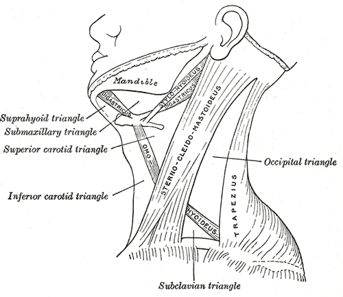
The sternocleidomastoid muscle is one of the largest and most superficial cervical muscles. The primary actions of the muscle are rotation of the head to the opposite side and flexion of the neck. The sternocleidomastoid is innervated by the accessory nerve. Etymology and location It is given the name ''sternocleidomastoid'' because it originates at the manubrium of the sternum (''sterno-'') and the clavicle (''cleido-'') and has an insertion at the mastoid process of the temporal bone of the skull. Structure The sternocleidomastoid muscle originates from two locations: the manubrium of the sternum and the clavicle. It travels obliquely across the side of the neck and inserts at the mastoid process of the temporal bone of the skull by a thin aponeurosis. The sternocleidomastoid is thick and narrow at its centre, and broader and thinner at either end. The sternal head is a round fasciculus, tendinous in front, fleshy behind, arising from the upper part of the front of the manubri ... [...More Info...] [...Related Items...] OR: [Wikipedia] [Google] [Baidu] |
Splenius Capitis
The splenius capitis () () is a broad, straplike muscle in the back of the neck. It pulls on the base of the skull from the vertebrae in the neck and upper thorax. It is involved in movements such as shaking the head. Structure It arises from the lower half of the nuchal ligament, from the spinous process of the seventh cervical vertebra, and from the spinous processes of the upper three or four thoracic vertebrae. The fibers of the muscle are directed upward and laterally and are inserted, under cover of the sternocleidomastoideus, into the mastoid process of the temporal bone, and into the rough surface on the occipital bone just below the lateral third of the superior nuchal line. The splenius capitis is deep to sternocleidomastoideus at the mastoid process, and to the trapezius for its lower portion. It is one of the muscles that forms the floor of the posterior triangle of the neck. The splenius capitis muscle is innervated by the posterior ramus of spinal nerves C3 and ... [...More Info...] [...Related Items...] OR: [Wikipedia] [Google] [Baidu] |
Mastoid Process
The mastoid part of the temporal bone is the posterior (back) part of the temporal bone, one of the bones of the skull. Its rough surface gives attachment to various muscles (via tendons) and it has openings for blood vessels. From its borders, the mastoid part articulates with two other bones. Etymology The word "mastoid" is derived from the Greek word for "breast", a reference to the shape of this bone. Surfaces Outer surface Its outer surface is rough and gives attachment to the occipitalis and posterior auricular muscles. It is perforated by numerous foramina (holes); for example, the mastoid foramen is situated near the posterior border and transmits a vein to the transverse sinus and a small branch of the occipital artery to the dura mater. The position and size of this foramen are very variable; it is not always present; sometimes it is situated in the occipital bone, or in the suture between the temporal and the occipital. Mastoid process The mastoid proce ... [...More Info...] [...Related Items...] OR: [Wikipedia] [Google] [Baidu] |
Articular Processes
The articular processes or zygapophyses (Greek ζυγον = "yoke" (because it links two vertebrae) + απο = "away" + φυσις = " process") of a vertebra are projections of the vertebra that serve the purpose of fitting with an adjacent vertebra. The actual region of contact is called the ''articular facet''.Moore, Keith L. et al. (2010) ''Clinically Oriented Anatomy'', 6th Ed, p.442 fig. 4.2 Articular processes spring from the junctions of the pedicles and laminæ, and there are two right and left, and two superior and inferior. These stick out of an end of a vertebra to lock with a zygapophysis on the next vertebra, to make the backbone more stable. * The superior processes or prezygapophysis project upward from a lower vertebra, and their articular surfaces are directed more or less backward (oblique coronal plane). * The inferior processes or postzygapophysis project downward from a higher vertebra, and their articular surfaces are directed more or less forward and out ... [...More Info...] [...Related Items...] OR: [Wikipedia] [Google] [Baidu] |
Semispinalis Capitis
The semispinalis muscles are a group of three muscles belonging to the transversospinales. These are the semispinalis capitis, the semispinalis cervicis and the semispinalis thoracis. The semispinalis capitis (''complexus'') is situated at the upper and back part of the neck, deep to the splenius, and medial to the longissimus cervicis and longissimus capitis. It arises by a series of tendons from the tips of the transverse processes of the upper six or seven thoracic and the seventh cervical vertebrae, and from the articular processes of the three cervical vertebrae above this ( C4-C6). The tendons, uniting, form a broad muscle, which passes upward, and is inserted between the superior and inferior nuchal lines of the occipital bone. It lies deep to the trapezius muscle and can be palpated as a firm round muscle mass just lateral to the cervical spinous processes. The semispinalis cervicis (or ''semispinalis colli''), arises by a series of tendinous and fleshy fibers fr ... [...More Info...] [...Related Items...] OR: [Wikipedia] [Google] [Baidu] |
Rib Cage
The rib cage, as an enclosure that comprises the ribs, vertebral column and sternum in the thorax of most vertebrates, protects vital organs such as the heart, lungs and great vessels. The sternum, together known as the thoracic cage, is a semi-rigid bony and cartilaginous structure which surrounds the thoracic cavity and supports the shoulder girdle to form the core part of the human skeleton. A typical human thoracic cage consists of 12 pairs of ribs and the adjoining costal cartilages, the sternum (along with the manubrium and xiphoid process), and the 12 thoracic vertebrae articulating with the ribs. Together with the skin and associated fascia and muscles, the thoracic cage makes up the thoracic wall and provides attachments for extrinsic skeletal muscles of the neck, upper limbs, upper abdomen and back. The rib cage intrinsically holds the muscles of respiration (diaphragm, intercostal muscles, etc.) that are crucial for active inhalation and forced exhalat ... [...More Info...] [...Related Items...] OR: [Wikipedia] [Google] [Baidu] |
Lumbodorsal Fascia
The thoracolumbar fascia (lumbodorsal fascia or thoracodorsal fascia) is a deep investing membrane throughout most of the posterior thorax and abdomen although it is a thin fibrous lamina in the thoracic region. Above, it is continuous with a similar investing layer on the back of the neck—the nuchal fascia. It is formed of longitudinal and transverse fibers that bridge the aponeuroses of internal oblique and transversus, costal angles and iliac crest laterally, to the vertebral column and sacrum medially. In doing so, they cover the paravertebral muscles. It is made up of three layers, anterior, middle, and posterior. The anterior and middle layers insert onto the transverse processes of the vertebral column while the posterior layer inserts onto the tips of the spinous processes, hence it is indirectly continuous with the interspinous ligaments. The anterior layer is the thinnest and the posterior layer is the thickest. Two spaces are formed between these three layers of ... [...More Info...] [...Related Items...] OR: [Wikipedia] [Google] [Baidu] |
Accessory Processes
The spinal column, a defining synapomorphy shared by nearly all vertebrates,Hagfish are believed to have secondarily lost their spinal column is a moderately flexible series of vertebrae (singular vertebra), each constituting a characteristic irregular bone whose complex structure is composed primarily of bone, and secondarily of hyaline cartilage. They show variation in the proportion contributed by these two tissue types; such variations correlate on one hand with the cerebral/caudal rank (i.e., location within the backbone), and on the other with phylogenetic differences among the vertebrate taxa. The basic configuration of a vertebra varies, but the bone is its ''body'', with the central part of the body constituting the ''centrum''. The upper (closer to) and lower (further from), respectively, the cranium and its central nervous system surfaces of the vertebra body support attachment to the intervertebral discs. The posterior part of a vertebra forms a vertebral arch (i ... [...More Info...] [...Related Items...] OR: [Wikipedia] [Google] [Baidu] |
Lateral Sacral Artery
The lateral sacral arteries is an artery in the pelvis that arises from the posterior division of the internal iliac artery. It later splits into two smaller branches, a superior and an inferior. Structure The lateral sacral artery is the second branch of the posterior division of the internal iliac artery. It is a parietal branch. Superior The superior, of large size, passes medialward, and, after anastomosing with branches from the middle sacral, enters the first or second anterior sacral foramen, supplies branches to the contents of the sacral canal, and, escaping by the corresponding posterior sacral foramen, is distributed to the skin and muscles on the dorsum of the sacrum, anastomosing with the superior gluteal. Inferior The inferior runs obliquely across the front of the piriformis and the sacral nerves to the medial side of the anterior sacral foramina, descends on the front of the sacrum, and anastomoses over the coccyx with the middle sacral and opposite la ... [...More Info...] [...Related Items...] OR: [Wikipedia] [Google] [Baidu] |


