|
Leukoedema
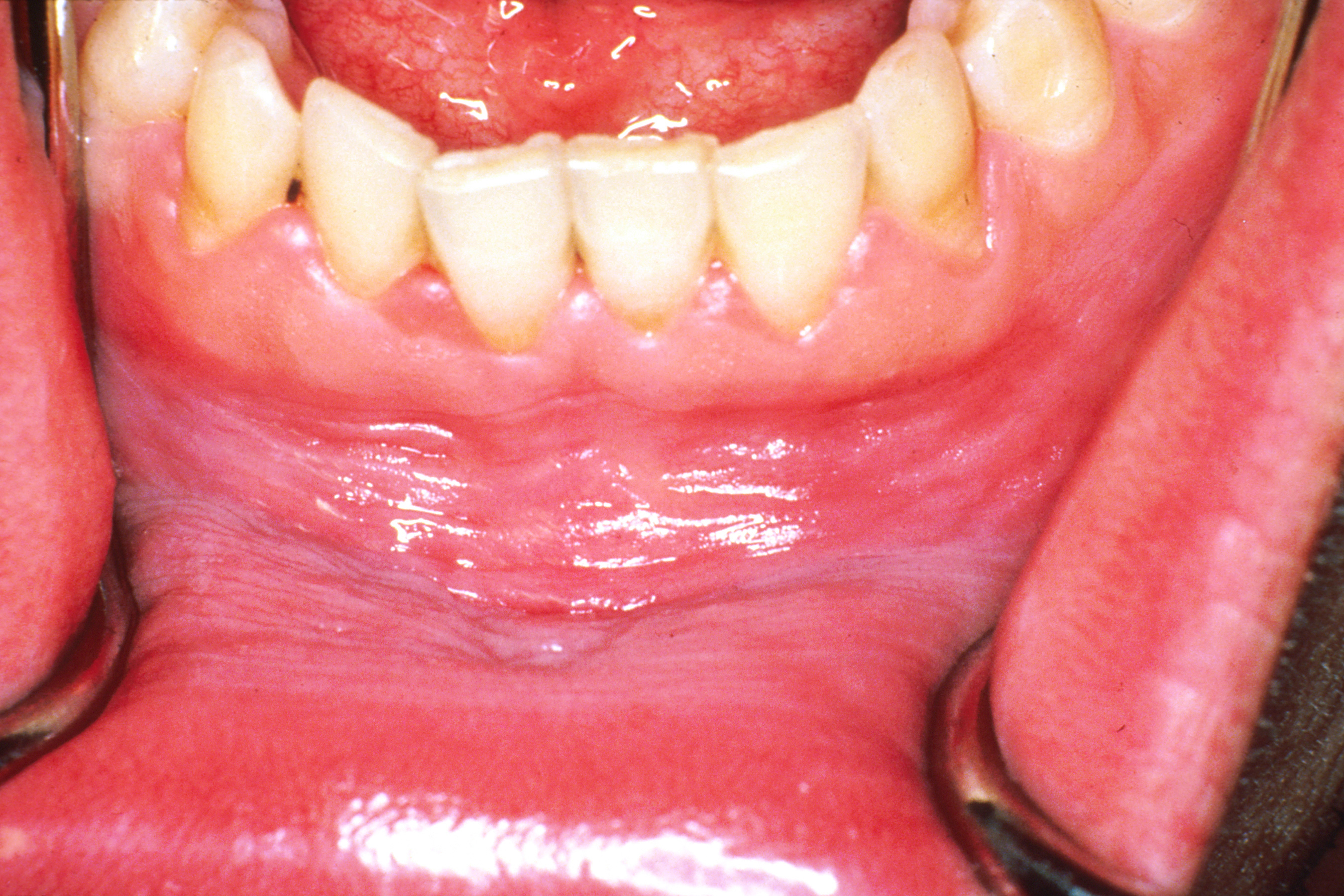
Leukoedema is a blue, grey or white appearance of mucosae, particularly the buccal mucosa (the inside of the cheeks); it may also occur on the mucosa of the larynx or vagina. It is a harmless and very common condition. Because it is so common, it has been argued that it may in fact represent a variation of the normal appearance rather than a disease, but empirical evidence suggests that leukoedema is an acquired condition caused by local irritation. It is found more commonly in black skinned people and tobacco users. The term is derived from the Greek words λευκός ''leukós'', "white" and οἴδημα ''oídēma'', "swelling". Signs and symptoms There is a diffuse, gray-white, milky opalescent appearance of the mucosa which usually occurs bilaterally on the buccal mucosa. Less often, the labial mucosa, the palate or the floor of mouth may be affected. The surface of the area is folded, creating a wrinkled, white streaked lesion. Apart from the appearance, the lesion is enti ... [...More Info...] [...Related Items...] OR: [Wikipedia] [Google] [Baidu] |
Leukoplakia
Oral leukoplakia is a ''potentially malignant disorder'' affecting the oral mucosa. It is defined as "essentially an oral mucosal white lesion that cannot be considered as any other definable lesion." Oral leukoplakia is a white patch or plaque that develops in the oral cavity and is strongly associated with smoking. Leukoplakia is a firmly attached white patch on a mucous membrane which is associated with increased risk of cancer. The edges of the lesion are typically abrupt and the lesion changes with time. Advanced forms may develop red patches. There are generally no other symptoms. It usually occurs within the mouth, although sometimes mucosa in other parts of the gastrointestinal tract, urinary tract, or genitals may be affected. The cause of leukoplakia is unknown. Risk factors for formation inside the mouth include smoking, chewing tobacco, excessive alcohol, and use of betel nuts. One specific type is common in HIV/AIDS. It is a precancerous lesion, a tissue alteration i ... [...More Info...] [...Related Items...] OR: [Wikipedia] [Google] [Baidu] |
Morsicatio Buccarum
Morsicatio buccarum is a condition characterized by chronic irritation or injury to the buccal mucosa (the lining of the inside of the cheek within the mouth), caused by repetitive chewing, biting or nibbling. Signs and symptoms The lesions are located on the mucosa, usually bilaterally in the central part of the anterior buccal mucosa and along the level of the occlusal plane (the level at which the upper and lower teeth meet). Sometimes the tongue or the labial mucosa (the inside lining of the lips) is affected by a similarly produced lesion, termed morsicatio linguarum and morsicatio labiorum respectively. There may be a coexistent linea alba, which corresponds to the occlusal plane, or crenated tongue. The lesions are white with thickening and shredding of mucosa commonly combined with intervening zones of erythema (redness) or ulceration. The surface is irregular, and people may occasionally have loose sections of mucosa that comes away. Causes The cause is chronic parafunc ... [...More Info...] [...Related Items...] OR: [Wikipedia] [Google] [Baidu] |
Oral Medicine
An oral medicine or stomatology doctor (or stomatologist) has received additional specialized training and experience in the diagnosis and management of oral mucosal abnormalities (growths, ulcers, infection, allergies, immune-mediated and autoimmune disorders) including oral cancer, salivary gland disorders, temporomandibular disorders (e.g.: problems with the TMJ) and facial pain (due to musculoskeletal or neurologic conditions), taste and smell disorders; and recognition of the oral manifestations of systemic and infectious diseases. It lies at the interface between medicine and dentistry. An oral medicine doctor is trained to diagnose and manage patients with disorders of the orofacial region, essentially as a "physician of the mouth." History The importance of the mouth in medicine has been recognized since the earliest known medical writings. For example, Hippocrates, Galen and others considered the tongue to be a "barometer" of health, and emphasized the diagnostic and prog ... [...More Info...] [...Related Items...] OR: [Wikipedia] [Google] [Baidu] |
Acanthosis
The epidermis is the outermost of the three layers that comprise the skin, the inner layers being the dermis and hypodermis. The epidermis layer provides a barrier to infection from environmental pathogens and regulates the amount of water released from the body into the atmosphere through transepidermal water loss. The epidermis is composed of multiple layers of flattened cells that overlie a base layer (stratum basale) composed of columnar cells arranged perpendicularly. The layers of cells develop from stem cells in the basal layer. The human epidermis is a familiar example of epithelium, particularly a stratified squamous epithelium. The word epidermis is derived through Latin , itself and . Something related to or part of the epidermis is termed epidermal. Structure Cellular components The epidermis primarily consists of keratinocytes ( proliferating basal and differentiated suprabasal), which comprise 90% of its cells, but also contains melanocytes, Langerhans ce ... [...More Info...] [...Related Items...] OR: [Wikipedia] [Google] [Baidu] |
Exfoliative Cytology
Cytopathology (from Greek , ''kytos'', "a hollow"; , ''pathos'', "fate, harm"; and , ''-logia'') is a branch of pathology that studies and diagnoses diseases on the cellular level. The discipline was founded by George Nicolas Papanicolaou in 1928. Cytopathology is generally used on samples of free cells or tissue fragments, in contrast to histopathology, which studies whole tissues. Cytopathology is frequently, less precisely, called "cytology", which means "the study of cells". Cytopathology is commonly used to investigate diseases involving a wide range of body sites, often to aid in the diagnosis of cancer but also in the diagnosis of some infectious diseases and other inflammatory conditions. For example, a common application of cytopathology is the Pap smear, a screening tool used to detect precancerous cervical lesions that may lead to cervical cancer. Cytopathologic tests are sometimes called smear tests because the samples may be smeared across a glass microscope slide ... [...More Info...] [...Related Items...] OR: [Wikipedia] [Google] [Baidu] |
Edema
Edema, also spelled oedema, and also known as fluid retention, dropsy, hydropsy and swelling, is the build-up of fluid in the body's Tissue (biology), tissue. Most commonly, the legs or arms are affected. Symptoms may include skin which feels tight, the area may feel heavy, and joint stiffness. Other symptoms depend on the underlying cause. Causes may include Chronic venous insufficiency, venous insufficiency, heart failure, kidney problems, hypoalbuminemia, low protein levels, liver problems, deep vein thrombosis, infections, angioedema, certain medications, and lymphedema. It may also occur after prolonged sitting or standing and during menstruation or pregnancy. The condition is more concerning if it starts suddenly, or pain or shortness of breath is present. Treatment depends on the underlying cause. If the underlying mechanism involves Hypernatremia, sodium retention, decreased salt intake and a diuretic may be used. Elevating the legs and support stockings may be useful ... [...More Info...] [...Related Items...] OR: [Wikipedia] [Google] [Baidu] |
Cytoplasm
In cell biology, the cytoplasm is all of the material within a eukaryotic cell, enclosed by the cell membrane, except for the cell nucleus. The material inside the nucleus and contained within the nuclear membrane is termed the nucleoplasm. The main components of the cytoplasm are cytosol (a gel-like substance), the organelles (the cell's internal sub-structures), and various cytoplasmic inclusions. The cytoplasm is about 80% water and is usually colorless. The submicroscopic ground cell substance or cytoplasmic matrix which remains after exclusion of the cell organelles and particles is groundplasm. It is the hyaloplasm of light microscopy, a highly complex, polyphasic system in which all resolvable cytoplasmic elements are suspended, including the larger organelles such as the ribosomes, mitochondria, the plant plastids, lipid droplets, and vacuoles. Most cellular activities take place within the cytoplasm, such as many metabolic pathways including glycolysis, and proces ... [...More Info...] [...Related Items...] OR: [Wikipedia] [Google] [Baidu] |
Squamous Cell
Epithelium or epithelial tissue is one of the four basic types of animal tissue, along with connective tissue, muscle tissue and nervous tissue. It is a thin, continuous, protective layer of compactly packed cells with a little intercellular matrix. Epithelial tissues line the outer surfaces of organs and blood vessels throughout the body, as well as the inner surfaces of cavities in many internal organs. An example is the epidermis, the outermost layer of the skin. There are three principal shapes of epithelial cell: squamous (scaly), columnar, and cuboidal. These can be arranged in a singular layer of cells as simple epithelium, either squamous, columnar, or cuboidal, or in layers of two or more cells deep as stratified (layered), or ''compound'', either squamous, columnar or cuboidal. In some tissues, a layer of columnar cells may appear to be stratified due to the placement of the nuclei. This sort of tissue is called pseudostratified. All glands are made up of epithelia ... [...More Info...] [...Related Items...] OR: [Wikipedia] [Google] [Baidu] |
Pyknosis
Pyknosis, or karyopyknosis, is the irreversible condensation of chromatin in the nucleus of a cell undergoing necrosis or apoptosis. It is followed by karyorrhexis, or fragmentation of the nucleus. Pyknosis (from Ancient Greek meaning "thick, closed or condensed") is also observed in the maturation of erythrocytes (a red blood cell) and the neutrophil (a type of white blood cell). The maturing metarubricyte (a stage in RBC maturation) will condense its nucleus before expelling it to become a reticulocyte. The maturing neutrophil will condense its nucleus into several connected lobes that stay in the cell until the end of its cell life. File:4_Bd_obs_4_680x512px.tif, Micrograph of an infarct in the biliary tract, with pyknotic nuclei (arrows) (400x). Pyknotic nuclei are often found in the zona reticularis of the adrenal gland. They are also found in the keratinocytes of the outermost layer in parakeratinised epithelium. Another use of the word pyknotic, introduced in mathemati ... [...More Info...] [...Related Items...] OR: [Wikipedia] [Google] [Baidu] |
Vacuole
A vacuole () is a membrane-bound organelle which is present in plant and fungal cells and some protist, animal, and bacterial cells. Vacuoles are essentially enclosed compartments which are filled with water containing inorganic and organic molecules including enzymes in solution, though in certain cases they may contain solids which have been engulfed. Vacuoles are formed by the fusion of multiple membrane vesicles and are effectively just larger forms of these. The organelle has no basic shape or size; its structure varies according to the requirements of the cell. Discovery Contractile vacuoles ("stars") were first observed by Spallanzani (1776) in protozoa, although mistaken for respiratory organs. Dujardin (1841) named these "stars" as ''vacuoles''. In 1842, Schleiden applied the term for plant cells, to distinguish the structure with cell sap from the rest of the protoplasm. In 1885, de Vries named the vacuole membrane as tonoplast. Function The function and signifi ... [...More Info...] [...Related Items...] OR: [Wikipedia] [Google] [Baidu] |
Stratum Spinosum
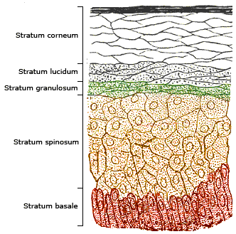
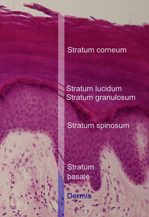
The stratum spinosum (or spinous layer/prickle cell layer) is a layer of the epidermis found between the stratum granulosum and stratum basale. This layer is composed of polyhedral keratinocytes. These are joined with desmosomes. Their spiny (Latin, spinosum) appearance is due to shrinking of the microfilaments between desmosomes that occurs when stained with H&E. Keratinization begins in the stratum spinosum, although the actual keratinocytes begin in the stratum basale. They have large pale-staining nuclei as they are active in synthesizing fibrilar proteins, known as cytokeratin, which build up within the cells aggregating together forming tonofibrils. The tonofibrils go on to form the desmosomes, which allow for strong connections to form between adjacent keratinocytes. The stratum spinosum also contains Langerhans cells. Clinical significance Diffuse hyperplasia of the stratum spinosum is termed acanthosis. Additional images Image:Normal Epidermis and Dermis with Intrade ... [...More Info...] [...Related Items...] OR: [Wikipedia] [Google] [Baidu] |
Hydropic Swell
Hydropic swelling is intracellular edema of keratinocytes, often seen with viral infections. See also * Skin lesion * Skin disease * List of skin diseases Many skin conditions affect the human integumentary system—the organ system covering the entire surface of the body and composed of skin, hair, nails, and related muscle and glands. The major function of this system is as a barrier against ... References Dermatologic terminology {{Med-sign-stub ... [...More Info...] [...Related Items...] OR: [Wikipedia] [Google] [Baidu] |


