|
Left Gastric Vein
The left gastric vein (or coronary vein) is a vein that derives from tributaries draining the lesser curvature of the stomach. Structure The left gastric vein runs from right to left along the lesser curvature of the stomach. It passes to the esophageal opening of the stomach, where it receives some esophageal veins. It then turns backward and passes from left to right behind the omental bursa. It drains into the portal vein near the superior border of the pancreas. Function The left gastric vein drains deoxygenated blood from the lesser curvature of the stomach. It also acts as collaterals between the portal vein and the systemic venous system of the lower esophagus ( azygous vein). Clinical significance Esophageal and paraesophageal varices are supplied primarily by the left gastric vein (due to flow reversal) and typically drain into the azygos/ hemiazygos venous system.Siegelman, E.: "Body MRI", page 47. Saunders, 2004 See also * Right gastric vein The right g ... [...More Info...] [...Related Items...] OR: [Wikipedia] [Google] [Baidu] |
Lesser Curvature Of The Stomach
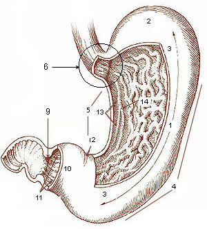
The curvatures of the stomach refer to the greater and lesser curvatures. The greater curvature of the stomach is four or five times as long as the lesser curvature. Greater curvature The greater curvature of the stomach forms the lower left or lateral border of the stomach. Surface Starting from the cardiac orifice at the incisura cardiaca, it forms an arch backward, upward, and to the left; the highest point of the convexity is on a level with the sixth left costal cartilage. From this level it may be followed downward and forward, with a slight convexity to the left as low as the cartilage of the ninth rib; it then turns to the right, to the end of the pylorus. Directly opposite the incisura angularis of the lesser curvature the greater curvature presents a dilatation, which is the left extremity of the pyloric part; this dilatation is limited on the right by a slight groove, the sulcus intermedius, which is about 2.5 cm, from the duodenopyloric constriction. The ... [...More Info...] [...Related Items...] OR: [Wikipedia] [Google] [Baidu] |
Portal Vein
The portal vein or hepatic portal vein (HPV) is a blood vessel that carries blood from the gastrointestinal tract, gallbladder, pancreas and spleen to the liver. This blood contains nutrients and toxins extracted from digested contents. Approximately 75% of total liver blood flow is through the portal vein, with the remainder coming from the hepatic artery proper. The blood leaves the liver to the heart in the hepatic veins. The portal vein is not a true vein, because it conducts blood to capillary beds in the liver and not directly to the heart. It is a major component of the hepatic portal system, one of only two portal venous systems in the body – with the hypophyseal portal system being the other. The portal vein is usually formed by the confluence of the superior mesenteric, splenic veins, inferior mesenteric, left, right gastric veins and the pancreatic vein. Conditions involving the portal vein cause considerable illness and death. An important example of such a condi ... [...More Info...] [...Related Items...] OR: [Wikipedia] [Google] [Baidu] |
Curvatures Of The Stomach
The curvatures of the stomach refer to the greater and lesser curvatures. The Greater curvature of the stomach, greater curvature of the stomach is four or five times as long as the Lesser curvature of the stomach, lesser curvature. Greater curvature The greater curvature of the stomach forms the lower left or lateral border of the stomach. Surface Starting from the cardiac orifice at the cardiac notch of stomach, incisura cardiaca, it forms an arch backward, upward, and to the left; the highest point of the convexity is on a level with the sixth left costal cartilage. From this level it may be followed downward and forward, with a slight convexity to the left as low as the cartilage of the ninth rib; it then turns to the right, to the end of the pylorus. Directly opposite the incisura angularis of the lesser curvature the greater curvature presents a dilatation, which is the left extremity of the pyloric part; this dilatation is limited on the right by a slight groove, the ... [...More Info...] [...Related Items...] OR: [Wikipedia] [Google] [Baidu] |
Stomach
The stomach is a muscular, hollow organ in the gastrointestinal tract of humans and many other animals, including several invertebrates. The stomach has a dilated structure and functions as a vital organ in the digestive system. The stomach is involved in the gastric phase of digestion, following chewing. It performs a chemical breakdown by means of enzymes and hydrochloric acid. In humans and many other animals, the stomach is located between the oesophagus and the small intestine. The stomach secretes digestive enzymes and gastric acid to aid in food digestion. The pyloric sphincter controls the passage of partially digested food ( chyme) from the stomach into the duodenum, where peristalsis takes over to move this through the rest of intestines. Structure In the human digestive system, the stomach lies between the oesophagus and the duodenum (the first part of the small intestine). It is in the left upper quadrant of the abdominal cavity. The top of the stomach lies ag ... [...More Info...] [...Related Items...] OR: [Wikipedia] [Google] [Baidu] |
Esophagus
The esophagus (American English) or oesophagus (British English; both ), non-technically known also as the food pipe or gullet, is an organ in vertebrates through which food passes, aided by peristaltic contractions, from the pharynx to the stomach. The esophagus is a fibromuscular tube, about long in adults, that travels behind the trachea and heart, passes through the diaphragm, and empties into the uppermost region of the stomach. During swallowing, the epiglottis tilts backwards to prevent food from going down the larynx and lungs. The word ''oesophagus'' is from Ancient Greek οἰσοφάγος (oisophágos), from οἴσω (oísō), future form of φέρω (phérō, “I carry”) + ἔφαγον (éphagon, “I ate”). The wall of the esophagus from the lumen outwards consists of mucosa, submucosa (connective tissue), layers of muscle fibers between layers of fibrous tissue, and an outer layer of connective tissue. The mucosa is a stratified squamous epithel ... [...More Info...] [...Related Items...] OR: [Wikipedia] [Google] [Baidu] |
Esophageal Vein
The esophageal veins drain blood from the esophagus to the azygos vein, in the thorax, and to the inferior thyroid vein in the neck. It also drains, although with less significance, to the hemiazygos vein, posterior intercostal vein and bronchial veins. In the abdomen, some drain to the left gastric vein which drains into the portal vein. See also * Esophageal varices Esophageal varices are extremely dilated sub-mucosal veins in the lower third of the esophagus. They are most often a consequence of portal hypertension, commonly due to cirrhosis. People with esophageal varices have a strong tendency to develop ... References External links * * () * https://web.archive.org/web/20080506103555/http://www.med.mun.ca/anatomyts/digest/avein6.htm Veins of the torso {{circulatory-stub ... [...More Info...] [...Related Items...] OR: [Wikipedia] [Google] [Baidu] |
Omental Bursa
The lesser sac, also known as the omental bursa, is a part of the peritoneal cavity that is formed by the lesser and greater omentum. Usually found in mammals, it is connected with the greater sac via the omental foramen or ''Foramen of Winslow''. In mammals, it is common for the lesser sac to contain considerable amounts of fat. Anatomic margins ;Anterior margin: listed from the top-to-bottom margin: Quadrate lobe of the liver, lesser omentum, stomach, gastrocolic ligament ;Lateral margin: listed from the most anterior to the most posterior margin: Gastrosplenic ligament, spleen, phrenicosplenic ligament ;Posterior margin: Left kidney and adrenal gland, pancreas ;Inferior margin: Greater omentum ;Superior margin: Liver If any of the marginal structures rupture their contents could leak into the lesser sac. If the stomach were to rupture on its anterior side though the leak would collect in the greater sac. The lesser sac is formed during embryogenesis from an infolding of ... [...More Info...] [...Related Items...] OR: [Wikipedia] [Google] [Baidu] |
Pancreas
The pancreas is an organ of the digestive system and endocrine system of vertebrates. In humans, it is located in the abdomen behind the stomach and functions as a gland. The pancreas is a mixed or heterocrine gland, i.e. it has both an endocrine and a digestive exocrine function. 99% of the pancreas is exocrine and 1% is endocrine. As an endocrine gland, it functions mostly to regulate blood sugar levels, secreting the hormones insulin, glucagon, somatostatin, and pancreatic polypeptide. As a part of the digestive system, it functions as an exocrine gland secreting pancreatic juice into the duodenum through the pancreatic duct. This juice contains bicarbonate, which neutralizes acid entering the duodenum from the stomach; and digestive enzymes, which break down carbohydrates, proteins, and fats in food entering the duodenum from the stomach. Inflammation of the pancreas is known as pancreatitis, with common causes including chronic alcohol use and gallstones. Becaus ... [...More Info...] [...Related Items...] OR: [Wikipedia] [Google] [Baidu] |
Blood
Blood is a body fluid in the circulatory system of humans and other vertebrates that delivers necessary substances such as nutrients and oxygen to the cells, and transports metabolic waste products away from those same cells. Blood in the circulatory system is also known as ''peripheral blood'', and the blood cells it carries, ''peripheral blood cells''. Blood is composed of blood cells suspended in blood plasma. Plasma, which constitutes 55% of blood fluid, is mostly water (92% by volume), and contains proteins, glucose, mineral ions, hormones, carbon dioxide (plasma being the main medium for excretory product transportation), and blood cells themselves. Albumin is the main protein in plasma, and it functions to regulate the colloidal osmotic pressure of blood. The blood cells are mainly red blood cells (also called RBCs or erythrocytes), white blood cells (also called WBCs or leukocytes) and platelets (also called thrombocytes). The most abundant cells in vertebrate blo ... [...More Info...] [...Related Items...] OR: [Wikipedia] [Google] [Baidu] |
Azygous Vein
The azygos vein is a vein running up the right side of the thoracic vertebral column draining itself towards the superior vena cava. It connects the systems of superior vena cava and inferior vena cava and can provide an alternative path for blood to the right atrium when either of the venae cavae is blocked. Structure The azygos vein transports deoxygenated blood from the posterior walls of the thorax and abdomen into the superior vena cava. It is formed by the union of the ascending lumbar veins with the right subcostal veins at the level of the 12th thoracic vertebra, ascending to the right of the descending aorta and thoracic duct, passing behind the right crus of diaphragm, anterior to the vertebral bodies of T12 to T5 and right posterior intercostal arteries. At the level of T4 vertebrae, it arches over the root of the right lung from behind to the front to join the superior vena cava. The trachea and oesophagus is located medially to the arch of the azygous vein. The "a ... [...More Info...] [...Related Items...] OR: [Wikipedia] [Google] [Baidu] |
Esophageal Varices
Esophageal varices are extremely dilated sub-mucosal veins in the lower third of the esophagus. They are most often a consequence of portal hypertension, commonly due to cirrhosis. People with esophageal varices have a strong tendency to develop severe bleeding which left untreated can be fatal. Esophageal varices are typically diagnosed through an esophagogastroduodenoscopy. Pathogenesis The upper two thirds of the esophagus are drained via the esophageal veins, which carry deoxygenated blood from the esophagus to the azygos vein, which in turn drains directly into the superior vena cava. These veins have no part in the development of esophageal varices. The lower one third of the esophagus is drained into the superficial veins lining the esophageal mucosa, which drain into the left gastric vein, which in turn drains directly into the portal vein. These superficial veins (normally only approximately 1 mm in diameter) become distended up to 1–2 cm in diameter in as ... [...More Info...] [...Related Items...] OR: [Wikipedia] [Google] [Baidu] |
Azygos Vein
The azygos vein is a vein running up the right side of the thoracic vertebral column draining itself towards the superior vena cava. It connects the systems of superior vena cava and inferior vena cava and can provide an alternative path for blood to the right atrium when either of the venae cavae is blocked. Structure The azygos vein transports deoxygenated blood from the posterior walls of the thorax and abdomen into the superior vena cava. It is formed by the union of the ascending lumbar veins with the right subcostal veins at the level of the 12th thoracic vertebra, ascending to the right of the descending aorta and thoracic duct, passing behind the right crus of diaphragm, anterior to the vertebral bodies of T12 to T5 and right posterior intercostal arteries. At the level of T4 vertebrae, it arches over the root of the right lung from behind to the front to join the superior vena cava. The trachea and oesophagus is located medially to the arch of the azygous vein. The ... [...More Info...] [...Related Items...] OR: [Wikipedia] [Google] [Baidu] |


