|
Lateral Nasal Branch Of Facial Artery
The lateral nasal branch of facial artery (lateral nasal artery) is derived from the facial artery as that vessel ascends along the side of the nose. Supplies It supplies the ala and dorsum of the nose, anastomosing with its fellow, with the septal and alar branches, with the dorsal nasal branch of the ophthalmic artery, and with the infraorbital branch of the internal maxillary The maxillary artery supplies deep structures of the face. It branches from the external carotid artery just deep to the neck of the mandible. Structure The maxillary artery, the larger of the two terminal branches of the external carotid artery, .... If the posterior lateral nasal artery is superficial in the nasal wall, a laceration may occur during an aggressive curettage. A sinus floor elevation procedure requires a separation and elevation of the sinus lining with subsequent introduction of space maintaining graft material. During the lining elevation this artery may be cut in the osseous nasal wal ... [...More Info...] [...Related Items...] OR: [Wikipedia] [Google] [Baidu] |
Facial Artery
The facial artery (external maxillary artery in older texts) is a branch of the external carotid artery that supplies structures of the superficial face. Structure The facial artery arises in the carotid triangle from the external carotid artery, a little above the lingual artery and, sheltered by the ramus of the mandible. It passes obliquely up beneath the digastric and stylohyoid muscles, over which it arches to enter a groove on the posterior surface of the submandibular gland. It then curves upward over the body of the mandible at the antero-inferior angle of the masseter; passes forward and upward across the cheek to the angle of the mouth, then ascends along the side of the nose, and ends at the medial commissure of the eye, under the name of the angular artery. The facial artery is remarkably tortuous. This is to accommodate itself to neck movements such as those of the pharynx in deglutition; and facial movements such as those of the mandible, lips, and cheeks. ... [...More Info...] [...Related Items...] OR: [Wikipedia] [Google] [Baidu] |
Ala Of Nose
The human nose is the most protruding part of the face. It bears the nostrils and is the first organ of the respiratory system. It is also the principal organ in the olfactory system. The shape of the nose is determined by the nasal bones and the nasal cartilages, including the nasal septum which separates the nostrils and divides the nasal cavity into two. On average the nose of a male is larger than that of a female. The nose has an important function in breathing. The nasal mucosa lining the nasal cavity and the paranasal sinuses carries out the necessary conditioning of inhaled air by warming and moistening it. Nasal conchae, shell-like bones in the walls of the cavities, play a major part in this process. Filtering of the air by nasal hair in the nostrils prevents large particles from entering the lungs. Sneezing is a reflex to expel unwanted particles from the nose that irritate the mucosal lining. Sneezing can transmit infections, because aerosols are created in w ... [...More Info...] [...Related Items...] OR: [Wikipedia] [Google] [Baidu] |
Dorsum (anatomy)
Standard anatomical terms of location are used to unambiguously describe the anatomy of animals, including humans. The terms, typically derived from Latin or Greek roots, describe something in its standard anatomical position. This position provides a definition of what is at the front ("anterior"), behind ("posterior") and so on. As part of defining and describing terms, the body is described through the use of anatomical planes and anatomical axes. The meaning of terms that are used can change depending on whether an organism is bipedal or quadrupedal. Additionally, for some animals such as invertebrates, some terms may not have any meaning at all; for example, an animal that is radially symmetrical will have no anterior surface, but can still have a description that a part is close to the middle ("proximal") or further from the middle ("distal"). International organisations have determined vocabularies that are often used as standard vocabularies for subdisciplines of anatom ... [...More Info...] [...Related Items...] OR: [Wikipedia] [Google] [Baidu] |
Human Nose
The human nose is the most protruding part of the face. It bears the nostrils and is the first organ of the respiratory system. It is also the principal organ in the olfactory system. The shape of the nose is determined by the nasal bones and the nasal cartilages, including the nasal septum which separates the nostrils and divides the nasal cavity into two. On average the nose of a male is larger than that of a female. The nose has an important function in breathing. The nasal mucosa lining the nasal cavity and the paranasal sinuses carries out the necessary conditioning of inhaled air by warming and moistening it. Nasal conchae, shell-like bones in the walls of the cavities, play a major part in this process. Filtering of the air by nasal hair in the nostrils prevents large particles from entering the lungs. Sneezing is a reflex to expel unwanted particles from the nose that irritate the mucosal lining. Sneezing can transmit infections, because aerosols are created in w ... [...More Info...] [...Related Items...] OR: [Wikipedia] [Google] [Baidu] |
Anastomosing
An anastomosis (, plural anastomoses) is a connection or opening between two things (especially cavities or passages) that are normally diverging or branching, such as between blood vessels, leaf veins, or streams. Such a connection may be normal (such as the foramen ovale in a fetus's heart) or abnormal (such as the patent foramen ovale in an adult's heart); it may be acquired (such as an arteriovenous fistula) or innate (such as the arteriovenous shunt of a metarteriole); and it may be natural (such as the aforementioned examples) or artificial (such as a surgical anastomosis). The reestablishment of an anastomosis that had become blocked is called a reanastomosis. Anastomoses that are abnormal, whether congenital or acquired, are often called fistulas. The term is used in medicine, biology, mycology, geology, and geography. Etymology Anastomosis: medical or Modern Latin, from Greek ἀναστόμωσις, anastomosis, "outlet, opening", Gr ana- "up, on, upon", stoma "mouth", ... [...More Info...] [...Related Items...] OR: [Wikipedia] [Google] [Baidu] |
Septal
In biology, a septum (Latin for ''something that encloses''; plural septa) is a wall, dividing a cavity or structure into smaller ones. A cavity or structure divided in this way may be referred to as septate. Examples Human anatomy * Interatrial septum, the wall of tissue that is a sectional part of the left and right atria of the heart * Interventricular septum, the wall separating the left and right ventricles of the heart * Lingual septum, a vertical layer of fibrous tissue that separates the halves of the tongue. *Nasal septum: the cartilage wall separating the nostrils of the nose * Alveolar septum: the thin wall which separates the alveoli from each other in the lungs * Orbital septum, a palpebral ligament in the upper and lower eyelids * Septum pellucidum or septum lucidum, a thin structure separating two fluid pockets in the brain * Uterine septum, a malformation of the uterus * Vaginal septum, a lateral or transverse partition inside the vagina * Intermuscular sep ... [...More Info...] [...Related Items...] OR: [Wikipedia] [Google] [Baidu] |
Alar
Daminozide—also known as aminozide, Alar, Kylar, SADH, B-995, B-nine, and DMASA,—is a plant growth regulator, a chemical sprayed on fruit to regulate growth, make harvest easier, and keep apples from falling off the trees before they ripen so they are red and firm for storage. It was produced in the U.S. by the Uniroyal Chemical Company, Inc, (now integrated into the Chemtura Corporation), which registered daminozide for use on fruits intended for human consumption in 1963. In addition to apples and ornamental plants, they also registered it for use on cherries, peaches, pears, Concord grapes, tomato transplants, and peanut vines. Alar was first approved for use in the U.S. in 1963. It was primarily used on apples until 1989, when the manufacturer voluntarily withdrew it after the U.S. Environmental Protection Agency proposed banning it based on concerns about cancer risks to consumers. On fruit trees, daminozide affects flow-bud initiation, fruit-set maturity, fruit firmness ... [...More Info...] [...Related Items...] OR: [Wikipedia] [Google] [Baidu] |
Dorsal Nasal Branch
The dorsal nasal artery is an artery of the face. It is one of the two terminal branches of the ophthalmic artery. It contributes arterial supply to the lacrimal sac, and outer surface of the nose. Structure Origin The dorsal nasal artery is one of the two terminal branches of the ophthalmic artery (the other being the supratrochlear artery). It arises in the superomedial orbit. Course and relations It passes anteriorly to exit the orbit between the trochlea (superiorly), the medial palpebral ligament (inferiorly). It gives a branch to the lacrimal sac before bifurcating into two branches: one branch anastomoses with the terminal (angular) part of the facial artery and is important for the blood supply of the face; the other travels along the dorsum of the nose to supply the outer surface of the nose, and forms anastomoses with its contralateral fellow, and with the lateral nasal branch of the facial artery. Distribution The dorsal nasal artery contributes arterial supply ... [...More Info...] [...Related Items...] OR: [Wikipedia] [Google] [Baidu] |
Ophthalmic Artery
The ophthalmic artery (OA) is an artery of the head. It is the first branch of the internal carotid artery distal to the cavernous sinus. Branches of the ophthalmic artery supply all the structures in the orbit around the eye, as well as some structures in the nose, face, and meninges. Occlusion of the ophthalmic artery or its branches can produce sight-threatening conditions. Structure The ophthalmic artery emerges from the internal carotid artery. This is usually just after the internal carotid artery emerges from the cavernous sinus. In some cases, the ophthalmic artery branches just before the internal carotid exits the cavernous sinus. The ophthalmic artery emerges along the medial side of the anterior clinoid process. It runs anteriorly, passing through the optic canal inferolaterally to the optic nerve. It can also pass superiorly to the optic nerve in a minority of cases. In the posterior third of the cone of the orbit, the ophthalmic artery turns sharply and medially t ... [...More Info...] [...Related Items...] OR: [Wikipedia] [Google] [Baidu] |
Infraorbital Branch
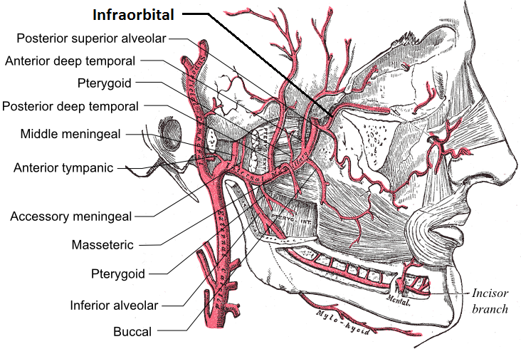
The infraorbital artery is an artery in the head that branches off the maxillary artery, emerging through the infraorbital foramen, just under the orbit of the eye. Course The infraorbital artery appears, from its direction, to be the continuation of the trunk of the maxillary artery, but often arises in conjunction with the posterior superior alveolar artery. It runs along the infraorbital groove and canal with the infraorbital nerve, and emerges on the face through the infraorbital foramen, beneath the infraorbital head of the levator labii superioris muscle. Branches While in the canal, it gives off * (a) orbital branches which assist in supplying the inferior rectus and inferior oblique and the lacrimal sac, and * (b) anterior superior alveolar arteries - branches which descend through the anterior alveolar canals to supply the upper incisor and canine teeth and the mucous membrane of the maxillary sinus. On the face, some branches pass upward to the medial angle of the orb ... [...More Info...] [...Related Items...] OR: [Wikipedia] [Google] [Baidu] |
Internal Maxillary
The maxillary artery supplies deep structures of the face. It branches from the external carotid artery just deep to the neck of the mandible. Structure The maxillary artery, the larger of the two terminal branches of the external carotid artery, arises behind the neck of the mandible, and is at first imbedded in the substance of the parotid gland; it passes forward between the ramus of the mandible and the sphenomandibular ligament, and then runs, either superficial or deep to the lateral pterygoid muscle, to the pterygopalatine fossa. It supplies the deep structures of the face, and may be divided into mandibular, pterygoid, and pterygopalatine portions. First portion The ''first'' or ''mandibular '' or ''bony'' portion passes horizontally forward, between the neck of the mandible and the sphenomandibular ligament, where it lies parallel to and a little below the auriculotemporal nerve; it crosses the inferior alveolar nerve, and runs along the lower border of the lateral ptery ... [...More Info...] [...Related Items...] OR: [Wikipedia] [Google] [Baidu] |


