|
Lateral Compartment Of Leg
The lateral compartment of the leg is a fascial compartment of the lower leg. It contains muscles which make eversion and plantarflexion of the foot. Muscles The lateral compartment of the leg contains: * Fibularis longus * Fibularis brevis Action * Foot evertors * Foot plantarflexion Nerve Supply The lateral compartment of the leg is supplied by the superficial fibular nerve (superficial peroneal nerve). Blood Supply Its proximal and distal arterial supply consists of perforating branches of the anterior tibial artery and fibular artery In anatomy, the fibular artery, also known as the peroneal artery, supplies blood to the lateral compartment of the leg. It arises from the tibial-fibular trunk. Structure The fibular artery arises from the bifurcation of tibial-fibular trunk int .... Additional images File:Lateral compartment of leg - animation.gif, Animation. Fibularis longus (blue) and fibularis brevis (red). See also * Fascial compartments of leg Re ... [...More Info...] [...Related Items...] OR: [Wikipedia] [Google] [Baidu] |
Fibular Artery
In anatomy, the fibular artery, also known as the peroneal artery, supplies blood to the lateral compartment of the leg. It arises from the tibial-fibular trunk. Structure The fibular artery arises from the bifurcation of tibial-fibular trunk into the fibular and posterior tibial arteries in the upper part of the leg proper, just below the knee. It runs towards the foot in the deep posterior compartment of the leg, just medial to the fibula. It supplies a perforating branch to both the lateral and anterior compartments of the leg; it also provides a nutrient artery to the fibula. Some sources claim that the fibular artery arises directly from the posterior tibial artery, but vascular and plastic surgeons note the clinical significance of the tibial-fibular trunk. The fibular artery is accompanied by small veins (venae comitantes) known as fibular veins. Branches Communication branch to posterior tibial artery. Perforating branch to anterior lateral malleolar artery. A ca ... [...More Info...] [...Related Items...] OR: [Wikipedia] [Google] [Baidu] |
Superficial Fibular Nerve
The superficial fibular nerve (also known as superficial peroneal nerve) innervates the fibularis longus and fibularis brevis muscles and the skin over the antero-lateral aspect of the leg along with the greater part of the dorsum of the foot (with the exception of the first web space, which is innervated by the deep fibular nerve). Structure Lateral side of the leg The superficial fibular nerve is the main nerve of the lateral compartment of the leg. It begins at the lateral side of the neck of fibula, and runs through the fibularis longus and fibularis brevis muscles. In the middle third of the leg, it descends between the fibularis longus and fibularis brevis, and then reaches the anterior border of the fibularis brevis to enter the groove between the fibularis brevis and the extensor digitorum longus under the deep fascia of leg. It becomes superficial at the junction of upper two-thirds and lower one-thirds of the leg by piercing the deep fascia. The superficial fibular nerve ... [...More Info...] [...Related Items...] OR: [Wikipedia] [Google] [Baidu] |
Fascial Compartments Of Leg
The fascial compartments of the leg are the four fascial compartments that separate and contain the muscles of the lower leg (from the knee to the ankle). The compartments are divided by septa formed from the fascia. The compartments usually have nerve and blood supplies separate from their neighbours. All of the muscles within a compartment will generally be supplied by the same nerve. Intermuscular septa The lower leg is divided into four compartments by the interosseous membrane of the leg, the anterior intermuscular septum, the transverse intermuscular septum and the posterior intermuscular septum. Each compartment contains connective tissue, nerves and blood vessels. The septa are formed from the fascia which is made up of a strong type of connective tissue. The fascia also separates the skeletal muscles from the subcutaneous tissue. Due to the great pressure placed on the leg, from the column of blood from the heart to the feet, the fascia is very thick in order to s ... [...More Info...] [...Related Items...] OR: [Wikipedia] [Google] [Baidu] |
Anatomical Terms Of Motion
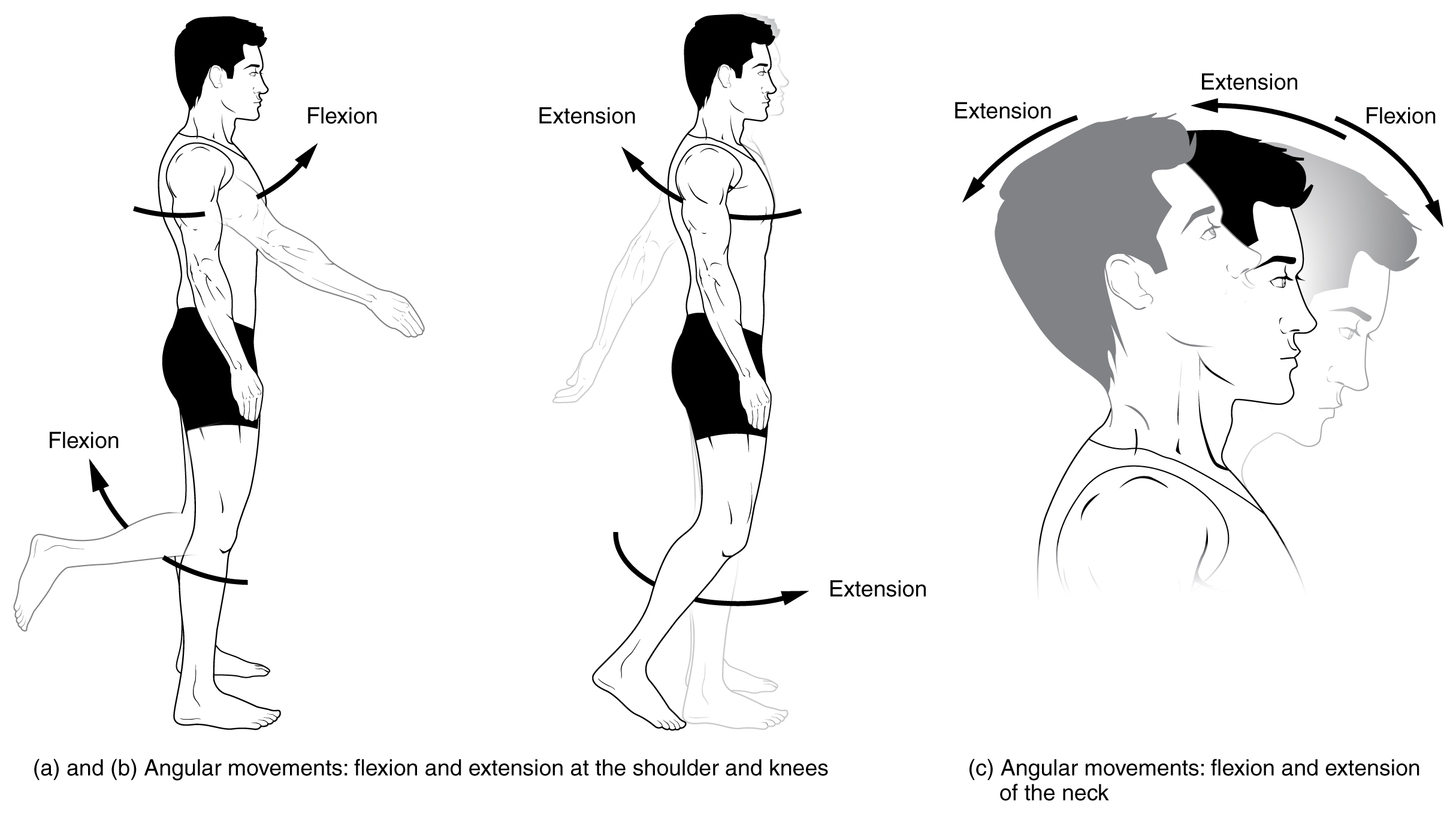
Motion, the process of movement, is described using specific anatomical terms. Motion includes movement of organs, joints, limbs, and specific sections of the body. The terminology used describes this motion according to its direction relative to the anatomical position of the body parts involved. Anatomists and others use a unified set of terms to describe most of the movements, although other, more specialized terms are necessary for describing unique movements such as those of the hands, feet, and eyes. In general, motion is classified according to the anatomical plane it occurs in. ''Flexion'' and ''extension'' are examples of ''angular'' motions, in which two axes of a joint are brought closer together or moved further apart. ''Rotational'' motion may occur at other joints, for example the shoulder, and are described as ''internal'' or ''external''. Other terms, such as ''elevation'' and ''depression'', describe movement above or below the horizontal plane. Many anatom ... [...More Info...] [...Related Items...] OR: [Wikipedia] [Google] [Baidu] |
Fibularis Longus
In human anatomy, the fibularis longus (also known as peroneus longus) is a superficial muscle in the lateral compartment of the leg. It acts to tilt the sole of the foot away from the midline of the body ( eversion) and to extend the foot downward away from the body (plantar flexion) at the ankle. The fibularis longus is the longest and most superficial of the three fibularis (peroneus) muscles. At its upper end, it is attached to the head of the fibula, and its "belly" runs down along most of this bone. The muscle becomes a tendon that wraps around and behind the lateral malleolus of the ankle, then continues under the foot to attach to the medial cuneiform and first metatarsal. It is supplied by the superficial fibular nerve. Structure The fibularis longus arises from the head and upper two-thirds of the lateral, or outward, surface of the fibula, from the deep surface of the fascia, and from the connective tissue between it and the muscles on the front and back of the leg. ... [...More Info...] [...Related Items...] OR: [Wikipedia] [Google] [Baidu] |
Fibularis Brevis
In human anatomy, the fibularis brevis (or peroneus brevis) is a muscle that lies underneath the fibularis longus within the lateral compartment of the leg. It acts to tilt the sole of the foot away from the midline of the body (eversion) and to extend the foot downward away from the body at the ankle (plantar flexion). Structure The fibularis brevis arises from the lower two-thirds of the lateral, or outward, surface of the fibula (inward in relation to the fibularis longus) and from the connective tissue between it and the muscles on the front and back of the leg. The muscle passes downward and ends in a tendon that runs behind the lateral malleolus of the ankle in a groove that it shares with the tendon of the fibularis longus; the groove is converted into a canal by the superior fibular retinaculum, and the tendons in it are contained in a common mucous sheath. The tendon then runs forward along the lateral side of the calcaneus, above the calcaneal tubercle and the tendon ... [...More Info...] [...Related Items...] OR: [Wikipedia] [Google] [Baidu] |
Lateral Compartment Of Leg - Fibularis Longus
Lateral is a geometric term of location which may refer to: Healthcare * Lateral (anatomy), an anatomical direction *Lateral cricoarytenoid muscle *Lateral release (surgery), a surgical procedure on the side of a kneecap Phonetics *Lateral consonant, an l-like consonant in which air flows along the sides of the tongue **Lateral release (phonetics), the release of a plosive consonant into a lateral consonant Other uses *''Lateral'', journal of the Cultural Studies Association *Lateral canal, a canal built beside another stream *Lateral hiring, recruiting that targets employees of another organization *Lateral mark, a sea mark used in maritime pilotage to indicate the edge of a channel * Lateral stability of aircraft during flight * Lateral pass, a type of pass in American and Canadian football *Lateral support (other), various meanings *Lateral thinking, the solution of problems through an indirect and creative approach *Lateral number, a proposed alternate term for i ... [...More Info...] [...Related Items...] OR: [Wikipedia] [Google] [Baidu] |
Lateral Compartment Of Leg - Fibularis Brevis
Lateral is a geometric term of location which may refer to: Healthcare * Lateral (anatomy), an anatomical direction *Lateral cricoarytenoid muscle *Lateral release (surgery), a surgical procedure on the side of a kneecap Phonetics *Lateral consonant, an l-like consonant in which air flows along the sides of the tongue **Lateral release (phonetics), the release of a plosive consonant into a lateral consonant Other uses *''Lateral'', journal of the Cultural Studies Association *Lateral canal, a canal built beside another stream *Lateral hiring, recruiting that targets employees of another organization *Lateral mark, a sea mark used in maritime pilotage to indicate the edge of a channel * Lateral stability of aircraft during flight * Lateral pass, a type of pass in American and Canadian football *Lateral support (other), various meanings *Lateral thinking, the solution of problems through an indirect and creative approach *Lateral number, a proposed alternate term for i ... [...More Info...] [...Related Items...] OR: [Wikipedia] [Google] [Baidu] |
Evertors
Motion, the process of movement, is described using specific anatomical terms. Motion includes movement of organs, joints, limbs, and specific sections of the body. The terminology used describes this motion according to its direction relative to the anatomical position of the body parts involved. Anatomists and others use a unified set of terms to describe most of the movements, although other, more specialized terms are necessary for describing unique movements such as those of the hands, feet, and eyes. In general, motion is classified according to the anatomical plane it occurs in. ''Flexion'' and ''extension'' are examples of ''angular'' motions, in which two axes of a joint are brought closer together or moved further apart. ''Rotational'' motion may occur at other joints, for example the shoulder, and are described as ''internal'' or ''external''. Other terms, such as ''elevation'' and ''depression'', describe movement above or below the horizontal plane. Many anatomic ... [...More Info...] [...Related Items...] OR: [Wikipedia] [Google] [Baidu] |
Plantarflexion
Motion, the process of movement, is described using specific anatomical terms. Motion includes movement of organs, joints, limbs, and specific sections of the body. The terminology used describes this motion according to its direction relative to the anatomical position of the body parts involved. Anatomists and others use a unified set of terms to describe most of the movements, although other, more specialized terms are necessary for describing unique movements such as those of the hands, feet, and eyes. In general, motion is classified according to the anatomical plane it occurs in. ''Flexion'' and ''extension'' are examples of ''angular'' motions, in which two axes of a joint are brought closer together or moved further apart. ''Rotational'' motion may occur at other joints, for example the shoulder, and are described as ''internal'' or ''external''. Other terms, such as ''elevation'' and ''depression'', describe movement above or below the horizontal plane. Many anatomic ... [...More Info...] [...Related Items...] OR: [Wikipedia] [Google] [Baidu] |
Anterior Tibial Artery
The anterior tibial artery is an artery of the leg. It carries blood to the anterior compartment of the leg and dorsal surface of the foot, from the popliteal artery. Structure Course The anterior tibial artery is a branch of the popliteal artery. It originates at the distal end of the popliteus muscle posterior to the tibia. The artery typically passes anterior to the popliteus muscle prior to passing between the tibia and fibula through an oval opening at the superior aspect of the interosseus membrane. The artery then descends between the tibialis anterior and extensor digitorum longus muscles. It is accompanied by the anterior tibial vein, and the deep peroneal nerve, along its course. It crosses the anterior aspect of the ankle joint, at which point it becomes the dorsalis pedis artery. Branches The branches of the anterior tibial artery are: *posterior tibial recurrent artery * anterior tibial recurrent artery * muscular branches * anterior medial malleolar ar ... [...More Info...] [...Related Items...] OR: [Wikipedia] [Google] [Baidu] |
Fascial Compartments Of Leg
The fascial compartments of the leg are the four fascial compartments that separate and contain the muscles of the lower leg (from the knee to the ankle). The compartments are divided by septa formed from the fascia. The compartments usually have nerve and blood supplies separate from their neighbours. All of the muscles within a compartment will generally be supplied by the same nerve. Intermuscular septa The lower leg is divided into four compartments by the interosseous membrane of the leg, the anterior intermuscular septum, the transverse intermuscular septum and the posterior intermuscular septum. Each compartment contains connective tissue, nerves and blood vessels. The septa are formed from the fascia which is made up of a strong type of connective tissue. The fascia also separates the skeletal muscles from the subcutaneous tissue. Due to the great pressure placed on the leg, from the column of blood from the heart to the feet, the fascia is very thick in order to s ... [...More Info...] [...Related Items...] OR: [Wikipedia] [Google] [Baidu] |


