|
Knee Reconstruction
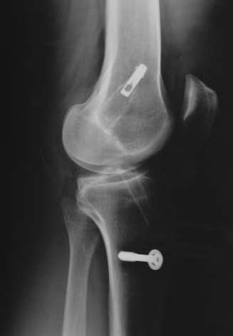
Anterior cruciate ligament reconstruction (ACL reconstruction) is a surgical tissue graft replacement of the anterior cruciate ligament, located in the knee, to restore its function after an injury. The torn ligament can either be removed from the knee (most common), or preserved (where the graft is passed inside the preserved ruptured native ligament) before reconstruction through an arthroscopic procedure. ACL repair is also a surgical option. This involves repairing the ACL by re-attaching it, instead of performing a reconstruction. Theoretical advantages of repair include faster recovery and a lack of donor site morbidity, but randomised controlled trials and long-term data regarding re-rupture rates using contemporary surgical techniques are lacking. Background The Anterior Cruciate Ligament is the ligament that keeps the knee stable. Anterior Cruciate Ligament damage is a very common injury, especially among athletes. Anterior Cruciate Ligament Reconstruction (ACL) sur ... [...More Info...] [...Related Items...] OR: [Wikipedia] [Google] [Baidu] |
Musculus Semitendinosus
The semitendinosus () is a long superficial muscle in the back of the thigh. It is so named because it has a very long tendon of insertion. It lies posteromedially in the thigh, superficial to the semimembranosus. Structure The semitendinosus, remarkable for the great length of its tendon of insertion, is situated at the posterior and medial aspect of the thigh. It arises from the lower and medial impression on the upper part of the tuberosity of the ischium, by a tendon common to it and the long head of the biceps femoris; it also arises from an aponeurosis which connects the adjacent surfaces of the two muscles to the extent of about 7.5 cm. from their origin. The muscle is fusiform and ends a little below the middle of the thigh in a long round tendon which lies along the medial side of the popliteal fossa; it then curves around the medial condyle of the tibia and passes over the medial collateral ligament of the knee-joint, from which it is separated by a bursa, and is ... [...More Info...] [...Related Items...] OR: [Wikipedia] [Google] [Baidu] |
Quadriceps Tendon
In human anatomy, the quadriceps tendon works with the Quadriceps muscle, quadriceps muscle to extend the leg. All four parts of the quadriceps muscle attach to the tuberosity of the tibia, shin via the patella (knee cap), where the quadriceps tendon becomes the patellar ligament. It attaches the quadriceps to the top of the patella, which in turn is connected to the shin from its bottom by the patellar ligament. A tendon connects muscle to bone, while a ligament connects bone to bone.Saladin, Kenneth S. Anatomy & Physiology: The Unity of Form and Function. 6th ed. New York: McGraw-Hill, 2012. Print. Injuries are common to this tendon, with tears, either partial or complete, being the most common. If the quadriceps tendon is completely torn, surgery will be required to regain function of the knee."Patellar Tendon Tear." OrthoInfo - AAOS. American Academy of Orthopaedic Surgeons, Aug. 2009. Web. 07 Dec. 2014. Without the quadriceps tendon, the knee cannot extend. Often, when the ... [...More Info...] [...Related Items...] OR: [Wikipedia] [Google] [Baidu] |
Patella
The patella, also known as the kneecap, is a flat, rounded triangular bone which articulates with the femur (thigh bone) and covers and protects the anterior articular surface of the knee joint. The patella is found in many tetrapods, such as mice, cats, birds and dogs, but not in whales, or most reptiles. In humans, the patella is the largest sesamoid bone (i.e., embedded within a tendon or a muscle) in the body. Babies are born with a patella of soft cartilage which begins to ossify into bone at about four years of age. Structure The patella is a sesamoid bone roughly triangular in shape, with the apex of the patella facing downwards. The apex is the most inferior (lowest) part of the patella. It is pointed in shape, and gives attachment to the patellar ligament. The front and back surfaces are joined by a thin margin and towards centre by a thicker margin. The tendon of the quadriceps femoris muscle attaches to the base of the patella., with the vastus intermedius muscl ... [...More Info...] [...Related Items...] OR: [Wikipedia] [Google] [Baidu] |
Newton (unit)
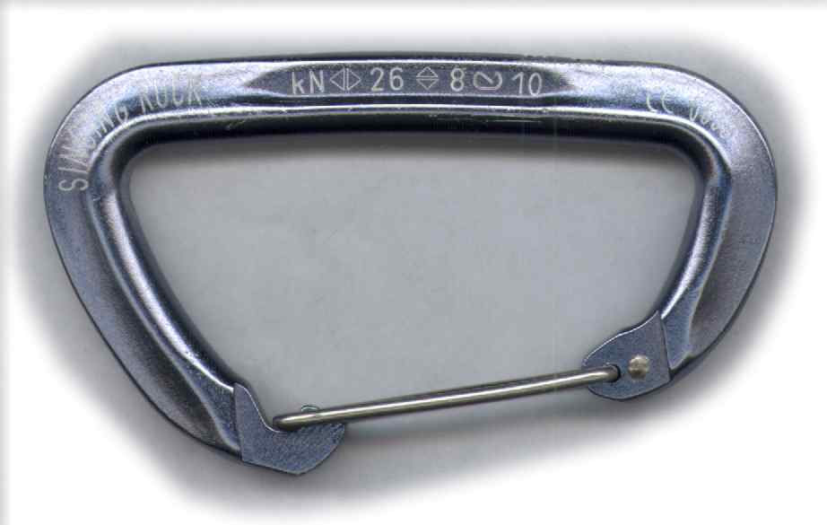
The newton (symbol: N) is the unit of force in the International System of Units (SI). It is defined as 1 kg⋅m/s, the force which gives a mass of 1 kilogram an acceleration of 1 metre per second per second. It is named after Isaac Newton in recognition of his work on classical mechanics, specifically Newton's second law of motion. Definition A newton is defined as 1 kg⋅m/s (it is a derived unit which is defined in terms of the SI base units). One newton is therefore the force needed to accelerate one kilogram of mass at the rate of one metre per second squared in the direction of the applied force. The units "metre per second squared" can be understood as measuring a rate of change in velocity per unit of time, i.e. an increase in velocity by 1 metre per second every second. In 1946, Conférence Générale des Poids et Mesures (CGPM) Resolution 2 standardized the unit of force in the MKS system of units to be the amount needed to accelerate 1 kilogram of mass at the ... [...More Info...] [...Related Items...] OR: [Wikipedia] [Google] [Baidu] |
Anatomical Terms Of Location
Standard anatomical terms of location are used to unambiguously describe the anatomy of animals, including humans. The terms, typically derived from Latin or Greek roots, describe something in its standard anatomical position. This position provides a definition of what is at the front ("anterior"), behind ("posterior") and so on. As part of defining and describing terms, the body is described through the use of anatomical planes and anatomical axes. The meaning of terms that are used can change depending on whether an organism is bipedal or quadrupedal. Additionally, for some animals such as invertebrates, some terms may not have any meaning at all; for example, an animal that is radially symmetrical will have no anterior surface, but can still have a description that a part is close to the middle ("proximal") or further from the middle ("distal"). International organisations have determined vocabularies that are often used as standard vocabularies for subdisciplines o ... [...More Info...] [...Related Items...] OR: [Wikipedia] [Google] [Baidu] |
Triceps Surae Muscle
The triceps surae consists of two muscles located at the calf – the two-headed gastrocnemius and the soleus. These muscles both insert into the calcaneus, the bone of the heel of the human foot, and form the major part of the muscle of the posterior leg, commonly known as the calf muscle. Structure The triceps surae is connected to the foot through the Achilles tendon, and has 3 heads deriving from the 2 major masses of muscle. * The superficial portion (the gastrocnemius) gives off 2 heads attaching to the base of the femur directly above the knee. * The deep (profundus) mass of muscle (the soleus) forms the remaining head which attaches to the superior posterior area of the tibia. The triceps surae is innervated by the tibial nerve, specifically, nerve roots L5–S2. Function Contraction of the triceps surae induce plantar flexion (sagittal plane) and stabilization of the ankle complex in the transverse plane. Functional activities include primarily movement in the ... [...More Info...] [...Related Items...] OR: [Wikipedia] [Google] [Baidu] |
Iliotibial Tract
The iliotibial tract or iliotibial band (ITB; also known as Maissiat's band or the IT band) is a longitudinal fibrous reinforcement of the fascia lata. The action of the muscles associated with the ITB (tensor fasciae latae and some fibers of gluteus maximus) flex, extend, abduct, and laterally and medially rotate the hip. The ITB contributes to lateral knee stabilization. During knee extension the ITB moves anterior to the lateral condyle of the femur, while ~30 degrees knee flexion, the ITB moves posterior to the lateral condyle. However, it has been suggested that this is only an illusion due to the changing tension in the anterior and posterior fibers during movement. It originates at the anterolateral iliac tubercle portion of the external lip of the iliac crest and inserts at the lateral condyle of the tibia at Gerdy's tubercle. The figure shows only the proximal part of the iliotibial tract. The part of the iliotibial band which lies beneath the tensor fasciae latae is ... [...More Info...] [...Related Items...] OR: [Wikipedia] [Google] [Baidu] |
Quadriceps Femoris Muscle
The quadriceps femoris muscle (, also called the quadriceps extensor, quadriceps or quads) is a large muscle group that includes the four prevailing muscles on the front of the thigh. It is the sole extensor muscle of the knee, forming a large fleshy mass which covers the front and sides of the femur. The name derives . Structure Parts The quadriceps femoris muscle is subdivided into four separate muscles (the 'heads'), with the first superficial to the other three over the femur (from the trochanters to the condyles): *The rectus femoris muscle occupies the middle of the thigh, covering most of the other three quadriceps muscles. It originates on the ilium. It is named for its straight course. *The vastus lateralis muscle is on the ''lateral side'' of the femur (i.e. on the outer side of the thigh). *The vastus medialis muscle is on the ''medial side'' of the femur (i.e. on the inner part thigh). *The vastus intermedius muscle lies between vastus lateralis and vastus medi ... [...More Info...] [...Related Items...] OR: [Wikipedia] [Google] [Baidu] |
Femur
The femur (; ), or thigh bone, is the proximal bone of the hindlimb in tetrapod vertebrates. The head of the femur articulates with the acetabulum in the pelvic bone forming the hip joint, while the distal part of the femur articulates with the tibia (shinbone) and patella (kneecap), forming the knee joint. By most measures the two (left and right) femurs are the strongest bones of the body, and in humans, the largest and thickest. Structure The femur is the only bone in the upper leg. The two femurs converge medially toward the knees, where they articulate with the proximal ends of the tibiae. The angle of convergence of the femora is a major factor in determining the femoral-tibial angle. Human females have thicker pelvic bones, causing their femora to converge more than in males. In the condition ''genu valgum'' (knock knee) the femurs converge so much that the knees touch one another. The opposite extreme is ''genu varum'' (bow-leggedness). In the general pop ... [...More Info...] [...Related Items...] OR: [Wikipedia] [Google] [Baidu] |
Tibia
The tibia (; ), also known as the shinbone or shankbone, is the larger, stronger, and anterior (frontal) of the two bones in the leg below the knee in vertebrates (the other being the fibula, behind and to the outside of the tibia); it connects the knee with the ankle. The tibia is found on the medial side of the leg next to the fibula and closer to the median plane. The tibia is connected to the fibula by the interosseous membrane of leg, forming a type of fibrous joint called a syndesmosis with very little movement. The tibia is named for the flute '' tibia''. It is the second largest bone in the human body, after the femur. The leg bones are the strongest long bones as they support the rest of the body. Structure In human anatomy, the tibia is the second largest bone next to the femur. As in other vertebrates the tibia is one of two bones in the lower leg, the other being the fibula, and is a component of the knee and ankle joints. The ossification or formation of the bo ... [...More Info...] [...Related Items...] OR: [Wikipedia] [Google] [Baidu] |
Gracilis Muscle
The gracilis muscle (; Latin for "slender") is the most superficial muscle on the medial side of the thigh. It is thin and flattened, broad above, narrow and tapering below. Structure It arises by a thin aponeurosis from the anterior margins of the lower half of the symphysis pubis and the upper half of the pubic arch. The muscle's fibers run vertically downward, ending in a rounded tendon. This tendon passes behind the medial condyle of the femur, curves around the medial condyle of the tibia where it becomes flattened, and inserts into the upper part of the medial surface of the body of the tibia, below the condyle. For this reason, the muscle is a lower limb adductor. At its insertion the tendon is situated immediately above that of the semitendinosus muscle, and its upper edge is overlapped by the tendon of the sartorius muscle, which it joins to form the pes anserinus. The pes anserinus is separated from the medial collateral ligament of the knee-joint by a bursa. ... [...More Info...] [...Related Items...] OR: [Wikipedia] [Google] [Baidu] |


