|
Kinaesthesis
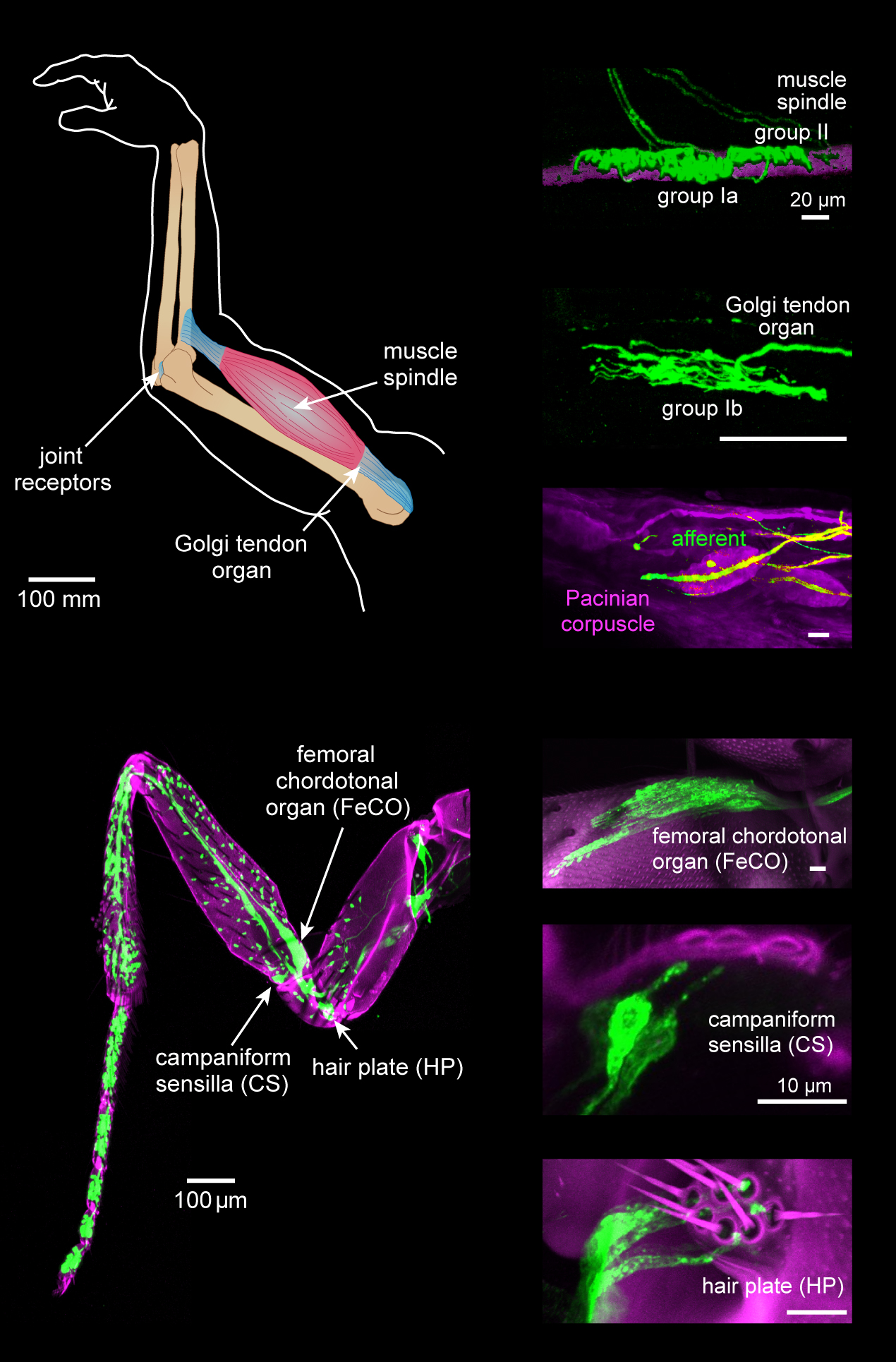
Proprioception ( ), also referred to as kinaesthesia (or kinesthesia), is the sense of self-movement, force, and body position. It is sometimes described as the "sixth sense". Proprioception is mediated by proprioceptors, mechanosensory neurons located within muscles, tendons, and joints. Most animals possess multiple subtypes of proprioceptors, which detect distinct kinematic parameters, such as joint position, movement, and load. Although all mobile animals possess proprioceptors, the structure of the sensory organs can vary across species. Proprioceptive signals are transmitted to the central nervous system, where they are integrated with information from other sensory systems, such as the visual system and the vestibular system, to create an overall representation of body position, movement, and acceleration. In many animals, sensory feedback from proprioceptors is essential for stabilizing body posture and coordinating body movement. System overview In vertebrates, limb ... [...More Info...] [...Related Items...] OR: [Wikipedia] [Google] [Baidu] |
Proprioception Image-01
Proprioception ( ), also referred to as kinaesthesia (or kinesthesia), is the sense of self-movement, force, and body position. It is sometimes described as the "sixth sense". Proprioception is mediated by proprioceptors, mechanosensory neurons located within muscles, tendons, and joints. Most animals possess multiple subtypes of proprioceptors, which detect distinct kinematic parameters, such as joint position, movement, and load. Although all mobile animals possess proprioceptors, the structure of the sensory organs can vary across species. Proprioceptive signals are transmitted to the central nervous system, where they are integrated with information from other Sensory nervous system, sensory systems, such as Visual perception, the visual system and the vestibular system, to create an overall representation of body position, movement, and acceleration. In many animals, sensory feedback from proprioceptors is essential for stabilizing body posture and coordinating body movement ... [...More Info...] [...Related Items...] OR: [Wikipedia] [Google] [Baidu] |
Pacinian Corpuscles
Pacinian corpuscle or lamellar corpuscle or Vater-Pacini corpuscle; is one of the four major types of mechanoreceptors (specialized nerve ending with adventitious tissue for mechanical sensation) found in mammalian skin. This type of mechanoreceptor is found in both glabrous (hairless) and hirsute (hairy) skins, viscera, joints and attached to periosteum of bone, primarily responsible for sensitivity to vibration. Few of them are also sensitive to quasi-static or low frequency pressure stimulus. Most of them respond only to sudden disturbances and are especially sensitive to vibration of few hundreds of Hz. The vibrational role may be used for detecting surface texture, e.g., rough vs. smooth. Most of the Pacinian corpuscles act as rapidly adapting mechanoreceptors. Groups of corpuscles respond to pressure changes, e.g. on grasping or releasing an object. Structure Pacinian corpuscles are larger and fewer in number than tactile corpuscle, Meissner's corpuscle, Merkel cells and bul ... [...More Info...] [...Related Items...] OR: [Wikipedia] [Google] [Baidu] |
Joint Capsule
In anatomy, a joint capsule or articular capsule is an envelope surrounding a synovial joint. Each joint capsule has two parts: an outer fibrous layer or membrane, and an inner synovial layer or membrane. Membranes Each capsule consists of two layers or membranes: * an outer (fibrous membrane, ''fibrous stratum'') composed of avascular white fibrous tissue * an inner ('' synovial membrane'', ''synovial stratum'') which is a secreting layer On the inside of the capsule, articular cartilage covers the end surfaces of the bones that articulate within that joint. The outer layer is hi ...[...More Info...] [...Related Items...] OR: [Wikipedia] [Google] [Baidu] |
Mechanosensation
Mechanosensation is the transduction of mechanical stimuli into neural signals. Mechanosensation provides the basis for the senses of light touch, hearing, proprioception, and pain. Mechanoreceptors found in the skin, called cutaneous mechanoreceptors, are responsible for the sense of touch. Tiny cells in the inner ear, called hair cells, are responsible for hearing and balance. States of neuropathic pain, such as hyperalgesia and allodynia, are also directly related to mechanosensation. A wide array of elements are involved in the process of mechanosensation, many of which are still not fully understood. Cutaneous mechanoreceptors Cutaneous mechanoreceptors are physiologically classified with respect to conduction velocity, which is directly related to the diameter and myelination of the axon. Rapidly adapting and slowly adapting mechanoreceptors Mechanoreceptors that possess a large diameter and high myelination are called ''low-threshold mechanoreceptors''. Fibers that respond ... [...More Info...] [...Related Items...] OR: [Wikipedia] [Google] [Baidu] |
Golgi Tendon Organ
The Golgi tendon organ (GTO) (also called Golgi organ, tendon organ, neurotendinous organ or neurotendinous spindle) is a proprioceptor – a type of sensory receptor that senses changes in muscle tension. It lies at the interface between a muscle and its tendon known as the musculotendinous junction also known as the myotendinous junction. It provides the sensory component of the Golgi tendon reflex. The Golgi tendon organ is one of several eponymous terms named after the Italian physician Camillo Golgi. Structure The body of the Golgi tendon organ is made up of braided strands of collagen (intrafusal fasciculi) that are less compact than elsewhere in the tendon and are encapsulated. The capsule is connected in series (along a single path) with a group of muscle fibers () at one end, and merge into the tendon proper at the other. Each capsule is about long, has a diameter of about , and is perforated by one or more afferent type Ib sensory nerve fibers ( Aɑ fiber), which ... [...More Info...] [...Related Items...] OR: [Wikipedia] [Google] [Baidu] |
Skeletal Muscle
Skeletal muscles (commonly referred to as muscles) are organs of the vertebrate muscular system and typically are attached by tendons to bones of a skeleton. The muscle cells of skeletal muscles are much longer than in the other types of muscle tissue, and are often known as muscle fibers. The muscle tissue of a skeletal muscle is striated – having a striped appearance due to the arrangement of the sarcomeres. Skeletal muscles are voluntary muscles under the control of the somatic nervous system. The other types of muscle are cardiac muscle which is also striated and smooth muscle which is non-striated; both of these types of muscle tissue are classified as involuntary, or, under the control of the autonomic nervous system. A skeletal muscle contains multiple muscle fascicle, fascicles – bundles of muscle fibers. Each individual fiber, and each muscle is surrounded by a type of connective tissue layer of fascia. Muscle fibers are formed from the cell fusion, fusion of ... [...More Info...] [...Related Items...] OR: [Wikipedia] [Google] [Baidu] |
Righting Reflex
The righting reflex, also known as the labyrinthine righting reflex, is a reflex that corrects the orientation of the body when it is taken out of its normal upright position. It is initiated by the vestibular system, which detects that the body is not erect and causes the head to move back into position as the rest of the body follows. The perception of head movement involves the body sensing linear acceleration or the force of gravity through the otoliths, and angular acceleration through the semicircular canals. The reflex uses a combination of visual system inputs, vestibular inputs, and somatosensory inputs to make postural adjustments when the body becomes displaced from its normal vertical position. These inputs are used to create what is called an efference copy. This means that the brain makes comparisons in the cerebellum between expected posture and perceived posture, and corrects for the difference. The reflex takes 6 or 7 weeks to perfect, but can be affected by various t ... [...More Info...] [...Related Items...] OR: [Wikipedia] [Google] [Baidu] |
Cerebellum
The cerebellum (Latin for "little brain") is a major feature of the hindbrain of all vertebrates. Although usually smaller than the cerebrum, in some animals such as the mormyrid fishes it may be as large as or even larger. In humans, the cerebellum plays an important role in motor control. It may also be involved in some cognition, cognitive functions such as attention and language as well as emotion, emotional control such as regulating fear and pleasure responses, but its movement-related functions are the most solidly established. The human cerebellum does not initiate movement, but contributes to Motor coordination, coordination, precision, and accurate timing: it receives input from sensory systems of the spinal cord and from other parts of the brain, and integrates these inputs to fine-tune motor activity. Cerebellar damage produces disorders in Fine motor skill, fine movement, Equilibrioception, equilibrium, Human positions, posture, and motor learning in humans. Anatomica ... [...More Info...] [...Related Items...] OR: [Wikipedia] [Google] [Baidu] |
Ventral Spinocerebellar Tract
The spinocerebellar tract is a nerve tract originating in the spinal cord and terminating in the same side (ipsilateral) of the cerebellum. Origins of proprioceptive information Proprioceptive information is obtained by Golgi tendon organs and muscle spindles. * Golgi tendon organs consist of a fibrous capsule enclosing tendon fascicles and bare nerve endings that respond to tension in the tendon by causing action potentials in type Ib afferents. These fibers are relatively large, myelinated, and quickly conducting. * Muscle spindles monitor the length within muscles and send information via faster Ia afferents. These axons are larger and faster than type Ib (from both nuclear bag fibers and nuclear chain fibers) and type II afferents (solely from nuclear chain fibers). All of these neurons are sensory (first order, or primary) and have their cell bodies in the dorsal root ganglia. They pass through Rexed laminae layers I-VI of the posterior grey column (dorsal horn) to form s ... [...More Info...] [...Related Items...] OR: [Wikipedia] [Google] [Baidu] |
Dorsal Spinocerebellar Tract
The spinocerebellar tract is a nerve tract originating in the spinal cord and terminating in the same side (ipsilateral) of the cerebellum. Origins of proprioceptive information Proprioceptive information is obtained by Golgi tendon organs and muscle spindles. * Golgi tendon organs consist of a fibrous capsule enclosing tendon fascicles and bare nerve endings that respond to tension in the tendon by causing action potentials in type Ib afferents. These fibers are relatively large, myelinated, and quickly conducting. * Muscle spindles monitor the length within muscles and send information via faster Ia afferents. These axons are larger and faster than type Ib (from both nuclear bag fibers and nuclear chain fibers) and type II afferents (solely from nuclear chain fibers). All of these neurons are sensory (first order, or primary) and have their cell bodies in the dorsal root ganglia. They pass through Rexed laminae layers I-VI of the posterior grey column (dorsal horn) to form ... [...More Info...] [...Related Items...] OR: [Wikipedia] [Google] [Baidu] |
Cerebrum
The cerebrum, telencephalon or endbrain is the largest part of the brain containing the cerebral cortex (of the two cerebral hemispheres), as well as several subcortical structures, including the hippocampus, basal ganglia, and olfactory bulb. In the human brain, the cerebrum is the uppermost region of the central nervous system. The cerebrum prenatal development, develops prenatally from the forebrain (prosencephalon). In mammals, the Dorsum (biology), dorsal telencephalon, or Pallium (neuroanatomy), pallium, develops into the cerebral cortex, and the ventral telencephalon, or Pallium (neuroanatomy), subpallium, becomes the basal ganglia. The cerebrum is also divided into approximately symmetric Lateralization of brain function, left and right cerebral hemispheres. With the assistance of the cerebellum, the cerebrum controls all voluntary actions in the human body. Structure The cerebrum is the largest part of the brain. Depending upon the position of the animal it lies eithe ... [...More Info...] [...Related Items...] OR: [Wikipedia] [Google] [Baidu] |
Dorsal Column-medial Lemniscus Pathway
Dorsal (from Latin ''dorsum'' ‘back’) may refer to: * Dorsal (anatomy), an anatomical term of location referring to the back or upper side of an organism or parts of an organism * Dorsal, positioned on top of an aircraft's fuselage * Dorsal consonant, a consonant articulated with the back of the tongue * Dorsal fin A dorsal fin is a fin located on the back of most marine and freshwater vertebrates within various taxa of the animal kingdom. Many species of animals possessing dorsal fins are not particularly closely related to each other, though through conv ..., the fin located on the back of a fish or aircraft * Dorsal transcription factor, a maternally synthesized transcription factor {{disambig de:Dorsale fr:Dorsale it:Dorsale ... [...More Info...] [...Related Items...] OR: [Wikipedia] [Google] [Baidu] |


