|
Juvenile Glaucoma
Primary juvenile glaucoma is glaucoma that develops due to ocular hypertension and is evident either at birth or within the first few years of life. It is caused due to abnormalities in the anterior chamber angle development that obstruct aqueous outflow in the absence of systemic anomalies or other ocular malformation. Presentation The typical infant who has congenital glaucoma usually is initially referred to an ophthalmologist because of apparent corneal edema. The commonly described triad of epiphora (excessive tearing), blepharospasm and photophobia may be missed until the corneal edema becomes apparent. Systemic associations Two of the more commonly encountered disorders that may be associated with congenital glaucoma are Aniridia and Sturge–Weber syndrome. Genetics JOAG is an autosomal dominant condition. The primary cause is the myocilin protein dysfunction. Myocilin gene mutations are identified in approximately 10% of patients affected by juvenile glaucoma. Diagnosis ... [...More Info...] [...Related Items...] OR: [Wikipedia] [Google] [Baidu] |
Buphthalmos
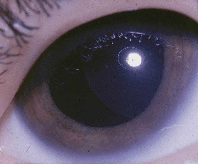
Buphthalmos (plural: buphthalmoses) is enlargement of the eyeball and is most commonly seen in infants and young children. It is sometimes referred to as buphthalmia (plural buphthalmias). It usually appears in the newborn period or the first 3 months of life. and in most cases indicates the presence of congenital (infantile) glaucoma, which is a disorder in which elevated pressures within the eye lead to structural eye damage and vision loss. Signs and symptoms Buphthalmos in itself is merely a clinical sign and does not generate symptoms. Patients with glaucoma often initially have no symptoms; later, they can exhibit excessive tearing (lacrimation) and extreme sensitivity to light (photophobia). On ophthalmologic exam, a doctor can detect increased intraocular pressure, distortion of the optic disc, and corneal edema, which manifests as haziness. Other symptoms include a prominent eyeball, Haab's striae tear in the Descemet's membrane of the cornea, an enlarged cornea, and myop ... [...More Info...] [...Related Items...] OR: [Wikipedia] [Google] [Baidu] |
Megalocornea
Megalocornea (MGCN, MGCN1) is an extremely rare nonprogressive condition in which the cornea has an enlarged diameter, reaching and exceeding 13 mm. It is thought to have two subforms, one with autosomal inheritance and the other X-linked (Xq21.3-q22). The X-linked form is more common and males generally constitute 90% of cases. It may be associated with Alport syndrome, craniosynostosis, dwarfism, Down syndrome, Parry–Romberg syndrome, Marfan syndrome, mucolipidosis, Frank–ter Haar syndrome, crouzon syndrome, megalocornea-mental retardation syndrome etc. Clinical features Eyes are usually highly myopic. There may be 'with the rule' astigmatism. Lens (anatomy), Lens may be luxated due to Zonule of Zinn, zonular streaching.In rare cases, it might be Megalocornea-intellectual disability syndrome, associated with intellectual disabilities. References External links Megalocornea- eMedicine ophthalmology; May 15, 2009; Thomas A Oetting, MD, Mark A Hendrix, MDAn Infant Wi ... [...More Info...] [...Related Items...] OR: [Wikipedia] [Google] [Baidu] |
Glaucoma
Glaucoma is a group of eye diseases that result in damage to the optic nerve (or retina) and cause vision loss. The most common type is open-angle (wide angle, chronic simple) glaucoma, in which the drainage angle for fluid within the eye remains open, with less common types including closed-angle (narrow angle, acute congestive) glaucoma and normal-tension glaucoma. Open-angle glaucoma develops slowly over time and there is no pain. Peripheral vision may begin to decrease, followed by central vision, resulting in blindness if not treated. Closed-angle glaucoma can present gradually or suddenly. The sudden presentation may involve severe eye pain, blurred vision, mid-dilated pupil, redness of the eye, and nausea. Vision loss from glaucoma, once it has occurred, is permanent. Eyes affected by glaucoma are referred to as being glaucomatous. Risk factors for glaucoma include increasing age, high pressure in the eye, a family history of glaucoma, and use of steroid medication. F ... [...More Info...] [...Related Items...] OR: [Wikipedia] [Google] [Baidu] |
Congenital Disorders Of Eyes
A birth defect, also known as a congenital disorder, is an abnormal condition that is present at birth regardless of its cause. Birth defects may result in disabilities that may be physical, intellectual, or developmental. The disabilities can range from mild to severe. Birth defects are divided into two main types: structural disorders in which problems are seen with the shape of a body part and functional disorders in which problems exist with how a body part works. Functional disorders include metabolic and degenerative disorders. Some birth defects include both structural and functional disorders. Birth defects may result from genetic or chromosomal disorders, exposure to certain medications or chemicals, or certain infections during pregnancy. Risk factors include folate deficiency, drinking alcohol or smoking during pregnancy, poorly controlled diabetes, and a mother over the age of 35 years old. Many are believed to involve multiple factors. Birth defects may be visib ... [...More Info...] [...Related Items...] OR: [Wikipedia] [Google] [Baidu] |
EMedicine
eMedicine is an online clinical medical knowledge base founded in 1996 by doctors Scott Plantz and Jonathan Adler, and computer engineer Jeffrey Berezin. The eMedicine website consists of approximately 6,800 medical topic review articles, each of which is associated with a clinical subspecialty "textbook". The knowledge base includes over 25,000 clinically multimedia files. Each article is authored by board certified specialists in the subspecialty to which the article belongs and undergoes three levels of physician peer-review, plus review by a Doctor of Pharmacy. The article's authors are identified with their current faculty appointments. Each article is updated yearly, or more frequently as changes in practice occur, and the date is published on the article. eMedicine.com was sold to WebMD in January, 2006 and is available as the Medscape Reference. History Plantz, Adler and Berezin evolved the concept for eMedicine.com in 1996 and deployed the initial site via Boston Med ... [...More Info...] [...Related Items...] OR: [Wikipedia] [Google] [Baidu] |
CYP1B1
Cytochrome P450 1B1 is an enzyme that in humans is encoded by the ''CYP1B1'' gene. Function CYP1B1 belongs to the cytochrome P450 superfamily of enzymes. The cytochrome P450 proteins are monooxygenases which catalyze many reactions involved in drug metabolism and synthesis of cholesterol, steroids, and other lipids. The enzyme encoded by this gene localizes to the endoplasmic reticulum ( ER) and metabolizes procarcinogens such as polycyclic aromatic hydrocarbons and 17beta-estradiol. Despite over 20 years of research on CYP1A1 and CYP1A2, CYP1B1 was not identified and sequenced until 1994. Nucleic and amino acid analysis showed approximately 40% identity with CYP1A1. Despite this similarity, these two enzymes have very different catalytic efficiencies and metabolites when incubated with common substrates, such as retinoic acid and arachidonic acid. Recently CYP1B1 has been shown to be physiologically important in fetal development, since mutations in CYP1B1 are linked with ... [...More Info...] [...Related Items...] OR: [Wikipedia] [Google] [Baidu] |
MYOC
Myocilin, trabecular meshwork inducible glucocorticoid response (TIGR), also known as MYOC, is a protein which in humans is encoded by the ''MYOC'' gene. Mutations in ''MYOC'' are a major cause of glaucoma. Gene location The cytogenetic location of human ''MYOC'' gene is on the long (q) arm of chromosome 1, specifically at position 24.3 (1q24.3). The gene's molecular location starts at 171,635,417 bp and ends at 171,652,63 bp on chromosome 1 (Annotation: GRCh38.p12(assembly). Protein characteristics Myocilin is a protein with a weight of 55 kDa (504 amino acid) and an overall acidic property is the first gene that has been linked to Primary Open Angle Glaucoma (POAG). Protein structure The protein is made up of the two folding domains, the leucine zipper-like domain at the N-terminal and an olfactomedin-like domain at the C-terminal. The domain at the N-terminal is known to have 77.6% homology to the myosin heavy chain of ''Dictyostelium discoideum'' and 25% homology wit ... [...More Info...] [...Related Items...] OR: [Wikipedia] [Google] [Baidu] |
Weill–Marchesani Syndrome
Weill–Marchesani syndrome is a rare genetic disorder characterized by short stature; an unusually short, broad head (brachycephaly) and other facial abnormalities; hand defects, including unusually short fingers (brachydactyly); and distinctive eye (ocular) abnormalities. It was named after ophthalmologists Georges Weill (1866–1952) and Oswald Marchesani (1900–1952) who first described it in 1932 and 1939, respectively. The eye manifestations typically include unusually small, round lenses of the eyes ( microspherophakia), which may be prone to dislocating (ectopia lentis), as well as other ocular defects. Due to such abnormalities, affected individuals may have varying degrees of visual impairment, ranging from nearsightedness myopia to blindness. Weill–Marchesani syndrome may have autosomal recessive inheritance involving the ''ADAMTS10'' gene, or autosomal An autosome is any chromosome that is not a sex chromosome. The members of an autosome pair in a diploid cell h ... [...More Info...] [...Related Items...] OR: [Wikipedia] [Google] [Baidu] |
Peters-plus Syndrome
Peters-plus syndrome or Krause–Kivlin syndrome is a hereditary syndrome defined by Peters' anomaly, dwarfism and intellectual disability. Signs and symptoms Features of this syndrome include Peters' anomaly, corneal opacity, central defect of Descemet's membrane, and shallow anterior chamber with synechiae between the iris and cornea. Craniofacial abnormalities commonly seen in patients with PPS include hypertelorism, ear malformations, micrognathia, round face and broad neck, and cleft lip and palate. Infants are commonly born small for gestational age and have delayed growth. It is associated with short limb dwarfism and mild to severe intellectual disability and autism spectrum disorder. Cause The pattern of inheritance of Peters-plus is autosomal recessive, where both parents are heterozygous they can produce a child with the syndrome. The B3GALTL (now called B3GLCT) gene codes for the enzyme beta 3-glucosyltransferase (B3Glc-T). The beta 3-glucosyltransferase enzyme is ... [...More Info...] [...Related Items...] OR: [Wikipedia] [Google] [Baidu] |
Axenfeld Syndrome
Axenfeld or Aksenfeld may refer to: * Israel Aksenfeld (aka Israel Axenfeld / Yisroel Aksenfeld, 1787-1866), a German writer * Karl Theodor Paul Polykarpus Axenfeld (1867-1930), a German ophthalmologist * Karl Theodor Georg Axenfeld (1869-1924), a German superintendent of the Kurmark *Edith Picht-Axenfeld Edith Picht-Axenfeld (Freiburg im Breisgau, 1 January 1914 – Hinterzarten, 19 April 2001) was a German pianist and harpsichordist. Career She started her concert career in 1935, and took part two years later in the III International Chopin Pian ... (1914-2001), a German pianist and harpsichordist * Morax-Axenfeld diplobacilli, a bacterium * Axenfeld syndrome, a rare autosomal dominant disorder {{disamb ... [...More Info...] [...Related Items...] OR: [Wikipedia] [Google] [Baidu] |
Cyclophotocoagulation
Cyclodestruction or cycloablation is a surgical procedure done in management of glaucoma. Cyclodestruction reduce intraocular pressure (IOP) of the eye by decreasing production of aqueous humor by the destruction of ciliary body. Until the development of safer and less destructive techniques like micropulse diode cyclophotocoagulation and endocyclophotocoagulation, cyclodestructive surgeries were mainly done in refractory glaucoma, or advanced glaucomatous eyes with poor visual prognosis. Types Cyclodestruction may be done by using diathermy, penetrating cyclodiathermy, cryotherapy, ultrasound, laser or by surgical excision. Cyclophotocoagulation Cyclophotocoagulation (CPC), the most common cyclodestructive procedure is done using laser beam of different wavelengths. Ruby laser (693 nm wavelength), Nd:YAG laser (1064 nm wavelength) or diode laser (810 nm wavelength) can be used to perform CPC. Commomon cyclophotocoagulation techniques include transscleral cyclophot ... [...More Info...] [...Related Items...] OR: [Wikipedia] [Google] [Baidu] |
Corneal Opacity
The human cornea is a transparent membrane which allows light to pass through it. The word corneal opacification literally means loss of normal transparency of cornea. The term corneal opacity is used particularly for the loss of transparency of cornea due to scarring. Transparency of the cornea is dependent on the uniform diameter and the regular spacing and arrangement of the collagen fibrils within the stroma. Alterations in the spacing of collagen fibrils in a variety of conditions including corneal edema, scars, and macular corneal dystrophy is clinically manifested as corneal opacity. The term corneal blindness is commonly used to describe blindness due to corneal opacity. Types Depending on the density, corneal opacity is graded as nebular, macular and leucomatous. Nebular corneal opacity Nebular corneal opacity is a faint opacity which results due to superficial scars involving Bowman's layer and superficial stroma. A nebular corneal opacity allows the details of the iri ... [...More Info...] [...Related Items...] OR: [Wikipedia] [Google] [Baidu] |


