|
Isothalamus
The isothalamus is a division used by some researchers in describing the thalamus. The isothalamus constitutes 90% or more of the thalamus, and despite the variety of functions it serves, follows a simple organizational scheme. The constituting neurons belong to two different neuronal genera. The first correspond to the ''thalamocortical neurons'' (or principal). They have a "tufted" (or radiate) morphology, as their dendritic arborisation is made up of straight dendritic distal branches starting from short and thick stems. The number of branches and the diameter of the arborisation are linked to the specific system of which they are a part of, and to the animal species. They have the rather rare property of having no initial axonal collaterals, which implies that one emitting thalamocortical neuron does not send information to its neighbor. They send long-range glutamatergic projections to the cerebral cortex where they end electively at the layer IV (or around) level. The other ... [...More Info...] [...Related Items...] OR: [Wikipedia] [Google] [Baidu] |
Thalamus
The thalamus (from Greek θάλαμος, "chamber") is a large mass of gray matter located in the dorsal part of the diencephalon (a division of the forebrain). Nerve fibers project out of the thalamus to the cerebral cortex in all directions, allowing hub-like exchanges of information. It has several functions, such as the relaying of sensory signals, including motor signals to the cerebral cortex and the regulation of consciousness, sleep, and alertness. Anatomically, it is a paramedian symmetrical structure of two halves (left and right), within the vertebrate brain, situated between the cerebral cortex and the midbrain. It forms during embryonic development as the main product of the diencephalon, as first recognized by the Swiss embryologist and anatomist Wilhelm His Sr. in 1893. Anatomy The thalamus is a paired structure of gray matter located in the forebrain which is superior to the midbrain, near the center of the brain, with nerve fibers projecting out to the ... [...More Info...] [...Related Items...] OR: [Wikipedia] [Google] [Baidu] |
Félix Vicq-d'Azyr
Félix Vicq d'Azyr (; 23 April 1748 – 20 June 1794) was a French physician and anatomist, the originator of comparative anatomy and discoverer of the theory of homology in biology. Biography Vicq d'Azyr was born in Valognes, Normandy, the son of a physician. He graduated in medicine at the University of Paris and became a renowned and brilliant animal and human anatomist and physician. From 1773 Vicq d'Azyr taught a celebrated course of anatomy at the Jardin du Roi, currently the Museum of Natural History, in Paris. In 1774 he was elected a member of the Académie des Sciences with the support of his friend Condorcet, the Perpetual Secretary. In this latter capacity, he was in charge of writing the eulogies of his colleagues. This he accomplished with great talent, thus winning a lifetime membership to the Académie française in 1788. On the outbreak of an epidemic in Guyenne he was charged with writing a report, of making propositions and with their execution. Pursuing an e ... [...More Info...] [...Related Items...] OR: [Wikipedia] [Google] [Baidu] |
Auditory System
The auditory system is the sensory system for the sense of hearing. It includes both the sensory organs (the ears) and the auditory parts of the sensory system. System overview The outer ear funnels sound vibrations to the eardrum, increasing the sound pressure in the middle frequency range. The middle-ear ossicles further amplify the vibration pressure roughly 20 times. The base of the stapes couples vibrations into the cochlea via the oval window, which vibrates the perilymph liquid (present throughout the inner ear) and causes the round window to bulb out as the oval window bulges in. Vestibular and tympanic ducts are filled with perilymph, and the smaller cochlear duct between them is filled with endolymph, a fluid with a very different ion concentration and voltage. Vestibular duct perilymph vibrations bend organ of Corti outer cells (4 lines) causing prestin to be released in cell tips. This causes the cells to be chemically elongated and shrunk ( somatic motor), and ... [...More Info...] [...Related Items...] OR: [Wikipedia] [Google] [Baidu] |
Primary Auditory Cortex
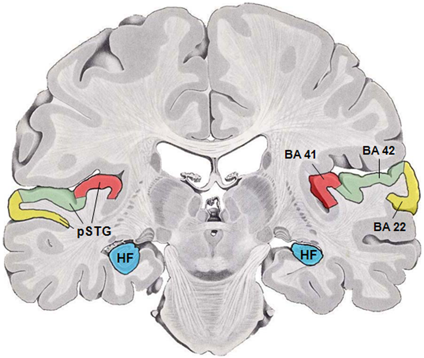
The auditory cortex is the part of the temporal lobe that processes auditory information in humans and many other vertebrates. It is a part of the auditory system, performing basic and higher functions in hearing, such as possible relations to language switching.Cf. Pickles, James O. (2012). ''An Introduction to the Physiology of Hearing'' (4th ed.). Bingley, UK: Emerald Group Publishing Limited, p. 238. It is located bilaterally, roughly at the upper sides of the temporal lobes – in humans, curving down and onto the medial surface, on the superior temporal plane, within the lateral sulcus and comprising parts of the transverse temporal gyri, and the superior temporal gyrus, including the planum polare and planum temporale (roughly Brodmann areas 41 and 42, and partially 22). The auditory cortex takes part in the spectrotemporal, meaning involving time and frequency, analysis of the inputs passed on from the ear. The cortex then filters and passes on the information to the ... [...More Info...] [...Related Items...] OR: [Wikipedia] [Google] [Baidu] |
Brachium Of The Inferior Colliculus
The inferior colliculus (IC) (Latin for ''lower hill'') is the principal midbrain nucleus of the auditory pathway and receives input from several peripheral brainstem nuclei in the auditory pathway, as well as inputs from the auditory cortex. The inferior colliculus has three subdivisions: the central nucleus, a dorsal cortex by which it is surrounded, and an external cortex which is located laterally. Its bimodal neurons are implicated in auditory-somatosensory interaction, receiving projections from somatosensory nuclei. This multisensory integration may underlie a filtering of self-effected sounds from vocalization, chewing, or respiration activities. The inferior colliculi together with the superior colliculi form the eminences of the corpora quadrigemina, and also part of the tectal region of the midbrain. The inferior colliculus lies caudal to its counterpart – the superior colliculus – above the trochlear nerve, and at the base of the projection of the medial genicu ... [...More Info...] [...Related Items...] OR: [Wikipedia] [Google] [Baidu] |
Inferior Colliculus
The inferior colliculus (IC) (Latin for ''lower hill'') is the principal midbrain nucleus of the auditory pathway and receives input from several peripheral brainstem nuclei in the auditory pathway, as well as inputs from the auditory cortex. The inferior colliculus has three subdivisions: the central nucleus, a dorsal cortex by which it is surrounded, and an external cortex which is located laterally. Its bimodal neurons are implicated in auditory-somatosensory interaction, receiving projections from somatosensory nuclei. This multisensory integration may underlie a filtering of self-effected sounds from vocalization, chewing, or respiration activities. The inferior colliculi together with the superior colliculi form the eminences of the corpora quadrigemina, and also part of the tectal region of the midbrain. The inferior colliculus lies caudal to its counterpart – the superior colliculus – above the trochlear nerve, and at the base of the projection of the medial genicu ... [...More Info...] [...Related Items...] OR: [Wikipedia] [Google] [Baidu] |
Nucleus Geniculatus Medialis
The medial geniculate nucleus (MGN) or medial geniculate body (MGB) is part of the auditory thalamus and represents the thalamic relay between the inferior colliculus (IC) and the auditory cortex (AC). It is made up of a number of sub-nuclei that are distinguished by their neuronal morphology and density, by their afferent and efferent connections, and by the coding properties of their neurons. It is thought that the MGN influences the direction and maintenance of attention. Divisions The MGN has three major divisions; ventral (VMGN), dorsal (DMGN) and medial (MMGN). Whilst the VMGN is specific to auditory information processing, the DMGN and MMGN also receive information from non-auditory pathways. Ventral subnucleus Cell types There are two main cell types in the ventral subnucleus of the medial geniculate body (VMGN): * Thalamocortical relay cells (or principal neurons): The dendritic input to these cells comes from two sets of dendritic trees oriented on opposite poles of th ... [...More Info...] [...Related Items...] OR: [Wikipedia] [Google] [Baidu] |
Insular Cortex
The insular cortex (also insula and insular lobe) is a portion of the cerebral cortex folded deep within the lateral sulcus (the fissure separating the temporal lobe from the parietal and frontal lobes) within each hemisphere of the mammalian brain. The insulae are believed to be involved in consciousness and play a role in diverse functions usually linked to emotion or the regulation of the body's homeostasis. These functions include compassion, empathy, taste, perception, motor control, self-awareness, cognitive functioning, interpersonal experience, and awareness of homeostatic emotions such as hunger, pain and fatigue. In relation to these, it is involved in psychopathology. The insular cortex is divided into two parts: the anterior insula and the posterior insula in which more than a dozen field areas have been identified. The cortical area overlying the insula toward the lateral surface of the brain is the operculum (meaning ''lid''). The opercula are formed from parts o ... [...More Info...] [...Related Items...] OR: [Wikipedia] [Google] [Baidu] |
Frontal Cortex
The frontal lobe is the largest of the four major lobes of the brain in mammals, and is located at the front of each cerebral hemisphere (in front of the parietal lobe and the temporal lobe). It is parted from the parietal lobe by a groove between tissues called the central sulcus and from the temporal lobe by a deeper groove called the lateral sulcus (Sylvian fissure). The most anterior rounded part of the frontal lobe (though not well-defined) is known as the frontal pole, one of the three poles of the cerebrum. The frontal lobe is covered by the frontal cortex. The frontal cortex includes the premotor cortex, and the primary motor cortex – parts of the motor cortex. The front part of the frontal cortex is covered by the prefrontal cortex. There are four principal gyri in the frontal lobe. The precentral gyrus is directly anterior to the central sulcus, running parallel to it and contains the primary motor cortex, which controls voluntary movements of specific body parts. T ... [...More Info...] [...Related Items...] OR: [Wikipedia] [Google] [Baidu] |
Lobotomy
A lobotomy, or leucotomy, is a form of neurosurgical treatment for psychiatric disorder or neurological disorder (e.g. epilepsy) that involves severing connections in the brain's prefrontal cortex. The surgery causes most of the connections to and from the prefrontal cortex, the anterior part of the frontal lobes of the brain, to be severed. In the past, this treatment was used for treating psychiatric disorders as a mainstream procedure in some countries. The procedure was controversial from its initial use, in part due to a lack of recognition of the severity and chronicity of severe and enduring psychiatric illnesses, so it was claimed to be an inappropriate treatment. Frontal lobe surgery, including lobotomy, is the second most common surgery for epilepsy to this day, and usually done on one side of the brain, unlike lobotomies for psychiatric disorder which were done on both sides of the brain. The originator of the procedure, Portuguese neurologist António Egas Moniz, ... [...More Info...] [...Related Items...] OR: [Wikipedia] [Google] [Baidu] |
Medial Dorsal Nucleus
The medial dorsal nucleus (or dorsomedial nucleus of thalamus) is a large nucleus in the thalamus. It is believed to play a role in memory. Structure It relays inputs from the amygdala and olfactory cortex and projects to the prefrontal cortex and the limbic system and in turn relays them to the prefrontal association cortex. As a result, it plays a crucial role in attention, planning, organization, abstract thinking, multi-tasking, and active memory. The connections of the medial dorsal nucleus have even been used to delineate the prefrontal cortex of the Göttingen minipig brain. By stereology the number of brain cells in the region has been estimated to around 6.43 million neurons in the adult human brain and 36.3 million glial cell Glia, also called glial cells (gliocytes) or neuroglia, are non-neuronal cells in the central nervous system (brain and spinal cord) and the peripheral nervous system that do not produce electrical impulses. They maintain homeostasis, form m ... [...More Info...] [...Related Items...] OR: [Wikipedia] [Google] [Baidu] |


