|
Iopromide
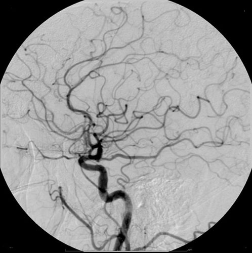
Iopromide is an iodinated contrast medium for X-ray imaging. It is marketed under the name Ultravist which is produced by Bayer Healthcare. It is a low osmolar, non-ionic contrast agent for intravascular use; i.e., it is injected into blood vessels. It is commonly used in radiographic studies such as intravenous urograms, brain computer tomography (CT) and CT pulmonary angiograms (CTPAs). Medical uses The radiocontrast agent is given intravenously in computed tomography (CT) scans, angiography and excretory urography. Contraindications Iopromide use is contraindicated in myelography, cerebral ventriculography and cisternography procedures. It is also contraindicated in those with hyperthyroidism, or with known allergy to the drug. Iopromide is also contraindicated in children with prolonged fasting, fluid restriction, on laxative Laxatives, purgatives, or aperients are substances that loosen stools and increase bowel movements. They are used to treat and prevent constipa ... [...More Info...] [...Related Items...] OR: [Wikipedia] [Google] [Baidu] |
Iodinated Contrast
Iodinated contrast is a form of intravenous radiocontrast agent containing iodine, which enhances the visibility of vascular structures and organs during radiographic procedures. Some pathologies, such as cancer, have particularly improved visibility with iodinated contrast. The radiodensity of iodinated contrast is 25–30 Hounsfield units (HU) per milligram of iodine per milliliter at a tube voltage of 100–120 kVp. Types Iodine-based contrast media are usually classified as ionic or nonionic. Both types are used most commonly in radiology due to their relatively harmless interaction with the body and its solubility. Contrast media are primarily used to visualize vessels and changes in tissues on radiography and CT (computerized tomography). Contrast media can also be used for tests of the urinary tract, uterus and fallopian tubes. It may cause the patient to feel as if they have had urinary incontinence. It also puts a metallic taste in the mouth of the patient. The iodine ... [...More Info...] [...Related Items...] OR: [Wikipedia] [Google] [Baidu] |
Intravascular
The blood vessels are the components of the circulatory system that transport blood throughout the human body. These vessels transport blood cells, nutrients, and oxygen to the tissues of the body. They also take waste and carbon dioxide away from the tissues. Blood vessels are needed to sustain life, because all of the body's tissues rely on their functionality. There are five types of blood vessels: the arteries, which carry the blood away from the heart; the arterioles; the capillaries, where the exchange of water and chemicals between the blood and the tissues occurs; the venules; and the veins, which carry blood from the capillaries back towards the heart. The word ''vascular'', meaning relating to the blood vessels, is derived from the Latin ''vas'', meaning vessel. Some structures – such as cartilage, the epithelium, and the lens and cornea of the eye – do not contain blood vessels and are labeled ''avascular''. Etymology * artery: late Middle English; from Latin ' ... [...More Info...] [...Related Items...] OR: [Wikipedia] [Google] [Baidu] |
Benzamides
Benzamide is a organic compound with the chemical formula of C6H5C(O)NH2. It is the simplest amide derivative of benzoic acid Benzoic acid is a white (or colorless) solid organic compound with the formula , whose structure consists of a benzene ring () with a carboxyl () substituent. It is the simplest aromatic carboxylic acid. The name is derived from gum benzoin, wh .... In powdered form, it appears as a white solid, while in crystalline form, it appears as colourless crystals. It is slightly soluble in water, and soluble in many organic solvents. It is a natural alkaloid found in the herbs of Berberis pruinosa. Chemical derivatives A number of substituted benzamides are commercial drugs, including: See also * References External links Physical characteristics {{Authority control Phenyl compounds ... [...More Info...] [...Related Items...] OR: [Wikipedia] [Google] [Baidu] |
Radiocontrast Agents
Radiocontrast agents are substances used to enhance the visibility of internal structures in X-ray-based imaging techniques such as computed tomography (contrast CT), projectional radiography, and fluoroscopy. Radiocontrast agents are typically iodine, or more rarely barium sulfate. The contrast agents absorb external X-rays, resulting in decreased exposure on the X-ray detector. This is different from radiopharmaceuticals used in nuclear medicine which emit radiation. Magnetic resonance imaging (MRI) functions through different principles and thus MRI contrast agents have a different mode of action. These compounds work by altering the magnetic properties of nearby hydrogen nuclei. Types and uses Radiocontrast agents used in X-ray examinations can be grouped in positive (iodinated agents, barium sulfate), and negative agents (air, carbon dioxide, methylcellulose). Iodine (circulatory system) Iodinated contrast contains iodine. It is the main type of radiocontrast used for i ... [...More Info...] [...Related Items...] OR: [Wikipedia] [Google] [Baidu] |
Laxative
Laxatives, purgatives, or aperients are substances that loosen stools and increase bowel movements. They are used to treat and prevent constipation. Laxatives vary as to how they work and the side effects they may have. Certain stimulant, lubricant and saline laxatives are used to evacuate the colon for rectal and bowel examinations, and may be supplemented by enemas under certain circumstances. Sufficiently high doses of laxatives may cause diarrhea. Some laxatives combine more than one active ingredient. Laxatives may be administered orally or rectally. Types Bulk-forming agents Bulk-forming laxatives, also known as roughage, are substances, such as fiber in food and hydrophilic agents in over-the-counter drugs, that add bulk and water to stools so that they can pass more easily through the intestines (lower part of the digestive tract). Properties * Site of action: small and large intestines * Onset of action: 12–72 hours * Examples: dietary fiber, Metamucil, Citru ... [...More Info...] [...Related Items...] OR: [Wikipedia] [Google] [Baidu] |
Cisternography
Cisternography is a medical imaging technique to examine the flow of cerebrospinal fluid (CSF) in the brain, and spinal cord. The gold standard for diagnosis of a cranial cerebrospinal fluid leak is CT cisternography. For the diagnosis of a spinal CSF leak radionuclide cisternography also known as radioisotope cisternography is used. The third type of cisternography is MR cisternography. Types Radionuclide Radionuclide cisternography may be used to diagnose a spinal cerebrospinal fluid leak. CSF pressure is measured and imaged over 24 hours. A radionuclide (radioisotope) is injected by lumbar puncture (spinal tap) into the cerebral spinal fluid to determine if there is abnormal CSF flow within the brain and spinal canal which can be altered by hydrocephalus, Arnold–Chiari malformation, syringomyelia, or an arachnoid cyst. It may also evaluate a suspected CSF leak (also known as a CSF fistula) from the CSF cavity into the nasal cavity. A leak can also be confirmed by the prese ... [...More Info...] [...Related Items...] OR: [Wikipedia] [Google] [Baidu] |
Cerebral Ventriculography
Pneumoencephalography (sometimes abbreviated PEG; also referred to as an "air study") was a common medical procedure in which most of the cerebrospinal fluid (CSF) was drained from around the brain by means of a lumbar puncture and replaced with air, oxygen, or helium to allow the structure of the brain to show up more clearly on an X-ray image. It was derived from ventriculography, an earlier and more primitive method where the air is injected through holes drilled in the skull. The procedure was introduced in 1919 by the American neurosurgeon Walter Dandy and was performed extensively until the late 1970s, when it was replaced by more-sophisticated and less-invasive modern neuroimaging techniques. Procedure Though pneumoencephalography was the single most important way of localizing brain lesions of its time, it was, nevertheless, extremely painful and generally not well tolerated by conscious patients. Pneumoencephalography was associated with a wide range of side-effects, incl ... [...More Info...] [...Related Items...] OR: [Wikipedia] [Google] [Baidu] |
Myelography
Myelography is a type of radiographic examination that uses a contrast medium to detect pathology of the spinal cord, including the location of a spinal cord injury, cysts, and tumors. Historically the procedure involved the injection of a radiocontrast agent into the cervical or lumbar spine, followed by several X-ray projections. Today, myelography has largely been replaced by the use of MRI scans, although the technique is still sometimes used under certain circumstances – though now usually in conjunction with CT rather than X-ray projections. Types Cervical myelography This procedure is used to look for the level of where spinal cord disease occurs or compression of the spinal cord at the neck region for those who are unable or unwilling to undergone MRI scan of the spine. Lumbar myelography This procedure is to look for the level of spinal cord disease such as lumbar nerve root compression, cauda equina syndrome, conus medullaris lesions, and spinal stenosis. This is do ... [...More Info...] [...Related Items...] OR: [Wikipedia] [Google] [Baidu] |
Computer Tomography
A computed tomography scan (CT scan; formerly called computed axial tomography scan or CAT scan) is a medical imaging technique used to obtain detailed internal images of the body. The personnel that perform CT scans are called radiographers or radiology technologists. CT scanners use a rotating X-ray tube and a row of detectors placed in a gantry to measure X-ray attenuations by different tissues inside the body. The multiple X-ray measurements taken from different angles are then processed on a computer using tomographic reconstruction algorithms to produce tomographic (cross-sectional) images (virtual "slices") of a body. CT scans can be used in patients with metallic implants or pacemakers, for whom magnetic resonance imaging (MRI) is contraindicated. Since its development in the 1970s, CT scanning has proven to be a versatile imaging technique. While CT is most prominently used in medical diagnosis, it can also be used to form images of non-living objects. The 1979 Nob ... [...More Info...] [...Related Items...] OR: [Wikipedia] [Google] [Baidu] |
CT Pulmonary Angiogram
A CT pulmonary angiogram (CTPA) is a medical diagnostic test that employs computed tomography (CT) angiography to obtain an image of the pulmonary arteries. Its main use is to diagnose pulmonary embolism (PE). It is a preferred choice of imaging in the diagnosis of PE due to its minimally invasive nature for the patient, whose only requirement for the scan is an intravenous line. Modern MDCT (multi-detector CT) scanners are able to deliver images of sufficient resolution within a short time period, such that CTPA has now supplanted previous methods of testing, such as direct pulmonary angiography, as the gold standard for diagnosis of pulmonary embolism. The patient receives an intravenous injection of an iodine-containing contrast agent at a high-rate using an injector pump. Images are acquired with the maximum intensity of radio-opaque contrast in the pulmonary arteries. This can be done using bolus tracking. A normal CTPA scan will show the contrast filling the pulmonary ves ... [...More Info...] [...Related Items...] OR: [Wikipedia] [Google] [Baidu] |
Intravenous Urogram
Pyelogram (or pyelography or urography) is a form of imaging of the renal pelvis and ureter. Types include: * Intravenous pyelogram – In which a contrast solution is introduced through a vein into the circulatory system. * Retrograde pyelogram – Any pyelogram in which contrast medium is introduced from the lower urinary tract and flows toward the kidney (i.e. in a "retrograde" direction, against the normal flow of urine). * Anterograde pyelogram (also antegrade pyelogram) – A pyelogram where a contrast medium passes from the kidneys toward the bladder, mimicking the normal flow of urine. * Gas pyelogram – A pyelogram that uses a gaseous rather than liquid contrast medium. It may also form without the injection of a gas, when gas producing micro-organisms infect the most upper parts of urinary system. Intravenous pyelogram An intravenous pyelogram (IVP), also called an intravenous urogram (IVU), is a radiological procedure used to visualize abnormalities of the urinary sy ... [...More Info...] [...Related Items...] OR: [Wikipedia] [Google] [Baidu] |
Intravascular
The blood vessels are the components of the circulatory system that transport blood throughout the human body. These vessels transport blood cells, nutrients, and oxygen to the tissues of the body. They also take waste and carbon dioxide away from the tissues. Blood vessels are needed to sustain life, because all of the body's tissues rely on their functionality. There are five types of blood vessels: the arteries, which carry the blood away from the heart; the arterioles; the capillaries, where the exchange of water and chemicals between the blood and the tissues occurs; the venules; and the veins, which carry blood from the capillaries back towards the heart. The word ''vascular'', meaning relating to the blood vessels, is derived from the Latin ''vas'', meaning vessel. Some structures – such as cartilage, the epithelium, and the lens and cornea of the eye – do not contain blood vessels and are labeled ''avascular''. Etymology * artery: late Middle English; from Latin ' ... [...More Info...] [...Related Items...] OR: [Wikipedia] [Google] [Baidu] |


