|
Internal Globus Pallidus
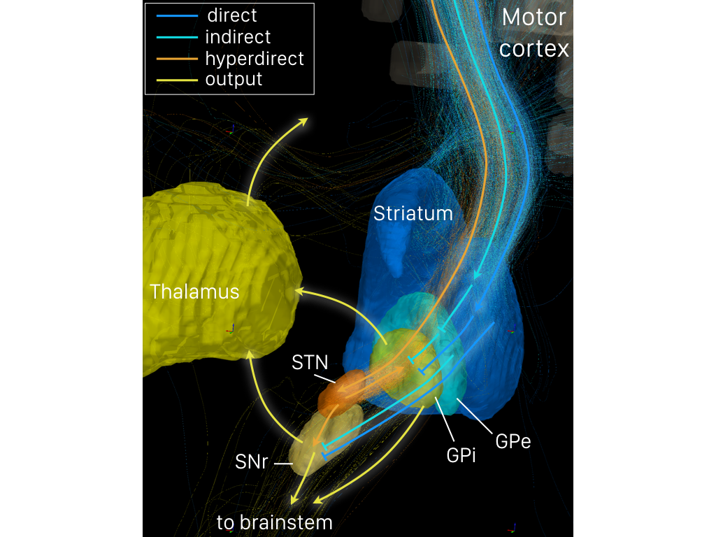
The internal globus pallidus (GPi or medial globus pallidus; in rodents its homologue is known as the entopeduncular nucleus) and the external globus pallidus (GPe) make up the globus pallidus. The GPi is one of the output nuclei of the basal ganglia (the other being the substantia nigra pars reticulata). The GABAergic neurons of the GPi send their axons to the ventral anterior nucleus (VA) and the ventral lateral nucleus (VL) in the dorsal thalamus, to the centromedian complex, and to the pedunculopontine complex. The efferent bundle is constituted first of the ansa and lenticular fasciculus, then crosses the internal capsule within and in parallel to the Edinger's comb system then arrives at the laterosuperior corner of the subthalamic nucleus and constitutes the field H2 of Forel, then H, and suddenly changes its direction to form field H1 that goes to the inferior part of the thalamus. The distribution of axonal islands is widespread in the lateral region of the thalamus. Th ... [...More Info...] [...Related Items...] OR: [Wikipedia] [Google] [Baidu] |
Globus Pallidus
The globus pallidus (GP), also known as paleostriatum or dorsal pallidum, is a subcortical structure of the brain. It consists of two adjacent segments, one external, known in rodents simply as the globus pallidus, and one internal, known in rodents as the entopeduncular nucleus. It is part of the telencephalon, but retains close functional ties with the subthalamus in the diencephalon – both of which are part of the extrapyramidal motor system. The globus pallidus is a major component of the basal ganglia, with principal inputs from the striatum, and principal direct outputs to the thalamus and the substantia nigra. The latter is made up of similar neuronal elements, has similar afferents from the striatum, similar projections to the thalamus, and has a similar synaptology. Neither receives direct cortical afferents, and both receive substantial additional inputs from the intralaminar thalamus. Globus pallidus is Latin for "pale globe". Structure Pallidal nuclei are ma ... [...More Info...] [...Related Items...] OR: [Wikipedia] [Google] [Baidu] |
Subthalamic Nucleus
The subthalamic nucleus (STN) is a small lens-shaped nucleus in the brain where it is, from a functional point of view, part of the basal ganglia system. In terms of anatomy, it is the major part of the subthalamus. As suggested by its name, the subthalamic nucleus is located ventral to the thalamus. It is also dorsal to the substantia nigra and medial to the internal capsule. It was first described by Jules Bernard Luys in 1865, and the term ''corpus Luysi'' or ''Luys' body'' is still sometimes used. Anatomy Structure The principal type of neuron found in the subthalamic nucleus has rather long, sparsely spiny dendrites. In the more centrally located neurons, the dendritic arbors have a more ellipsoidal shape. The dimensions of these arbors (1200 μm, 600 μm, and 300 μm) are similar across many species—including rat, cat, monkey and human—which is unusual. However, the number of neurons increases with brain size as well as the external dimensions of th ... [...More Info...] [...Related Items...] OR: [Wikipedia] [Google] [Baidu] |
Deep Brain Stimulation
Deep brain stimulation (DBS) is a neurosurgical procedure involving the placement of a medical device called a neurostimulator, which sends electrical impulses, through implanted electrodes, to specific targets in the brain (the brain nucleus) for the treatment of movement disorders, including Parkinson's disease, essential tremor, dystonia, and other conditions such as obsessive-compulsive disorder (OCD) and epilepsy. While its underlying principles and mechanisms are not fully understood, DBS directly changes brain activity in a controlled manner. DBS has been approved by the Food and Drug Administration as a treatment for essential tremor and Parkinson's disease (PD) since 1997. DBS was approved for dystonia in 2003, obsessive–compulsive disorder (OCD) in 2009, and epilepsy in 2018. DBS has been studied in clinical trials as a potential treatment for chronic pain for various affective disorders, including major depression. It is one of few neurosurgical procedures that ... [...More Info...] [...Related Items...] OR: [Wikipedia] [Google] [Baidu] |
Tardive Dyskinesia
Tardive dyskinesia (TD) is a disorder that results in involuntary repetitive body movements, which may include grimacing, sticking out the tongue or smacking the lips. Additionally, there may be rapid jerking movements or slow writhing movements. In about 20% of people with TD, the disorder interferes with daily functioning. Tardive dyskinesia occurs in some people as a result of long-term use of dopamine-receptor-blocking medications such as antipsychotics and metoclopramide. These medications are usually used for mental illness but may also be given for gastrointestinal or neurological problems. The condition typically develops only after months to years of use. The diagnosis is based on the symptoms after ruling out other potential causes. Efforts to prevent the condition include either using the lowest possible dose or discontinuing use of neuroleptics. Treatment includes stopping the neuroleptic medication if possible or switching to clozapine. Other medications such a ... [...More Info...] [...Related Items...] OR: [Wikipedia] [Google] [Baidu] |
Tourette Syndrome
Tourette syndrome or Tourette's syndrome (abbreviated as TS or Tourette's) is a common neurodevelopmental disorder that begins in childhood or adolescence. It is characterized by multiple movement (motor) tics and at least one vocal (phonic) tic. Common tics are blinking, coughing, throat clearing, sniffing, and facial movements. These are typically preceded by an unwanted urge or sensation in the affected muscles known as a premonitory urge, can sometimes be suppressed temporarily, and characteristically change in location, strength, and frequency. Tourette's is at the more severe end of a spectrum of tic disorders. The tics often go unnoticed by casual observers. Tourette's was once regarded as a rare and bizarre syndrome and has popularly been associated with coprolalia (the utterance of obscene words or socially inappropriate and derogatory remarks). It is no longer considered rare; about 1% of school-age children and adolescents are estimated to have Tourette's, and ... [...More Info...] [...Related Items...] OR: [Wikipedia] [Google] [Baidu] |
Parkinson's Disease
Parkinson's disease (PD), or simply Parkinson's, is a long-term degenerative disorder of the central nervous system that mainly affects the motor system. The symptoms usually emerge slowly, and as the disease worsens, non-motor symptoms become more common. The most obvious early symptoms are tremor, rigidity, slowness of movement, and difficulty with walking. Cognitive and behavioral problems may also occur with depression, anxiety, and apathy occurring in many people with PD. Parkinson's disease dementia becomes common in the advanced stages of the disease. Those with Parkinson's can also have problems with their sleep and sensory systems. The motor symptoms of the disease result from the death of cells in the substantia nigra, a region of the midbrain, leading to a dopamine deficit. The cause of this cell death is poorly understood, but involves the build-up of misfolded proteins into Lewy bodies in the neurons. Collectively, the main motor symptoms are also known as ... [...More Info...] [...Related Items...] OR: [Wikipedia] [Google] [Baidu] |
Indirect Pathway
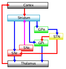
The indirect pathway, sometimes known as the indirect pathway of movement, is a neuronal circuit through the basal ganglia and several associated nuclei within the central nervous system (CNS) which helps to prevent unwanted muscle contractions from competing with voluntary movements. It operates in conjunction with the direct pathway. Overview of Neuronal Connections and Normal Function The indirect pathway passes through the caudate, putamen, and globus pallidus, which are parts of the basal ganglia. It traverses the subthalamic nucleus, a part of the diencephalon, and enters the substantia nigra, a part of the midbrain. In a resting individual, a specific region of the globus pallidus, known as the internus, and a portion of the substantia nigra, known as the pars reticulata, send spontaneous inhibitory signals to the ventrolateral nucleus (VL) of the thalamus, through the release of GABA, an inhibitory neurotransmitter. Inhibition of the excitatory neurons within VL, whi ... [...More Info...] [...Related Items...] OR: [Wikipedia] [Google] [Baidu] |
Direct Pathway
The direct pathway, sometimes known as the direct pathway of movement, is a neural pathway within the central nervous system (CNS) through the basal ganglia which facilitates the initiation and execution of voluntary movement. It works in conjunction with the indirect pathway. Both of these pathways are part of the cortico-basal ganglia-thalamo-cortical loop. Overview of neuronal connections and normal function The direct pathway passes through the caudate nucleus, putamen, and globus pallidus, which are parts of the basal ganglia. It also involves another basal ganglia component the substantia nigra, a part of the midbrain. In a resting individual, a specific region of the globus pallidus, the internal globus pallidus (GPi), and a part of the substantia nigra, the pars reticulata (SNpr), send spontaneous inhibitory signals to the ventral lateral nucleus (VL) of the thalamus, through the release of GABA, an inhibitory neurotransmitter. Inhibition of the inhibitory neurons that p ... [...More Info...] [...Related Items...] OR: [Wikipedia] [Google] [Baidu] |
Synapse
In the nervous system, a synapse is a structure that permits a neuron (or nerve cell) to pass an electrical or chemical signal to another neuron or to the target effector cell. Synapses are essential to the transmission of nervous impulses from one neuron to another. Neurons are specialized to pass signals to individual target cells, and synapses are the means by which they do so. At a synapse, the plasma membrane of the signal-passing neuron (the ''presynaptic'' neuron) comes into close apposition with the membrane of the target (''postsynaptic'') cell. Both the presynaptic and postsynaptic sites contain extensive arrays of molecular machinery that link the two membranes together and carry out the signaling process. In many synapses, the presynaptic part is located on an axon and the postsynaptic part is located on a dendrite or soma. Astrocytes also exchange information with the synaptic neurons, responding to synaptic activity and, in turn, regulating neurotransmission. Syna ... [...More Info...] [...Related Items...] OR: [Wikipedia] [Google] [Baidu] |
Cerebral Cortex
The cerebral cortex, also known as the cerebral mantle, is the outer layer of neural tissue of the cerebrum of the brain in humans and other mammals. The cerebral cortex mostly consists of the six-layered neocortex, with just 10% consisting of allocortex. It is separated into two cortices, by the longitudinal fissure that divides the cerebrum into the left and right cerebral hemispheres. The two hemispheres are joined beneath the cortex by the corpus callosum. The cerebral cortex is the largest site of neural integration in the central nervous system. It plays a key role in attention, perception, awareness, thought, memory, language, and consciousness. The cerebral cortex is part of the brain responsible for cognition. In most mammals, apart from small mammals that have small brains, the cerebral cortex is folded, providing a greater surface area in the confined volume of the cranium. Apart from minimising brain and cranial volume, cortical folding is crucial for the brain ... [...More Info...] [...Related Items...] OR: [Wikipedia] [Google] [Baidu] |
Striatopallidal Fibres
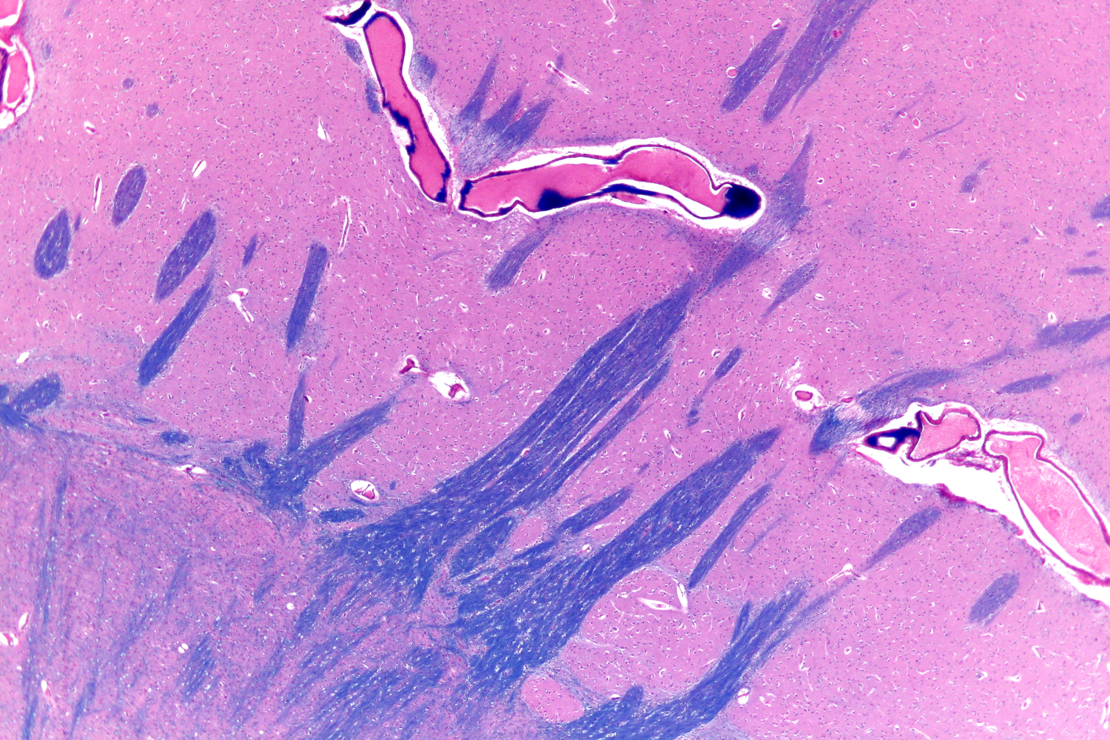
The striatopallidal fibres, also Wilson's pencils, pencil fibres of Wilson, and pencils of Wilson, are prominent myelinated fibres that connect the striatum to the globus pallidus. Their distinctive appearance allows the putamen to be identified on light microscopy. See also *Lentiform nucleus The lentiform nucleus, or lenticular nucleus, comprises the putamen and the globus pallidus within the basal ganglia. With the caudate nucleus, it forms the dorsal striatum. It is a large, lens-shaped mass of gray matter just lateral to the inter ... References {{Reflist, 2 Basal ganglia ... [...More Info...] [...Related Items...] OR: [Wikipedia] [Google] [Baidu] |
Striatum
The striatum, or corpus striatum (also called the striate nucleus), is a nucleus (a cluster of neurons) in the subcortical basal ganglia of the forebrain. The striatum is a critical component of the motor and reward systems; receives glutamatergic and dopaminergic inputs from different sources; and serves as the primary input to the rest of the basal ganglia. Functionally, the striatum coordinates multiple aspects of cognition, including both motor and action planning, decision-making, motivation, reinforcement, and reward perception. The striatum is made up of the caudate nucleus and the lentiform nucleus. The lentiform nucleus is made up of the larger putamen, and the smaller globus pallidus. Strictly speaking the globus pallidus is part of the striatum. It is common practice, however, to implicitly exclude the globus pallidus when referring to striatal structures. In primates, the striatum is divided into a ventral striatum, and a dorsal striatum, subdivisions that are ... [...More Info...] [...Related Items...] OR: [Wikipedia] [Google] [Baidu] |


