|
Internal Arcuate Fibres
In neuroanatomy, the internal arcuate fibers or internal arcuate tract are the axons of second-order sensory neurons that compose the gracile and cuneate nuclei of the medulla oblongata. These second-order neurons begin in the gracile and cuneate nuclei in the medulla. They receive input from first-order sensory neurons, which provide sensation to many areas of the body and have cell bodies in the dorsal root ganglia of the dorsal root of the spinal nerves. Upon decussation (crossing over) from one side of the medulla to the other, also known as the sensory decussation, they are then called the medial lemniscus. The internal arcuate fibers are part of the second-order neurons of the posterior column-medial lemniscus system, and are important for relaying the sensation of fine touch and proprioception to the thalamus and ultimately to the cerebral cortex The cerebral cortex, also known as the cerebral mantle, is the outer layer of neural tissue of the cerebrum of the b ... [...More Info...] [...Related Items...] OR: [Wikipedia] [Google] [Baidu] |
Leo Testut
Leo or Léo may refer to: Acronyms * Law enforcement officer * Law enforcement organisation * ''Louisville Eccentric Observer'', a free weekly newspaper in Louisville, Kentucky * Michigan Department of Labor and Economic Opportunity Arts and entertainment Music * Leo (band), a Missouri-based rock band that was founded in Cleveland, Ohio * L.E.O. (band), a band by musician Bleu and collaborators Film * ''Leo'' (2000 film), a Spanish film by José Luis Borau * ''Leo'' (2002 film), a British-American drama film * ''Leo'', a 2007 Swedish film by Josef Fares * ''Leo'' (2012 film), a Kenyan film * Leo the Lion (MGM), mascot of the Metro-Goldwyn-Mayer movie studio Television * Leo Awards, a British Columbian television award * "Leo", an episode of ''Being Erica'' * Léo, fictional lion in the animation ''Animal Crackers'' * ''Léo'', 2018 Quebec television series created by Fabien Cloutier Companies * Leo Namibia, former name for the TN Mobile phone network in Namibia * Leo ... [...More Info...] [...Related Items...] OR: [Wikipedia] [Google] [Baidu] |
Olivary Body
In anatomy, the olivary bodies or simply olives (Latin ''oliva'' and ''olivae'', singular and plural, respectively) are a pair of prominent oval structures in the medulla oblongata, the lower portion of the brainstem. They contain the olivary nuclei. Structure The olivary body is located on the anterior surface of the medulla lateral to the pyramid, from which it is separated by the antero-lateral sulcus and the fibers of the hypoglossal nerve. Behind (dorsally), it is separated from the postero-lateral sulcus by the ventral spinocerebellar fasciculus. In the depression between the upper end of the olive and the pons lies the vestibulocochlear nerve. In humans, it measures about 1.25 cm. in length, and between its upper end and the pons there is a slight depression to which the roots of the facial nerve are attached. The external arcuate fibers wind across the lower part of the pyramid and olive and enter the inferior peduncle. Olivary nuclei The olive consists of two pa ... [...More Info...] [...Related Items...] OR: [Wikipedia] [Google] [Baidu] |
Proprioception
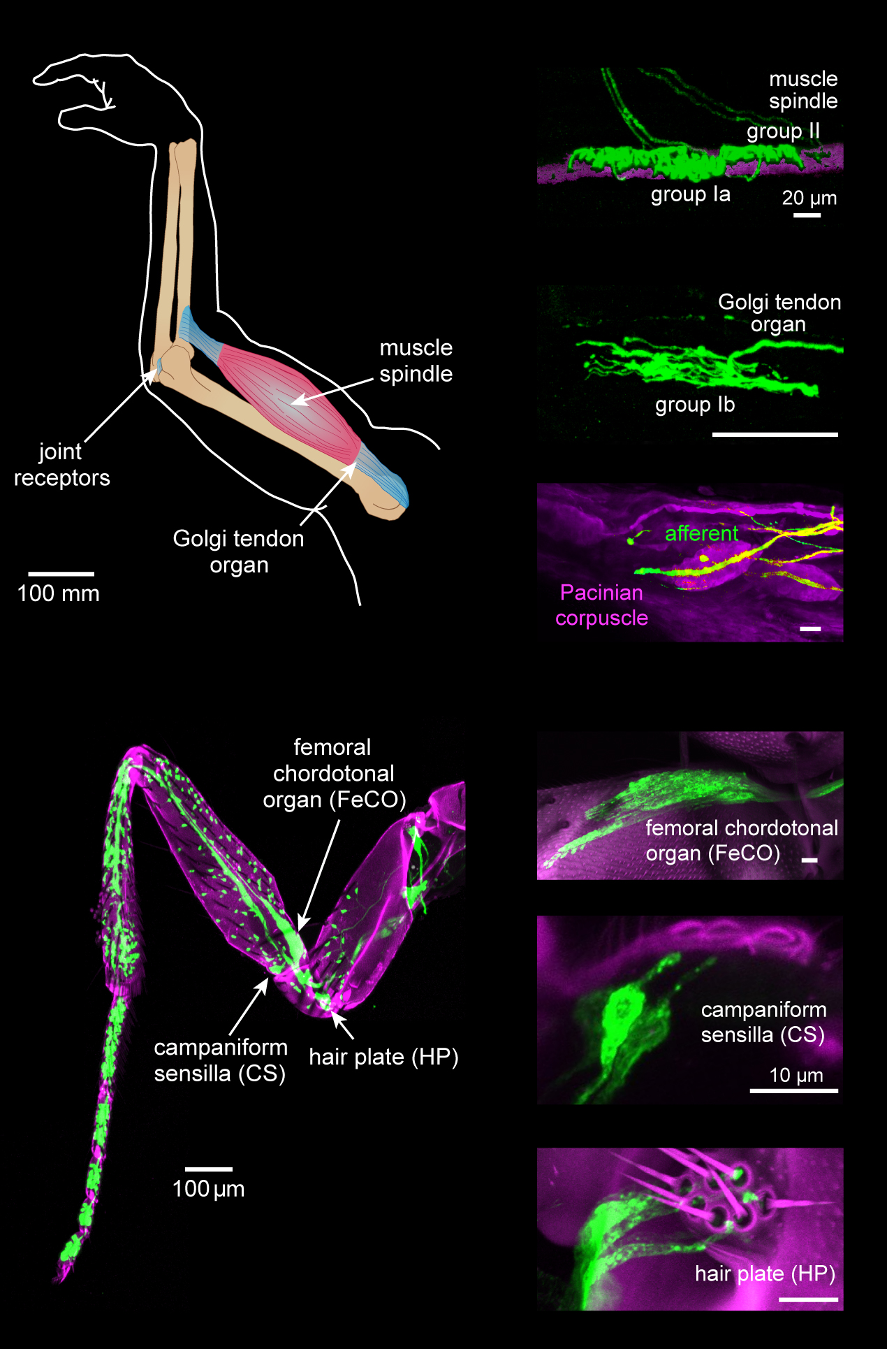
Proprioception ( ), also referred to as kinaesthesia (or kinesthesia), is the sense of self-movement, force, and body position. It is sometimes described as the "sixth sense". Proprioception is mediated by proprioceptors, mechanosensory neurons located within muscles, tendons, and joints. Most animals possess multiple subtypes of proprioceptors, which detect distinct kinematic parameters, such as joint position, movement, and load. Although all mobile animals possess proprioceptors, the structure of the sensory organs can vary across species. Proprioceptive signals are transmitted to the central nervous system, where they are integrated with information from other sensory systems, such as the visual system and the vestibular system, to create an overall representation of body position, movement, and acceleration. In many animals, sensory feedback from proprioceptors is essential for stabilizing body posture and coordinating body movement. System overview In vertebrates, limb ve ... [...More Info...] [...Related Items...] OR: [Wikipedia] [Google] [Baidu] |
Touch
In physiology, the somatosensory system is the network of neural structures in the brain and body that produce the perception of touch (haptic perception), as well as temperature (thermoception), body position (proprioception), and pain. It is a subset of the sensory nervous system, which also represents visual, auditory, olfactory, and gustatory stimuli. Somatosensation begins when mechano- and thermosensitive structures in the skin or internal organs sense physical stimuli such as pressure on the skin (see mechanotransduction, nociception). Activation of these structures, or receptors, leads to activation of peripheral sensory neurons that convey signals to the spinal cord as patterns of action potentials. Sensory information is then processed locally in the spinal cord to drive reflexes, and is also conveyed to the brain for conscious perception of touch and proprioception. Note, somatosensory information from the face and head enters the brain through peripheral senso ... [...More Info...] [...Related Items...] OR: [Wikipedia] [Google] [Baidu] |
Posterior Column-medial Lemniscus System , a relative future tense
{{disambiguation ...
Posterior may refer to: * Posterior (anatomy), the end of an organism opposite to its head ** Buttocks, as a euphemism * Posterior horn (other) * Posterior probability, the conditional probability that is assigned when the relevant evidence is taken into account * Posterior tense Relative tense and absolute tense are distinct possible uses of the grammatical category of Grammatical tense, tense. Absolute tense means the grammatical expression of time reference (usually past tense, past, present tense, present or future tense ... [...More Info...] [...Related Items...] OR: [Wikipedia] [Google] [Baidu] |
Medial Lemniscus
In neuroanatomy, the medial lemniscus, also known as Reil's band or Reil's ribbon (for German anatomist Johann Christian Reil), is a large ascending bundle of heavily myelinated axons that decussate (cross) in the brainstem, specifically in the medulla oblongata. The medial lemniscus is formed by the crossings of the internal arcuate fibers. The internal arcuate fibers are composed of axons of nucleus gracilis and nucleus cuneatus. The axons of the nucleus gracilis and nucleus cuneatus in the medial lemniscus have cell bodies that lie contralaterally. The medial lemniscus is part of the dorsal column–medial lemniscus pathway, which ascends from the skin to the thalamus, which is important for somatosensation from the skin and joints, therefore, lesion of the medial lemnisci causes an impairment of vibratory and touch-pressure sense. Etymology Lemniscus means "ribbon", so named because the medial lemniscus "spirals" or "turns" as it ascends. Path After neurons carrying propri ... [...More Info...] [...Related Items...] OR: [Wikipedia] [Google] [Baidu] |
Sensory Decussation
In neuroanatomy, the sensory decussation or decussation of the lemnisci is a decussation (i.e. crossover) of axons from the gracile nucleus and cuneate nucleus, which are responsible for fine touch, vibration, proprioception and two-point discrimination of the body. The fibres of this decussation are called the internal arcuate fibres and are found at the superior aspect of the closed medulla superior to the motor decussation. It is part of the second neuron in the posterior column–medial lemniscus pathway. Structure At the level of the closed medulla in the posterior white column, two large nuclei namely the gracile nucleus and the cuneate nucleus can be found. The two nuclei receive the impulse from the two ascending tracts: fasciculus gracilis and fasciculus cuneatus. After the two tracts terminate upon these nuclei, the heavily myelinated fibres arise and ascend anteromedially around the periaqueductal gray as internal arcuate fibres. These fibres decussate (cross) ... [...More Info...] [...Related Items...] OR: [Wikipedia] [Google] [Baidu] |
Decussate
Decussation is used in biological contexts to describe a crossing (due to the shape of the Roman numeral for ten, an uppercase 'X' (), ). In Latin anatomical terms, the form is used, e.g. . Similarly, the anatomical term chiasma is named after the Greek uppercase 'Χ' (chi). Whereas a decussation refers to a crossing within the central nervous system, various kinds of crossings in the peripheral nervous system are called chiasma. Examples include: * In the brain, where nerve fibers obliquely cross from one lateral side of the brain to the other, that is to say they cross at a level other than their origin. See for examples Decussation of pyramids and sensory decussation. In neuroanatomy, the term ''chiasma'' is reserved for crossing of- or within nerves such as in the optic chiasm. * In botanical leaf taxology, the word ''decussate'' describes an opposite pattern of leaves which has successive pairs at right angles to each other (i.e. rotated 90 degrees along the stem when ... [...More Info...] [...Related Items...] OR: [Wikipedia] [Google] [Baidu] |
Spinal Nerve
A spinal nerve is a mixed nerve, which carries motor, sensory, and autonomic signals between the spinal cord and the body. In the human body there are 31 pairs of spinal nerves, one on each side of the vertebral column. These are grouped into the corresponding cervical, thoracic, lumbar, sacral and coccygeal regions of the spine. There are eight pairs of cervical nerves, twelve pairs of thoracic nerves, five pairs of lumbar nerves, five pairs of sacral nerves, and one pair of coccygeal nerves. The spinal nerves are part of the peripheral nervous system. Structure Each spinal nerve is a mixed nerve, formed from the combination of nerve fibers from its dorsal and ventral roots. The dorsal root is the afferent sensory root and carries sensory information to the brain. The ventral root is the efferent motor root and carries motor information from the brain. The spinal nerve emerges from the spinal column through an opening (intervertebral foramen) between adjacent vertebrae. ... [...More Info...] [...Related Items...] OR: [Wikipedia] [Google] [Baidu] |
Dorsal Root Ganglia
A dorsal root ganglion (or spinal ganglion; also known as a posterior root ganglion) is a cluster of neurons (a ganglion) in a dorsal root of a spinal nerve. The cell bodies of sensory neurons known as first-order neurons are located in the dorsal root ganglia. The axons of dorsal root ganglion neurons are known as afferents. In the peripheral nervous system, afferents refer to the axons that relay sensory information into the central nervous system (i.e. the brain and the spinal cord). Structure The neurons comprising the dorsal root ganglion are of the pseudo-unipolar type, meaning they have a cell body (soma) with two branches that act as a single axon, often referred to as a ''distal process'' and a ''proximal process''. Unlike the majority of neurons found in the central nervous system, an action potential in posterior root ganglion neuron may initiate in the ''distal process'' in the periphery, bypass the cell body, and continue to propagate along the ''proximal pro ... [...More Info...] [...Related Items...] OR: [Wikipedia] [Google] [Baidu] |
Cuneate Nucleus
In neuroanatomy, the dorsal column nuclei are a pair of nuclei in the dorsal columns in the brainstem. The name refers collectively to the cuneate nucleus and gracile nucleus, which are present at the bottom of the medulla oblongata. Both nuclei contain second-order neurons of the dorsal column–medial lemniscus pathway, which carries fine touch and proprioceptive information from the body to the brain. Fibres reach the thalamus. Structure Nerve pathways The dorsal column nuclei each have an associated nerve tract in the spinal cord, the gracile fasciculus and the cuneate fasciculus. Both dorsal column nuclei contain synapses from afferent nerve fibers that have travelled in the spinal cord. They then send on second-order neurons of the dorsal column–medial lemniscal pathway. Neurons of the dorsal column nuclei eventually reach the midbrain and the thalamus. They send axons that form the internal arcuate fibers. These cross over at the sensory decussation to form ... [...More Info...] [...Related Items...] OR: [Wikipedia] [Google] [Baidu] |
Gracile Nucleus
In neuroanatomy, the dorsal column nuclei are a pair of nuclei in the dorsal columns in the brainstem. The name refers collectively to the cuneate nucleus and gracile nucleus, which are present at the bottom of the medulla oblongata. Both nuclei contain second-order neurons of the dorsal column–medial lemniscus pathway, which carries fine touch and proprioceptive information from the body to the brain. Fibres reach the thalamus. Structure Nerve pathways The dorsal column nuclei each have an associated nerve tract in the spinal cord, the gracile fasciculus and the cuneate fasciculus. Both dorsal column nuclei contain synapses from afferent nerve fibers that have travelled in the spinal cord. They then send on second-order neurons of the dorsal column–medial lemniscal pathway. Neurons of the dorsal column nuclei eventually reach the midbrain and the thalamus. They send axons that form the internal arcuate fibers. These cross over at the sensory decussation to form th ... [...More Info...] [...Related Items...] OR: [Wikipedia] [Google] [Baidu] |


