|
Interleukin 33
Interleukin 33 (IL-33) is a protein that in humans is encoded by the ''IL33'' gene. Interleukin 33 is a member of the IL-1 family that potently drives production of T helper-2 (Th2)-associated cytokines (e.g., IL-4). IL33 is a ligand for ST2 (IL1RL1), an IL-1 family receptor that is highly expressed on Th2 cells, mast cells and group 2 innate lymphocytes. IL-33 is expressed by a wide variety of cell types, including fibroblasts, mast cells, dendritic cells, macrophages, osteoblasts, endothelial cells, and epithelial cells. Structure IL-33 is a member of the IL-1 superfamily of cytokines, a determination based in part on the molecules β-trefoil structure, a conserved structure type described in other IL-1 cytokines, including IL-1α, IL-1β, IL-1Ra and IL-18. In this structure, the 12 β-strands of the β-trefoil are arranged in three pseudorepeats of four β-strand units, of which the first and last β-strands are antiparallel staves in a six-stranded β-barrel, wh ... [...More Info...] [...Related Items...] OR: [Wikipedia] [Google] [Baidu] |
Protein
Proteins are large biomolecules and macromolecules that comprise one or more long chains of amino acid residues. Proteins perform a vast array of functions within organisms, including catalysing metabolic reactions, DNA replication, responding to stimuli, providing structure to cells and organisms, and transporting molecules from one location to another. Proteins differ from one another primarily in their sequence of amino acids, which is dictated by the nucleotide sequence of their genes, and which usually results in protein folding into a specific 3D structure that determines its activity. A linear chain of amino acid residues is called a polypeptide. A protein contains at least one long polypeptide. Short polypeptides, containing less than 20–30 residues, are rarely considered to be proteins and are commonly called peptides. The individual amino acid residues are bonded together by peptide bonds and adjacent amino acid residues. The sequence of amino acid residue ... [...More Info...] [...Related Items...] OR: [Wikipedia] [Google] [Baidu] |
Interleukin 18
Interleukin-18 (IL-18), also known as interferon-gamma inducing factor) is a protein which in humans is encoded by the ''IL18'' gene. The protein encoded by this gene is a proinflammatory cytokine. Many cell types, both hematopoietic cells and non-hematopoietic cells, have the potential to produce IL-18. It was first described in 1989 as a factor that induced interferon-γ (IFN-γ) production in mouse spleen cells. Originally, IL-18 production was recognized in Kupffer cells, liver-resident macrophages. However, IL-18 is constitutively expressed in non-hematopoietic cells, such as intestinal epithelial cells, keratinocytes, and endothelial cells. IL-18 can modulate both innate and adaptive immunity and its dysregulation can cause autoimmune or inflammatory diseases. Processing Cytokines usually contain the signal peptide which is necessary for their extracellular release. In this case, ''IL18'' gene, similar to other IL-1 family members, lacks this signal peptide. Furthermore, ... [...More Info...] [...Related Items...] OR: [Wikipedia] [Google] [Baidu] |
Interleukin 5
Interleukin 5 (IL5) is an interleukin produced by type-2 T helper cells and mast cells. Function Through binding to the interleukin-5 receptor, interleukin 5 stimulates B cell growth and increases immunoglobulin secretion - primarily IgA. It is also a key mediator in eosinophil activation. Structure IL-5 is a 115-amino acid (in human, 133 in the mouse) -long TH2 cytokine that is part of the hematopoietic family. Unlike other members of this cytokine family (namely interleukin 3 and GM-CSF), this glycoprotein in its active form is a homodimer. Tissue expression The IL-5 gene is located on chromosome 11 in the mouse, and chromosome 5 in humans, in close proximity to the genes encoding IL-3, IL-4, and granulocyte-macrophage colony-stimulating factor (GM-CSF), which are often co-expressed in TH2 cells. Interleukin-5 is also expressed by eosinophils and has been observed in the mast cells of asthmatic airways by immunohistochemistry. IL-5 expression is regulated by several ... [...More Info...] [...Related Items...] OR: [Wikipedia] [Google] [Baidu] |
MAP Kinase
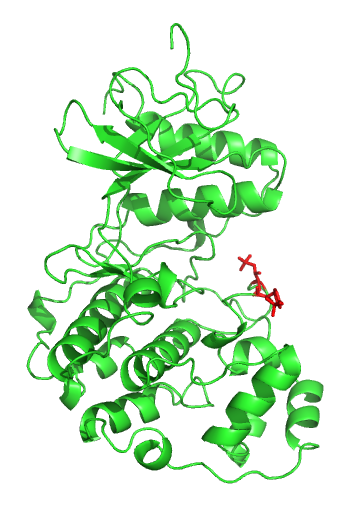
A mitogen-activated protein kinase (MAPK or MAP kinase) is a type of protein kinase that is specific to the amino acids serine and threonine (i.e., a serine/threonine-specific protein kinase). MAPKs are involved in directing cellular responses to a diverse array of stimuli, such as mitogens, osmotic stress, heat shock and proinflammatory cytokines. They regulate cell functions including proliferation, gene expression, differentiation, mitosis, cell survival, and apoptosis. MAP kinases are found in eukaryotes only, but they are fairly diverse and encountered in all animals, fungi and plants, and even in an array of unicellular eukaryotes. MAPKs belong to the CMGC (CDK/MAPK/GSK3/CLK) kinase group. The closest relatives of MAPKs are the cyclin-dependent kinases (CDKs). Discovery The first mitogen-activated protein kinase to be discovered was ERK1 (MAPK3) in mammals. Since ERK1 and its close relative ERK2 (MAPK1) are both involved in growth factor signaling, the family ... [...More Info...] [...Related Items...] OR: [Wikipedia] [Google] [Baidu] |
IL1RAP
Interleukin-1 receptor accessory protein is a protein that in humans is encoded by the ''IL1RAP'' gene. Interleukin 1 induces synthesis of acute phase and proinflammatory proteins during infection, tissue damage, or stress, by forming a complex at the cell membrane with an interleukin 1 receptor and an accessory protein. This gene encodes an interleukin 1 receptor accessory protein. Alternative splicing of this gene results in two transcript variants encoding two different isoforms, one membrane-bound and one soluble. Interactions IL1RAP has been shown to interact with TOLLIP and Interleukin 1 receptor, type I Interleukin 1 receptor, type I (IL1R1) also known as CD121a (Cluster of Differentiation 121a), is an interleukin receptor. IL1R1 also denotes its human gene. The protein encoded by this gene is a cytokine receptor that belongs to the interleukin-1 .... References Further reading * * * * * * * * * * * * * * * * * * External links * * {{gene-3-stub Proteins ... [...More Info...] [...Related Items...] OR: [Wikipedia] [Google] [Baidu] |
NF-κB
Nuclear factor kappa-light-chain-enhancer of activated B cells (NF-κB) is a protein complex that controls transcription of DNA, cytokine production and cell survival. NF-κB is found in almost all animal cell types and is involved in cellular responses to stimuli such as stress, cytokines, free radicals, heavy metals, ultraviolet irradiation, oxidized LDL, and bacterial or viral antigens. NF-κB plays a key role in regulating the immune response to infection. Incorrect regulation of NF-κB has been linked to cancer, inflammatory and autoimmune diseases, septic shock, viral infection, and improper immune development. NF-κB has also been implicated in processes of synaptic plasticity and memory. Discovery NF-κB was discovered by Ranjan Sen in the lab of Nobel laureate David Baltimore via its interaction with an 11-base pair sequence in the immunoglobulin light-chain enhancer in B cells. Later work by Alexander Poltorak and Bruno Lemaitre in mice and ''Drosophila'' frui ... [...More Info...] [...Related Items...] OR: [Wikipedia] [Google] [Baidu] |
SUV39H1
Histone-lysine N-methyltransferase SUV39H1 is an enzyme that in humans is encoded by the ''SUV39H1'' gene. Function This gene is a member of the suppressor of variegation 3-9 homolog family and encodes a protein with a chromodomain and a C-terminal SET domain. This nuclear protein moves to the centromeres during mitosis and functions as a histone methyltransferase, methylating Lys-9 of histone H3. Overall, it plays a vital role in heterochromatin organization, chromosome segregation, and mitotic progression. In mouse embryonic stem cells, Suv39h1 expression is repressed by OCT4 protein through the induction of an antisense long non-coding RNA. Interactions SUV39H1 has been shown to interact with: * CBX1, * CBX5, * DNMT3A, * HDAC1, * HDAC3, * HDAC9, * Histone deacetylase 2, * MBD1, * RUNX1, * Retinoblastoma protein, and * SBF1. * PIN1 Peptidyl-prolyl cis-trans isomerase NIMA-interacting 1 is an enzyme that in humans is encoded by the ''PIN1'' gene. Pin 1, or ... [...More Info...] [...Related Items...] OR: [Wikipedia] [Google] [Baidu] |
Histone Methyltransferase
Histone methyltransferases (HMT) are histone-modifying enzymes (e.g., histone-lysine N-methyltransferases and histone-arginine N-methyltransferases), that catalyze the transfer of one, two, or three methyl groups to lysine and arginine residues of histone proteins. The attachment of methyl groups occurs predominantly at specific lysine or arginine residues on histones H3 and H4. Two major types of histone methyltranferases exist, lysine-specific (which can be SET (Su(var)3-9, Enhancer of Zeste, Trithorax) domain containing or non-SET domain containing) and arginine-specific. In both types of histone methyltransferases, S-Adenosyl methionine (SAM) serves as a cofactor and methyl donor group. The genomic DNA of eukaryotes associates with histones to form chromatin. The level of chromatin compaction depends heavily on histone methylation and other post-translational modifications of histones. Histone methylation is a principal epigenetic modification of chromatin that determines ge ... [...More Info...] [...Related Items...] OR: [Wikipedia] [Google] [Baidu] |
Venule
A venule is a very small blood vessel in the microcirculation that allows blood to return from the capillary beds to drain into the larger blood vessels, the veins. Venules range from 7μm to 1mm in diameter. Veins contain approximately 70% of total blood volume, while about 25% is contained in the venules. Many venules unite to form a vein. Structure Venule walls have three layers: An inner endothelium composed of squamous endothelial cells that act as a membrane, a middle layer of muscle and elastic tissue and an outer layer of fibrous connective tissue. The middle layer is poorly developed so that venules have thinner walls than arterioles. They are porous so that fluid and blood cells can move easily from the bloodstream through their walls. Short portal venules between the neural and anterior pituitary lobes provide an avenue for rapid hormonal exchange via the blood. Specifically within and between the pituitary lobes is anatomical evidence for confluent interlobe ve ... [...More Info...] [...Related Items...] OR: [Wikipedia] [Google] [Baidu] |
Endothelial
The endothelium is a single layer of squamous endothelial cells that line the interior surface of blood vessels and lymphatic vessels. The endothelium forms an interface between circulating blood or lymph in the lumen and the rest of the vessel wall. Endothelial cells form the barrier between vessels and tissue and control the flow of substances and fluid into and out of a tissue. Endothelial cells in direct contact with blood are called vascular endothelial cells whereas those in direct contact with lymph are known as lymphatic endothelial cells. Vascular endothelial cells line the entire circulatory system, from the heart to the smallest capillaries. These cells have unique functions that include fluid filtration, such as in the glomerulus of the kidney, blood vessel tone, hemostasis, neutrophil recruitment, and hormone trafficking. Endothelium of the interior surfaces of the heart chambers is called endocardium. An impaired function can lead to serious health issues throug ... [...More Info...] [...Related Items...] OR: [Wikipedia] [Google] [Baidu] |
Cell Nucleus
The cell nucleus (pl. nuclei; from Latin or , meaning ''kernel'' or ''seed'') is a membrane-bound organelle found in eukaryotic cells. Eukaryotic cells usually have a single nucleus, but a few cell types, such as mammalian red blood cells, have no nuclei, and a few others including osteoclasts have many. The main structures making up the nucleus are the nuclear envelope, a double membrane that encloses the entire organelle and isolates its contents from the cellular cytoplasm; and the nuclear matrix, a network within the nucleus that adds mechanical support. The cell nucleus contains nearly all of the cell's genome. Nuclear DNA is often organized into multiple chromosomes – long stands of DNA dotted with various proteins, such as histones, that protect and organize the DNA. The genes within these chromosomes are structured in such a way to promote cell function. The nucleus maintains the integrity of genes and controls the activities of the cell by regulating gene expres ... [...More Info...] [...Related Items...] OR: [Wikipedia] [Google] [Baidu] |
Basophil
Basophils are a type of white blood cell. Basophils are the least common type of granulocyte, representing about 0.5% to 1% of circulating white blood cells. However, they are the largest type of granulocyte. They are responsible for inflammatory reactions during immune response, as well as in the formation of acute and chronic allergic diseases, including anaphylaxis, asthma, atopic dermatitis and hay fever. They also produce compounds that coordinate immune responses, including histamine and serotonin that induce inflammation, heparin that prevents blood clotting, although there are less than that found in mast cell granules. Mast cells were once thought to be basophils that migrated from blood into their resident tissues (connective tissue), but they are now known to be different types of cells. Basophils were discovered in 1879 by German physician Paul Ehrlich, who one year earlier had found a cell type present in tissues that he termed ''mastzellen'' (now mast cells). Ehrl ... [...More Info...] [...Related Items...] OR: [Wikipedia] [Google] [Baidu] |


