|
Infundibulopelvic Ligament
The suspensory ligament of the ovary, also infundibulopelvic ligament (commonly abbreviated IP ligament or simply IP), is a fold of peritoneum that extends out from the ovary to the wall of the pelvis. Some sources consider it a part of the broad ligament of uterus while other sources just consider it a "termination" of the ligament. It is not considered a true ligament in that it does not physically support any anatomical structures; however it is an important landmark and it houses the ovarian vessels. The suspensory ligament is directed upward over the iliac vessels. Structure It contains the ovarian artery, ovarian vein, ovarian nerve plexus, at |
Uterus
The uterus (from Latin ''uterus'', plural ''uteri'') or womb () is the organ in the reproductive system of most female mammals, including humans that accommodates the embryonic and fetal development of one or more embryos until birth. The uterus is a hormone-responsive sex organ that contains glands in its lining that secrete uterine milk for embryonic nourishment. In the human, the lower end of the uterus, is a narrow part known as the isthmus that connects to the cervix, leading to the vagina. The upper end, the body of the uterus, is connected to the fallopian tubes, at the uterine horns, and the rounded part above the openings to the fallopian tubes is the fundus. The connection of the uterine cavity with a fallopian tube is called the uterotubal junction. The fertilized egg is carried to the uterus along the fallopian tube. It will have divided on its journey to form a blastocyst that will implant itself into the lining of the uterus – the endometrium, where it will ... [...More Info...] [...Related Items...] OR: [Wikipedia] [Google] [Baidu] |
Ovarian Plexus
The ovarian plexus arises from the renal plexus, and is distributed to the ovary, and fundus of the uterus. It is carried in the suspensory ligament of the ovary. at eMedicine
eMedicine is an online clinical medical knowledge base founded in 1996 by doctors Scott Plantz and Jonathan Adler, and computer engineer Jeffrey Berezin. The eMedicine website consists of approximately 6,800 medical topic review articles, each of ... Dictionary
References External links Ne ...[...More Info...] [...Related Items...] OR: [Wikipedia] [Google] [Baidu] |
Ligament Of Ovary
The ovarian ligament (also called the utero-ovarian ligament or proper ovarian ligament) is a fibrous ligament that connects the ovary to the lateral surface of the uterus. Structure The ovarian ligament is composed of muscular and fibrous tissue; it extends from the uterine extremity of the ovary to the lateral aspect of the uterus, just below the point where the uterine tube and uterus meet. The ligament runs in the broad ligament of the uterus, which is a fold of peritoneum rather than a fibrous ligament. Specifically, it is located in the parametrium. Development Embryologically, each ovary (which forms from the gonadal ridge) is connected to a band of mesoderm, the gubernaculum. This strip of mesoderm remains in connection with the ovary throughout its development, and eventually spans this distance by attachment within the labia majora. During the latter parts of urogenital development, the gubernaculum forms a long fibrous band of connective tissue stretching from the ... [...More Info...] [...Related Items...] OR: [Wikipedia] [Google] [Baidu] |
Development Of The Reproductive System
The development of the reproductive system is the part of embryonic growth that results in the sex organs and contributes to sexual differentiation. Due to its large overlap with development of the urinary system, the two systems are typically described together as the urogenital or genitourinary system. The reproductive organs develop from the intermediate mesoderm and are preceded by more primitive structures that are superseded before birth. These embryonic structures are the mesonephric ducts (also known as ''Wolffian ducts'') and the paramesonephric ducts, (also known as ''Müllerian ducts''). The mesonephric duct gives rise to the male seminal vesicles, epididymes and vas deferens. The paramesonephric duct gives rise to the female fallopian tubes, uterus, cervix, and upper part of the vagina. Mesonephric ducts The mesonephric duct originates from a part of the pronephric duct. Origin In the outer part of the intermediate mesoderm, immediately under the ectoderm, in ... [...More Info...] [...Related Items...] OR: [Wikipedia] [Google] [Baidu] |
Prenatal Development
Prenatal development () includes the development of the embryo and of the fetus during a viviparous animal's gestation. Prenatal development starts with fertilization, in the germinal stage of embryonic development, and continues in fetal development until birth. In human pregnancy, prenatal development is also called antenatal development. The development of the human embryo follows fertilization, and continues as fetal development. By the end of the tenth week of gestational age the embryo has acquired its basic form and is referred to as a fetus. The next period is that of fetal development where many organs become fully developed. This fetal period is described both topically (by organ) and chronologically (by time) with major occurrences being listed by gestational age. The very early stages of embryonic development are the same in all mammals, but later stages of development, and the length of gestation varies. Terminology In the human: Different terms are used to ... [...More Info...] [...Related Items...] OR: [Wikipedia] [Google] [Baidu] |
Intermediate Mesoderm
Intermediate mesoderm or intermediate mesenchyme is a narrow section of the mesoderm (one of the three primary germ layers) located between the paraxial mesoderm and the lateral plate of the developing embryo. The intermediate mesoderm develops into vital parts of the urogenital system (kidneys, gonads and respective tracts). Early formation Factors regulating the formation of the intermediate mesoderm are not fully understood. It is believed that bone morphogenic proteins, or BMPs, specify regions of growth along the dorsal-ventral axis of the mesoderm and plays a central role in formation of the intermediate mesoderm. Vg1/Nodal signalling is an identified regulator of intermediate mesoderm formation acting through BMP signalling. Excess Vg1/Nodal signalling during early gastrulation stages results in expansion of the intermediate mesoderm at the expense of the adjacent paraxial mesoderm, whereas inhibition of Vg1/Nodal signalling represses intermediate mesoderm formation. A li ... [...More Info...] [...Related Items...] OR: [Wikipedia] [Google] [Baidu] |
Mesonephros
The mesonephros ( el, middle kidney) is one of three excretory system, excretory organs that develop in vertebrates. It serves as the main excretory organ of aquatic vertebrates and as a temporary kidney in reptiles, birds, and mammals. The mesonephros is included in the Wolffian body after Caspar Friedrich Wolff who described it in 1759. (The Wolffian body is composed of: mesonephros + paramesonephrotic blastema) Structure The mesonephros acts as a structure similar to the human kidney, kidney that, in humans, functions between the sixth and tenth weeks of embryological life. Despite the similarity in structure, function, and terminology, however, the mesonephric nephrons do not form any part of the mature kidney or nephrons. In humans, the mesonephros consists of units which are similar in structure and function to nephrons of the adult kidney. Each of these consists of a glomerulus, a tuft of capillary, capillaries which arises from lateral branches of dorsal aorta and drains i ... [...More Info...] [...Related Items...] OR: [Wikipedia] [Google] [Baidu] |
Pelvis
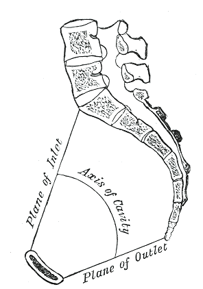
The pelvis (plural pelves or pelvises) is the lower part of the trunk, between the abdomen and the thighs (sometimes also called pelvic region), together with its embedded skeleton (sometimes also called bony pelvis, or pelvic skeleton). The pelvic region of the trunk includes the bony pelvis, the pelvic cavity (the space enclosed by the bony pelvis), the pelvic floor, below the pelvic cavity, and the perineum, below the pelvic floor. The pelvic skeleton is formed in the area of the back, by the sacrum and the coccyx and anteriorly and to the left and right sides, by a pair of hip bones. The two hip bones connect the spine with the lower limbs. They are attached to the sacrum posteriorly, connected to each other anteriorly, and joined with the two femurs at the hip joints. The gap enclosed by the bony pelvis, called the pelvic cavity, is the section of the body underneath the abdomen and mainly consists of the reproductive organs (sex organs) and the rectum, while the pelvic f ... [...More Info...] [...Related Items...] OR: [Wikipedia] [Google] [Baidu] |
Retroperitoneal
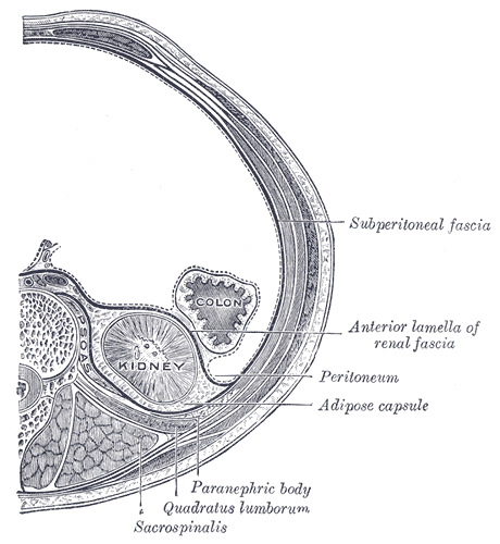
The retroperitoneal space (retroperitoneum) is the anatomical space (sometimes a potential space) behind (''retro'') the peritoneum. It has no specific delineating anatomical structures. Organs are retroperitoneal if they have peritoneum on their anterior side only. Structures that are not suspended by mesentery in the abdominal cavity and that lie between the parietal peritoneum and abdominal wall are classified as retroperitoneal. This is different from organs that are not retroperitoneal, which have peritoneum on their posterior side and are suspended by mesentery in the abdominal cavity. The retroperitoneum can be further subdivided into the following: *Perirenal (or perinephric) space *Anterior pararenal (or paranephric) space *Posterior pararenal (or paranephric) space Retroperitoneal structures Structures that lie behind the peritoneum are termed "retroperitoneal". Organs that were once suspended within the abdominal cavity by mesentery but migrated posterior to the ... [...More Info...] [...Related Items...] OR: [Wikipedia] [Google] [Baidu] |
Gray240
Grey (more common in British English) or gray (more common in American English) is an intermediate color between black and white. It is a neutral or achromatic color, meaning literally that it is "without color", because it can be composed of black and white. It is the color of a cloud-covered sky, of ash and of lead. The first recorded use of ''grey'' as a color name in the English language was in 700 CE.Maerz and Paul ''A Dictionary of Color'' New York:1930 McGraw-Hill Page 196 ''Grey'' is the dominant spelling in European and Commonwealth English, while ''gray'' has been the preferred spelling in American English; both spellings are valid in both varieties of English. In Europe and North America, surveys show that grey is the color most commonly associated with neutrality, conformity, boredom, uncertainty, old age, indifference, and modesty. Only one percent of respondents chose it as their favorite color. Etymology ''Grey'' comes from the Middle English or , ... [...More Info...] [...Related Items...] OR: [Wikipedia] [Google] [Baidu] |
Lymphatic Vessel
The lymphatic vessels (or lymph vessels or lymphatics) are thin-walled vessels (tubes), structured like blood vessels, that carry lymph. As part of the lymphatic system, lymph vessels are complementary to the cardiovascular system. Lymph vessels are lined by endothelial cells, and have a thin layer of smooth muscle, and adventitia that binds the lymph vessels to the surrounding tissue. Lymph vessels are devoted to the propulsion of the lymph from the lymph capillaries, which are mainly concerned with the absorption of interstitial fluid from the tissues. Lymph capillaries are slightly bigger than their counterpart capillaries of the vascular system. Lymph vessels that carry lymph to a lymph node are called afferent lymph vessels, and those that carry it from a lymph node are called efferent lymph vessels, from where the lymph may travel to another lymph node, may be returned to a vein, or may travel to a larger lymph duct. Lymph ducts drain the lymph into one of the subclavia ... [...More Info...] [...Related Items...] OR: [Wikipedia] [Google] [Baidu] |
EMedicine
eMedicine is an online clinical medical knowledge base founded in 1996 by doctors Scott Plantz and Jonathan Adler, and computer engineer Jeffrey Berezin. The eMedicine website consists of approximately 6,800 medical topic review articles, each of which is associated with a clinical subspecialty "textbook". The knowledge base includes over 25,000 clinically multimedia files. Each article is authored by board certified specialists in the subspecialty to which the article belongs and undergoes three levels of physician peer-review, plus review by a Doctor of Pharmacy. The article's authors are identified with their current faculty appointments. Each article is updated yearly, or more frequently as changes in practice occur, and the date is published on the article. eMedicine.com was sold to WebMD in January, 2006 and is available as the Medscape Reference. History Plantz, Adler and Berezin evolved the concept for eMedicine.com in 1996 and deployed the initial site via Boston Med ... [...More Info...] [...Related Items...] OR: [Wikipedia] [Google] [Baidu] |


