|
Infraspinatus Muscle
In human anatomy, the infraspinatus muscle is a thick triangular muscle, which occupies the chief part of the infraspinatous fossa.'' Gray's Anatomy'', see infobox. As one of the four muscles of the rotator cuff, the main function of the infraspinatus is to externally rotate the humerus and stabilize the shoulder joint. Structure It attaches medially to the infraspinous fossa of the scapula and laterally to the middle facet of the greater tubercle of the humerus. The muscle arises by fleshy fibers from the medial two-thirds of the infraspinatous fossa, and by tendinous fibers from the ridges on its surface; it also arises from the infraspinatous fascia which covers it, and separates it from the teres major and teres minor. The fibers converge to a tendon, which glides over the lateral border of the spine of the scapula and passing across the posterior part of the capsule of the shoulder-joint, is inserted into the middle impression on the greater tubercle of the humerus. The t ... [...More Info...] [...Related Items...] OR: [Wikipedia] [Google] [Baidu] |
Vertebral Column
The vertebral column, also known as the backbone or spine, is part of the axial skeleton. The vertebral column is the defining characteristic of a vertebrate in which the notochord (a flexible rod of uniform composition) found in all chordata, chordates has been replaced by a segmented series of bone: vertebrae separated by intervertebral discs. Individual vertebrae are named according to their region and position, and can be used as anatomical landmarks in order to guide procedures such as Lumbar puncture, lumbar punctures. The vertebral column houses the spinal canal, a cavity that encloses and protects the spinal cord. There are about 50,000 species of animals that have a vertebral column. The human vertebral column is one of the most-studied examples. Many different diseases in humans can affect the spine, with spina bifida and scoliosis being recognisable examples. The general structure of human vertebrae is fairly typical of that found in mammals, reptiles, and birds. Th ... [...More Info...] [...Related Items...] OR: [Wikipedia] [Google] [Baidu] |
Infraspinatous Fascia
The infraspinatous fascia is a dense fibrous membrane, covering the Infraspinatous muscle and fixed to the circumference of the infraspinatous fossa; it affords attachment, by its deep surface, to some fibers of that muscle. It is intimately attached to the deltoid fascia along the over-lapping border of the Deltoideus The deltoid muscle is the muscle forming the rounded contour of the human shoulder. It is also known as the 'common shoulder muscle', particularly in other animals such as the domestic cat. Anatomically, the deltoid muscle appears to be made up o .... References Fascia {{musculoskeletal-stub ... [...More Info...] [...Related Items...] OR: [Wikipedia] [Google] [Baidu] |
Muscles Of The Upper Limb
Skeletal muscles (commonly referred to as muscles) are organs of the vertebrate muscular system and typically are attached by tendons to bones of a skeleton. The muscle cells of skeletal muscles are much longer than in the other types of muscle tissue, and are often known as muscle fibers. The muscle tissue of a skeletal muscle is striated – having a striped appearance due to the arrangement of the sarcomeres. Skeletal muscles are voluntary muscles under the control of the somatic nervous system. The other types of muscle are cardiac muscle which is also striated and smooth muscle which is non-striated; both of these types of muscle tissue are classified as involuntary, or, under the control of the autonomic nervous system. A skeletal muscle contains multiple fascicles – bundles of muscle fibers. Each individual fiber, and each muscle is surrounded by a type of connective tissue layer of fascia. Muscle fibers are formed from the fusion of developmental myoblasts in a p ... [...More Info...] [...Related Items...] OR: [Wikipedia] [Google] [Baidu] |
Coracoid Process
The coracoid process (from Greek κόραξ, raven) is a small hook-like structure on the lateral edge of the superior anterior portion of the scapula (hence: coracoid, or "like a raven's beak"). Pointing laterally forward, it, together with the acromion, serves to stabilize the shoulder joint. It is palpable in the deltopectoral groove between the deltoid and pectoralis major muscles. Structure The coracoid process is a thick curved process attached by a broad base to the upper part of the neck of the scapula; it runs at first upward and medialward; then, becoming smaller, it changes its direction, and projects forward and lateralward. Anatomically it is divided into intervals of: base of coracoid process, angle of coracoid process, shaft and the apex of the coracoid process. The coracoglenoid notch is an indentation localized between the coracoid process and the glenoid. As the coracoid process projects laterally, it house underneath it the subcoracoid space. The ''ascend ... [...More Info...] [...Related Items...] OR: [Wikipedia] [Google] [Baidu] |
Pectoralis Minor Muscle
Pectoralis minor muscle () is a thin, triangular muscle, situated at the upper part of the chest, beneath the pectoralis major in the human body. Structure Attachments Pectoralis minor muscle arises from the upper margins and outer surfaces of the third, fourth, and fifth ribs, near their costal cartilages and from the aponeuroses covering the intercostalis. The fibers pass superior and lateral and converge to form a flat tendon. This tendon inserts onto the medial border and upper surface of the coracoid process of the scapula. Relations Pectoralis minor muscle forms part of the anterior wall of the axilla. It is covered anteriorly (superficially) by the clavipectoral fascia. The medial pectoral nerve pierces the pectoralis minor and the clavipectoral fascia. In attaching to the coracoid process, the pectoralis minor forms a 'bridge' - structures passing into the upper limb from the thorax will pass directly underneath.http://www.teachmeanatomy.com/muscles-of-the-pector ... [...More Info...] [...Related Items...] OR: [Wikipedia] [Google] [Baidu] |
Pectoralis Major Muscle
The pectoralis major () is a thick, fan-shaped or triangular convergent muscle, situated at the chest of the human body. It makes up the bulk of the chest muscles and lies under the breast. Beneath the pectoralis major is the pectoralis minor, a thin, triangular muscle. The pectoralis major's primary functions are flexion, adduction, and internal rotation of the humerus. The pectoral major may colloquially be referred to as "pecs", "pectoral muscle", or "chest muscle", because it is the largest and most superficial muscle in the chest area. Structure It arises from the anterior surface of the sternal half of the clavicle from breadth of the half of the anterior surface of the sternum, as low down as the attachment of the cartilage of the sixth or seventh rib; from the cartilages of all the true ribs, with the exception, frequently, of the first or seventh, and from the aponeurosis of the abdominal external oblique muscle. From this extensive origin the fibers converge toward the ... [...More Info...] [...Related Items...] OR: [Wikipedia] [Google] [Baidu] |
Thieme Medical Publishers
Thieme Medical Publishers is a German medical and science publisher in the Thieme Publishing Group. It produces professional journals, textbooks, atlases, monographs and reference books in both German and English covering a variety of medical specialties, including neurosurgery, orthopaedics, endocrinology, urology, radiology, anatomy, chemistry, otolaryngology, ophthalmology, audiology and speech-language pathology, complementary and alternative medicine. Thieme has more than 1,000 employees and maintains offices in seven cities worldwide, including New York City, Beijing, Delhi, Stuttgart, and three other cities in Germany. History Georg Thieme Verlag was founded in 1886 in Leipzig, Germany, by Georg Thieme when he was 26 years old. Thieme remains privately held and family-owned. The company received some early success in 1896 by publishing Wilhelm Röntgen's famous picture of his wife's hand in what is still one of Thieme's and Germany's oldest journals, the ''Deutsche ... [...More Info...] [...Related Items...] OR: [Wikipedia] [Google] [Baidu] |
Head Of The Humerus
The humerus (; ) is a long bone in the arm that runs from the shoulder to the elbow. It connects the scapula and the two bones of the lower arm, the radius and ulna, and consists of three sections. The humeral upper extremity consists of a rounded head, a narrow neck, and two short processes (tubercles, sometimes called tuberosities). The body is cylindrical in its upper portion, and more prismatic below. The lower extremity consists of 2 epicondyles, 2 processes (trochlea & capitulum), and 3 fossae (radial fossa, coronoid fossa, and olecranon fossa). As well as its true anatomical neck, the constriction below the greater and lesser tubercles of the humerus is referred to as its surgical neck due to its tendency to fracture, thus often becoming the focus of surgeons. Etymology The word "humerus" is derived from la, humerus, umerus meaning upper arm, shoulder, and is linguistically related to Gothic ''ams'' shoulder and Greek ''ōmos''. Structure Upper extremity The upper or pro ... [...More Info...] [...Related Items...] OR: [Wikipedia] [Google] [Baidu] |
Bursa (anatomy)
( grc-gre, Προῦσα, Proûsa, Latin: Prusa, ota, بورسه, Arabic:بورصة) is a city in northwestern Turkey and the administrative center of Bursa Province. The fourth-most populous city in Turkey and second-most populous in the Marmara Region, Bursa is one of the industrial centers of the country. Most of Turkey's automotive production takes place in Bursa. As of 2019, the Metropolitan Province was home to 3,056,120 inhabitants, 2,161,990 of whom lived in the 3 city urban districts ( Osmangazi, Yildirim and Nilufer) plus Gursu and Kestel, largely conurbated. Bursa was the first major and second overall capital of the Ottoman State between 1335 and 1363. The city was referred to as (, meaning "God's Gift" in Ottoman Turkish, a name of Persian origin) during the Ottoman period, while a more recent nickname is ("") in reference to the parks and gardens located across its urban fabric, as well as to the vast and richly varied forests of the surrounding re ... [...More Info...] [...Related Items...] OR: [Wikipedia] [Google] [Baidu] |
Shoulder-joint
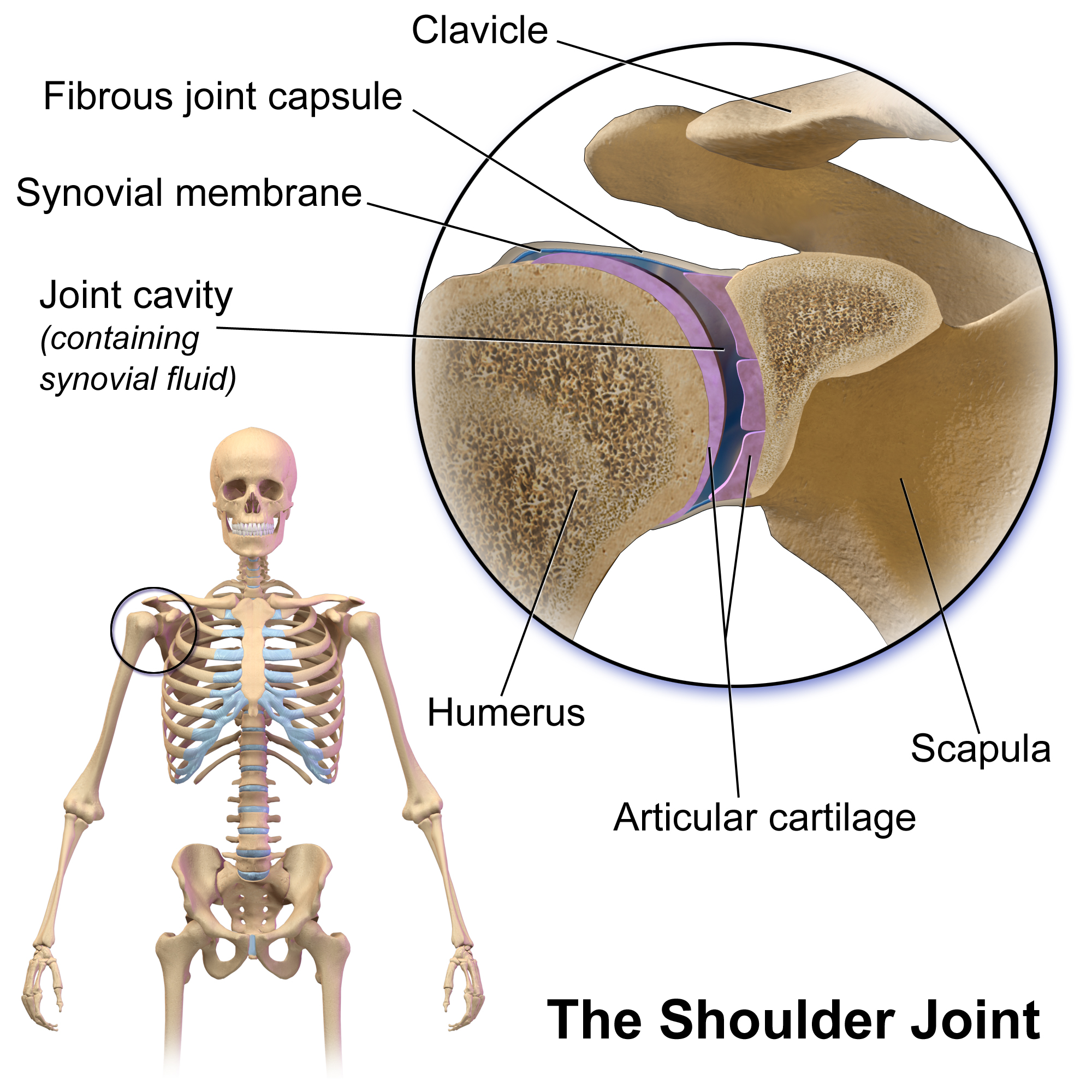
The shoulder joint (or glenohumeral joint from Greek ''glene'', eyeball, + -''oid'', 'form of', + Latin ''humerus'', shoulder) is structurally classified as a synovial ball-and-socket joint and functionally as a diarthrosis and multiaxial joint. It involves an articulation between the glenoid fossa of the scapula (shoulder blade) and the head of the humerus (upper arm bone). Due to the very loose joint capsule that gives a limited interface of the humerus and scapula, it is the most mobile joint of the human body. Structure The shoulder joint is a ball-and-socket joint between the scapula and the humerus. The socket of the glenoid fossa of the scapula is itself quite shallow, but it is made deeper by the addition of the glenoid labrum. The glenoid labrum is a ring of cartilaginous fibre attached to the circumference of the cavity. This ring is continuous with the tendon of the biceps brachii above. Spaces Significant joint spaces are: * The normal glenohumeral space is 4–5 ... [...More Info...] [...Related Items...] OR: [Wikipedia] [Google] [Baidu] |
Teres Minor
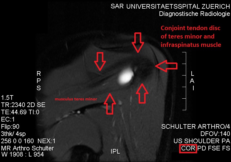
The teres minor (Latin ''teres'' meaning 'rounded') is a narrow, elongated muscle of the rotator cuff. The muscle originates from the lateral border and adjacent posterior surface of the corresponding right or left scapula and inserts at both the greater tubercle of the humerus and the posterior surface of the joint capsule. The primary function of the teres minor is to modulate the action of the deltoid, preventing the humeral head from sliding upward as the arm is abducted. It also functions to rotate the humerus laterally. The teres minor is innervated by the axillary nerve. Structure It arises from the dorsal surface of the axillary border of the scapula for the upper two-thirds of its extent, and from two aponeurotic laminae, one of which separates it from the infraspinatus muscle, the other from the teres major muscle. Its fibers run obliquely upwards and laterally; the upper ones end in a tendon which is inserted into the lowest of the three impressions on the greater tub ... [...More Info...] [...Related Items...] OR: [Wikipedia] [Google] [Baidu] |
Teres Major
The teres major muscle is a muscle of the upper limb. It attaches to the scapula and the humerus and is one of the seven scapulohumeral muscles. It is a thick but somewhat flattened muscle. The teres major muscle (from Latin ''teres'', meaning "rounded") is positioned above the latissimus dorsi muscle and assists in the Anatomical terms of motion#Flexion and extension, extension and Anatomical terms of motion#Rotation, medial rotation of the humerus. This muscle is commonly confused as a rotator cuff muscle, but it is not because it does not attach to the capsule of the shoulder joint, unlike the teres minor muscle for example. Structure The teres major muscle originates on the dorsal surface of the Scapula, inferior angle and the lower part of the Scapula, lateral border of the scapula. The fibers of teres major insert into the medial lip of the Bicipital groove, intertubercular sulcus of the humerus. Relations The tendon, at its insertion, lies behind that of the latissimus ... [...More Info...] [...Related Items...] OR: [Wikipedia] [Google] [Baidu] |


