|
Inferior Thyroid Arteries
The inferior thyroid artery is an artery in the neck. It arises from the thyrocervical trunk and passes upward, in front of the vertebral artery and longus colli muscle. It then turns medially behind the carotid sheath and its contents, and also behind the sympathetic trunk, the middle cervical ganglion resting upon the vessel. Reaching the lower border of the thyroid gland it divides into two branches, which supply the postero-inferior parts of the gland, and anastomose with the superior thyroid artery, and with the corresponding artery of the opposite side. Structure The branches of the inferior thyroid artery are the inferior laryngeal, the oesophageal, the tracheal, the ascending cervical and the pharyngeal arteries. The inferior laryngeal artery climbs the trachea to the back part of the larynx under cover of the inferior pharyngeal constrictor muscle. It is accompanied by the recurrent nerve, and supplies the muscles and mucous membrane of this part, anastomosing with th ... [...More Info...] [...Related Items...] OR: [Wikipedia] [Google] [Baidu] |
Thyrocervical Trunk
The thyrocervical trunk is an artery of the neck. It is a branch of the subclavian artery. It arises from the first portion of this vessel, between the origin of the subclavian artery and the inner border of the scalenus anterior muscle. It is located distally to the vertebral artery and proximally to the costocervical trunk. It gives off a number of branches to the neck. It helps to supply blood to the muscles of the neck. Structure The thyrocervical trunk is a branch of the subclavian artery. It arises from the first portion of this vessel, between the origin of the subclavian artery and the inner border of the scalenus anterior muscle. It is located distally to the vertebral artery and proximally to the costocervical trunk. It is short and thick. Branches The thyrocervical trunk divides soon after its origin into a number of branches. * Inferior thyroid artery. * Suprascapular artery. * Transverse cervical artery. This is present in about 1/3 of cases. In the rest, the ... [...More Info...] [...Related Items...] OR: [Wikipedia] [Google] [Baidu] |
Inferior Pharyngeal Constrictor Muscle
The inferior pharyngeal constrictor muscle is a skeletal muscle of the neck. It is the thickest of the three outer pharyngeal muscles. It arises from the sides of the cricoid cartilage and the thyroid cartilage. It is supplied by the vagus nerve (CN X). It is active during swallowing, and partially during breathing and speech. It may be affected by Zenker's diverticulum. Structure The inferior pharyngeal constrictor muscle is composed of two parts. The first part (and more superior) arises from the thyroid cartilage (thyropharyngeal part), and the second part arises from the cricoid cartilage (cricopharyngeal part). * On the ''thyroid cartilage'', it arises from the oblique line on the side of the lamina, from the surface behind this nearly as far as the posterior border and from the inferior horn of the thyroid cartilage. * From the ''cricoid cartilage'', it arises in the interval between the cricothyroid muscle in front, and the articular facet for the inferior horn of the t ... [...More Info...] [...Related Items...] OR: [Wikipedia] [Google] [Baidu] |
Spinal Cord
The spinal cord is a long, thin, tubular structure made up of nervous tissue, which extends from the medulla oblongata in the brainstem to the lumbar region of the vertebral column (backbone). The backbone encloses the central canal of the spinal cord, which contains cerebrospinal fluid. The brain and spinal cord together make up the central nervous system (CNS). In humans, the spinal cord begins at the occipital bone, passing through the foramen magnum and then enters the spinal canal at the beginning of the cervical vertebrae. The spinal cord extends down to between the first and second lumbar vertebrae, where it ends. The enclosing bony vertebral column protects the relatively shorter spinal cord. It is around long in adult men and around long in adult women. The diameter of the spinal cord ranges from in the cervical and lumbar regions to in the thoracic area. The spinal cord functions primarily in the transmission of nerve signals from the motor cortex to the body, ... [...More Info...] [...Related Items...] OR: [Wikipedia] [Google] [Baidu] |
Intervertebral Foramina
The intervertebral foramen (also called neural foramen, and often abbreviated as IV foramen or IVF) is a :wikt:foramen, foramen between two spinal vertebrae. Cervical vertebrae, Cervical, thoracic vertebrae, thoracic, and lumbar vertebrae all have intervertebral foramina. The foramina, or openings, are present between every pair of vertebrae in these areas. A number of structures pass through the foramen. These are the root of each spinal nerve, the spinal artery of the segmental artery, communicating veins between the internal and external plexuses, meningeal branches of spinal nerve, recurrent meningeal (sinu-vertebral) nerves, and transforaminal ligaments. When the spinal vertebrae are articulated with each other, the bodies form a strong pillar that supports the head and trunk, and the vertebral foramen constitutes a canal for the protection of the medulla spinalis (spinal cord). The size of the foramina is variable due to placement, pathology, spinal loading, and posture ... [...More Info...] [...Related Items...] OR: [Wikipedia] [Google] [Baidu] |
Spinal Canal
The spinal canal (or vertebral canal or spinal cavity) is the canal that contains the spinal cord within the vertebral column. The spinal canal is formed by the vertebrae through which the spinal cord passes. It is a process of the dorsal body cavity. This canal is enclosed within the foramen of the vertebrae. In the intervertebral spaces, the canal is protected by the ligamentum flavum posteriorly and the posterior longitudinal ligament anteriorly. Structure The outermost layer of the meninges, the dura mater, is closely associated with the arachnoid mater which in turn is loosely connected to the innermost layer, the pia mater. The meninges divide the spinal canal into the epidural space and the subarachnoid space. The pia mater is closely attached to the spinal cord. A subdural space is generally only present due to trauma and/or pathological situations. The subarachnoid space is filled with cerebrospinal fluid and contains the vessels that supply the spinal cord, namely ... [...More Info...] [...Related Items...] OR: [Wikipedia] [Google] [Baidu] |
Vertebral Arteries
The vertebral arteries are major arteries of the neck. Typically, the vertebral arteries originate from the subclavian arteries. Each vessel courses superiorly along each side of the neck, merging within the skull to form the single, midline basilar artery. As the supplying component of the ''vertebrobasilar vascular system'', the vertebral arteries supply blood to the upper spinal cord, brainstem, cerebellum, and posterior part of brain. Structure The vertebral arteries usually arise from the posterosuperior aspect of the central subclavian arteries on each side of the body, then enter deep to the transverse process at the level of the 6th cervical vertebrae (C6), or occasionally (in 7.5% of cases) at the level of C7. They then proceed superiorly, in the transverse foramen of each cervical vertebra. Once they have passed through the transverse foramen of C1 (also known as the atlas), the vertebral arteries travel across the posterior arch of C1 and through the suboccipital ... [...More Info...] [...Related Items...] OR: [Wikipedia] [Google] [Baidu] |
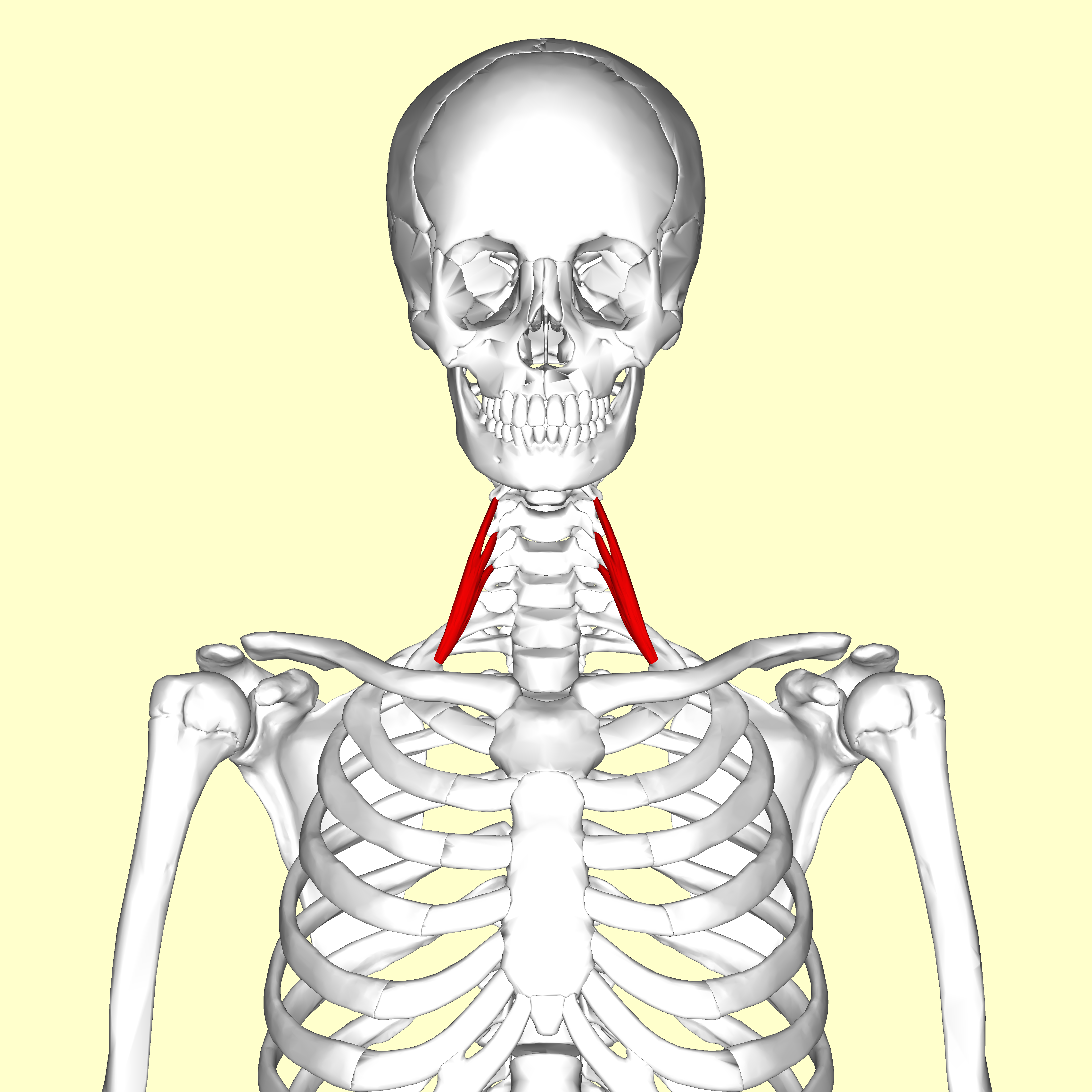
Longus Capitis
The longus capitis muscle (Latin for ''long muscle of the head'', alternatively rectus capitis anticus major), is broad and thick above, narrow below, and arises by four tendinous slips, from the anterior tubercles of the transverse processes of the third, fourth, fifth, and sixth cervical vertebræ, and ascends, converging toward its fellow of the opposite side, to be inserted into the inferior surface of the basilar part of the occipital bone The occipital bone () is a neurocranium, cranial dermal bone and the main bone of the occiput (back and lower part of the skull). It is trapezoidal in shape and curved on itself like a shallow dish. The occipital bone overlies the occipital lobe .... It is innervated by a branch of cervical plexus. Longus capitis has several actions: acting unilaterally, to: *flex the head and neck laterally *rotate the head ipsilaterally acting bilaterally: *flex the head and neck Additional images File:Gray129.png, Occipital bone. Outer surface. ... [...More Info...] [...Related Items...] OR: [Wikipedia] [Google] [Baidu] |
Anterior Scalene Muscle
The scalene muscles are a group of three pairs of muscles in the lateral neck, namely the anterior scalene, middle scalene, and posterior scalene. They are innervated by the third to the eight cervical spinal nerves (C3-C8). The anterior and middle scalene muscles lift the first rib and bend the neck to the same side; the posterior scalene lifts the second rib and tilts the neck to the same side. The muscles are named . Structure The scalene muscles originate from the transverse processes from the cervical vertebrae of C2 to C7 and insert onto the first and second ribs. Anterior scalene The anterior scalene muscle ( la, scalenus anterior), lies deeply at the side of the neck, behind the sternocleidomastoid muscle. It arises from the anterior tubercles of the transverse processes of the third, fourth, fifth, and sixth cervical vertebrae, and descending, almost vertically, is inserted by a narrow, flat tendon into the scalene tubercle on the inner border of the first rib, and i ... [...More Info...] [...Related Items...] OR: [Wikipedia] [Google] [Baidu] |
Cervical Vertebrae
In tetrapods, cervical vertebrae (singular: vertebra) are the vertebrae of the neck, immediately below the skull. Truncal vertebrae (divided into thoracic and lumbar vertebrae in mammals) lie caudal (toward the tail) of cervical vertebrae. In sauropsid species, the cervical vertebrae bear cervical ribs. In lizards and saurischian dinosaurs, the cervical ribs are large; in birds, they are small and completely fused to the vertebrae. The vertebral transverse processes of mammals are homologous to the cervical ribs of other amniotes. Most mammals have seven cervical vertebrae, with the only three known exceptions being the manatee with six, the two-toed sloth with five or six, and the three-toed sloth with nine. In humans, cervical vertebrae are the smallest of the true vertebrae and can be readily distinguished from those of the thoracic or lumbar regions by the presence of a foramen (hole) in each transverse process, through which the vertebral artery, vertebral veins, an ... [...More Info...] [...Related Items...] OR: [Wikipedia] [Google] [Baidu] |
Esophageal Branches Of The Aorta
The esophageal arteries four or five in number, arise from the front of the aorta, and pass obliquely downward to the esophagus, forming a chain of anastomoses along that tube, anastomosing with the esophageal branches of the inferior thyroid arteries above, and with ascending branches from the left inferior phrenic and left gastric arteries below. These arteries supply the middle third of the esophagus. References External links * - "Branches of the ascending aorta, arch of the aorta, and the descending aorta In human anatomy, the descending aorta is part of the aorta, the largest artery in the body. The descending aorta begins at the aortic arch and runs down through the chest and abdomen. The descending aorta anatomically consists of two portions o ...." Arteries of the thorax {{circulatory-stub ... [...More Info...] [...Related Items...] OR: [Wikipedia] [Google] [Baidu] |
Esophagus
The esophagus (American English) or oesophagus (British English; both ), non-technically known also as the food pipe or gullet, is an organ in vertebrates through which food passes, aided by peristaltic contractions, from the pharynx to the stomach. The esophagus is a fibromuscular tube, about long in adults, that travels behind the trachea and heart, passes through the diaphragm, and empties into the uppermost region of the stomach. During swallowing, the epiglottis tilts backwards to prevent food from going down the larynx and lungs. The word ''oesophagus'' is from Ancient Greek οἰσοφάγος (oisophágos), from οἴσω (oísō), future form of φέρω (phérō, “I carry”) + ἔφαγον (éphagon, “I ate”). The wall of the esophagus from the lumen outwards consists of mucosa, submucosa (connective tissue), layers of muscle fibers between layers of fibrous tissue, and an outer layer of connective tissue. The mucosa is a stratified squamous epithel ... [...More Info...] [...Related Items...] OR: [Wikipedia] [Google] [Baidu] |
Bronchial Arteries
In human anatomy, the bronchial arteries supply the lungs with nutrition and oxygenated blood. Although there is much variation, there are usually two bronchial arteries that run to the left lung, and one to the right lung and are a vital part of the respiratory system. Structure There are typically two left and one right bronchial arteries. The ''left bronchial arteries'' (superior and inferior) usually arise directly from the thoracic aorta. The single ''right bronchial artery'' usually arises from one of the following: * 1) the thoracic aorta at a common trunk with the right 3rd posterior intercostal artery * 2) the superior bronchial artery on the left side * 3) any number of the right intercostal arteries mostly the third right posterior. Function The bronchial arteries supply blood to the bronchi and connective tissue of the lungs. They travel with and branch with the bronchi, ending about at the level of the respiratory bronchioles. They anastomose with the branch ... [...More Info...] [...Related Items...] OR: [Wikipedia] [Google] [Baidu] |


