|
Image-guided Radiation Therapy
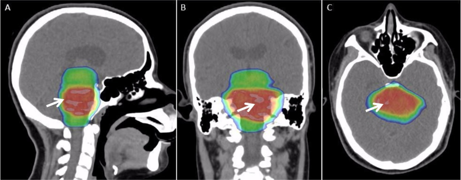
Image-guided radiation therapy is the process of frequent imaging, during a course of radiation treatment, used to direct the treatment, position the patient, and compare to the pre-therapy imaging from the treatment plan. Immediately prior to, or during, a treatment fraction, the patient is localized in the treatment room in the same position as planned from the reference imaging dataset. An example of IGRT would include comparison of a cone beam computed tomography (CBCT) dataset, acquired on the treatment machine, with the computed tomography (CT) dataset from planning. IGRT would also include matching planar kilovoltage (kV) radiographs or megavoltage (MV) images with digital reconstructed radiographs (DRRs) from the planning CT. This process is distinct from the use of imaging to delineate targets and organs in the planning process of radiation therapy. However, there is a connection between the imaging processes as IGRT relies directly on the imaging modalities from planni ... [...More Info...] [...Related Items...] OR: [Wikipedia] [Google] [Baidu] |
Medical Imaging
Medical imaging is the technique and process of imaging the interior of a body for clinical analysis and medical intervention, as well as visual representation of the function of some organs or tissues (physiology). Medical imaging seeks to reveal internal structures hidden by the skin and bones, as well as to diagnose and treat disease. Medical imaging also establishes a database of normal anatomy and physiology to make it possible to identify abnormalities. Although imaging of removed organs and tissues can be performed for medical reasons, such procedures are usually considered part of pathology instead of medical imaging. Measurement and recording techniques that are not primarily designed to produce images, such as electroencephalography (EEG), magnetoencephalography (MEG), electrocardiography (ECG), and others, represent other technologies that produce data susceptible to representation as a parameter graph versus time or maps that contain data about the measureme ... [...More Info...] [...Related Items...] OR: [Wikipedia] [Google] [Baidu] |
Cobalt-60
Cobalt-60 (60Co) is a synthetic radioactive isotope of cobalt with a half-life of 5.2713 years. It is produced artificially in nuclear reactors. Deliberate industrial production depends on neutron activation of bulk samples of the monoisotopic and mononuclidic cobalt isotope . (PDF also located aCanadian Nuclear FAQ Measurable quantities are also produced as a by-product of typical nuclear power plant operation and may be detected externally when leaks occur. In the latter case (in the absence of added cobalt) the incidentally produced is largely the result of multiple stages of neutron activation of iron isotopes in the reactor's steel structures via the creation of its precursor. The simplest case of the latter would result from the activation of . undergoes beta decay to the stable isotope nickel-60 (). The activated nickel nucleus emits two gamma rays with energies of 1.17 and 1.33 MeV, hence the overall equation of the nuclear reaction (activation and decay) is: ... [...More Info...] [...Related Items...] OR: [Wikipedia] [Google] [Baidu] |
Radiation Therapy
Radiation therapy or radiotherapy, often abbreviated RT, RTx, or XRT, is a therapy using ionizing radiation, generally provided as part of cancer treatment to control or kill malignant cells and normally delivered by a linear accelerator. Radiation therapy may be curative in a number of types of cancer if they are localized to one area of the body. It may also be used as part of adjuvant therapy, to prevent tumor recurrence after surgery to remove a primary malignant tumor (for example, early stages of breast cancer). Radiation therapy is synergistic with chemotherapy, and has been used before, during, and after chemotherapy in susceptible cancers. The subspecialty of oncology concerned with radiotherapy is called radiation oncology. A physician who practices in this subspecialty is a radiation oncologist. Radiation therapy is commonly applied to the cancerous tumor because of its ability to control cell growth. Ionizing radiation works by damaging the DNA of cancerous tissue ... [...More Info...] [...Related Items...] OR: [Wikipedia] [Google] [Baidu] |
Positron Emission Tomography
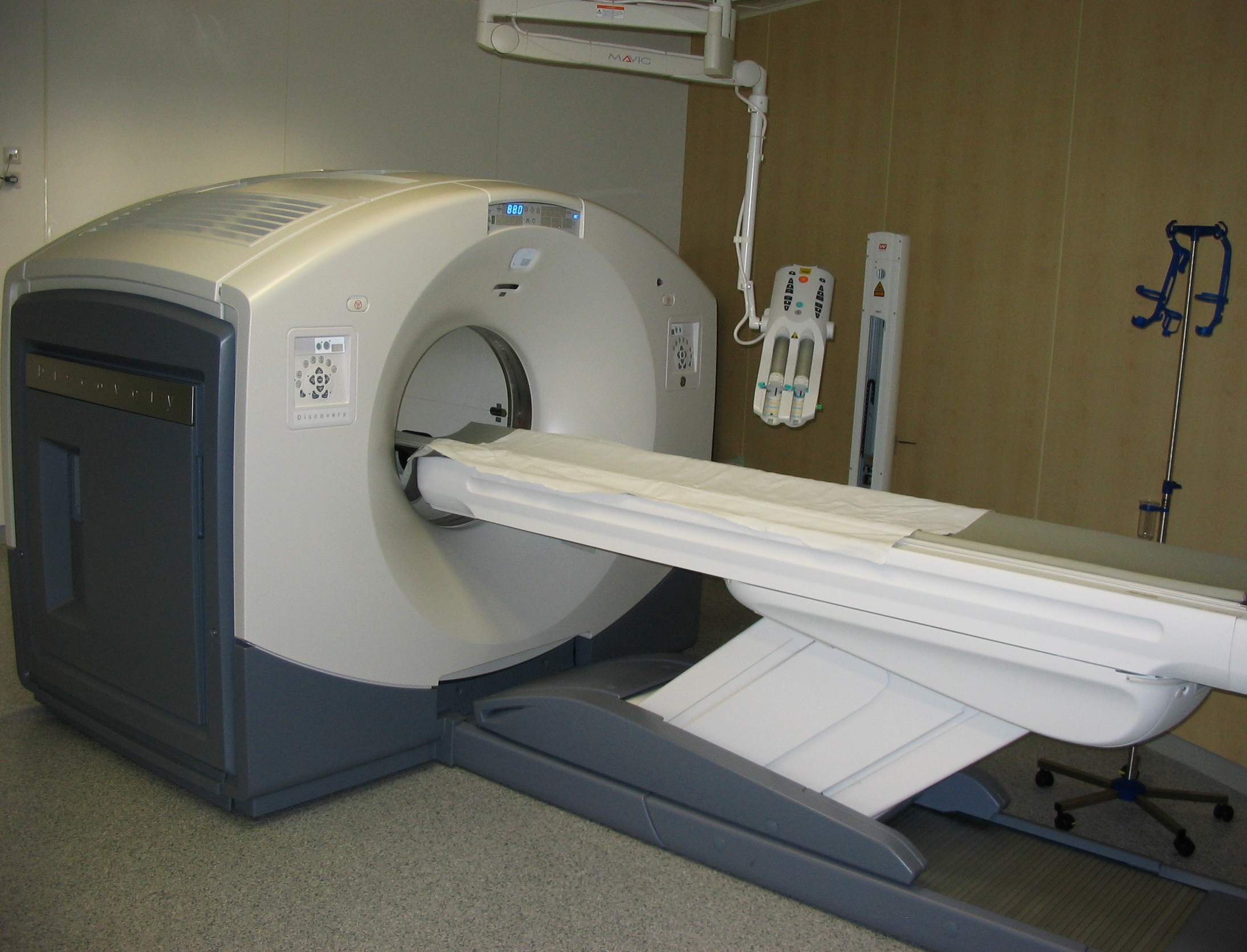
Positron emission tomography (PET) is a functional imaging technique that uses radioactive substances known as radiotracers to visualize and measure changes in metabolic processes, and in other physiological activities including blood flow, regional chemical composition, and absorption. Different tracers are used for various imaging purposes, depending on the target process within the body. For example, -FDG is commonly used to detect cancer, NaF is widely used for detecting bone formation, and oxygen-15 is sometimes used to measure blood flow. PET is a common imaging technique, a medical scintillography technique used in nuclear medicine. A radiopharmaceutical — a radioisotope attached to a drug — is injected into the body as a tracer. When the radiopharmaceutical undergoes beta plus decay, a positron is emitted, and when the positron collides with an ordinary electron, the two particles annihilate and gamma rays are emitted. These gamma rays are detecte ... [...More Info...] [...Related Items...] OR: [Wikipedia] [Google] [Baidu] |
Medical Radiography
Radiography is an imaging technique using X-rays, gamma rays, or similar ionizing radiation and non-ionizing radiation to view the internal form of an object. Applications of radiography include medical radiography ("diagnostic" and "therapeutic") and industrial radiography. Similar techniques are used in airport security (where "body scanners" generally use backscatter X-ray). To create an image in conventional radiography, a beam of X-rays is produced by an X-ray generator and is projected toward the object. A certain amount of the X-rays or other radiation is absorbed by the object, dependent on the object's density and structural composition. The X-rays that pass through the object are captured behind the object by a detector (either photographic film or a digital detector). The generation of flat two dimensional images by this technique is called projectional radiography. In computed tomography (CT scanning) an X-ray source and its associated detectors rotate around ... [...More Info...] [...Related Items...] OR: [Wikipedia] [Google] [Baidu] |
International Commission On Radiation Units And Measurements
The International Commission on Radiation Units and Measurements (ICRU) is a standardization body set up in 1925 by the International Congress of Radiology, originally as the X-Ray Unit Committee until 1950. Its objective "is to develop concepts, definitions and recommendations for the use of quantities and their units for ionizing radiation and its interaction with matter, in particular with respect to the biological effects induced by radiation". The ICRU is a sister organisation to the International Commission on Radiological Protection (ICRP). In general terms the ICRU defines the units, and the ICRP recommends how they are used for radiation protection. Development During the first two decades of its existence, its formal meetings were held during the International Congress of Radiology, but from 1950 onwards, when its mandate was extended, it has met annually. Until 1953, the president of the ICRU was a national of the country that was hosting the ICR, but in that year it ... [...More Info...] [...Related Items...] OR: [Wikipedia] [Google] [Baidu] |
Fluoroscopy
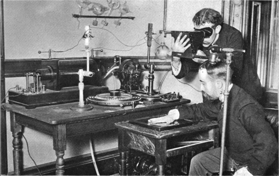
Fluoroscopy () is an imaging technique that uses X-rays to obtain real-time moving images of the interior of an object. In its primary application of medical imaging, a fluoroscope () allows a physician to see the internal structure and function of a patient, so that the pumping action of the heart or the motion of swallowing, for example, can be watched. This is useful for both diagnosis and therapy and occurs in general radiology, interventional radiology, and image-guided surgery. In its simplest form, a fluoroscope consists of an X-ray source and a fluorescent screen, between which a patient is placed. However, since the 1950s most fluoroscopes have included X-ray image intensifiers and cameras as well, to improve the image's visibility and make it available on a remote display screen. For many decades, fluoroscopy tended to produce live pictures that were not recorded, but since the 1960s, as technology improved, recording and playback became the norm. Fluoroscopy is simi ... [...More Info...] [...Related Items...] OR: [Wikipedia] [Google] [Baidu] |
Cyberknife (device)
The CyberKnife System is a radiation therapy device manufactured by Accuray Incorporated. The system is used to deliver radiosurgery for the treatment of benign tumors, malignant tumors and other medical conditions. Device The device combines a compact linear accelerator mounted on a robotic manipulator and an integrated image guidance system. The image guidance system acquires stereoscopic kV images during treatment, tracks tumor motion and guides the robotic manipulator to precisely and accurately align the treatment beam to the moving tumor. The system is designed for stereotactic radiosurgery (SRS) and stereotactic body radiation therapy (SBRT). The system is also used for select 3D conformal radiotherapy (3D-CRT) and intensity modulated radiation therapy (IMRT). History The system was invented by Stanford University and Peter and Russell Schonberg of Schonberg Research Corporation. It was a development of the first 3D irradiation treatment realized with a linear accelera ... [...More Info...] [...Related Items...] OR: [Wikipedia] [Google] [Baidu] |
Compton Effect
Compton scattering, discovered by Arthur Holly Compton, is the scattering of a high frequency photon after an interaction with a charged particle, usually an electron. If it results in a decrease in energy (increase in wavelength) of the photon (which may be an X-ray or gamma ray photon), it is called the Compton effect. Part of the energy of the photon is transferred to the recoiling electron. Inverse Compton scattering occurs when a charged particle transfers part of its energy to a photon. Introduction Compton scattering is an example of elastic scattering of light by a free charged particle, where the wavelength of the scattered light is different from that of the incident radiation. In Compton's original experiment (see Fig. 1), the energy of the X ray photon (≈17 keV) was significantly larger than the binding energy of the atomic electron, so the electrons could be treated as being free after scattering. The amount by which the light's wavelength changes is called the ... [...More Info...] [...Related Items...] OR: [Wikipedia] [Google] [Baidu] |
Alvin J
Alvin may refer to: Places Canada *Alvin, British Columbia United States *Alvin, Colorado *Alvin, Georgia *Alvin, Illinois * Alvin, Michigan *Alvin, Texas *Alvin, Wisconsin, a town *Alvin (community), Wisconsin, an unincorporated community Other uses * Alvin (given name) * Alvin (crater), a crater on Mars * Alvin (digital cultural heritage platform), a Swedish platform for digitised cultural heritage * Alvin (horse), a Canadian Standardbred racehorse * 13677 Alvin, an asteroid * DSV ''Alvin'', a deep-submergence vehicle * Alvin, a fictional planet on ''ALF'' (TV series) * Alvin Seville, of the fictional animated characters Alvin and the Chipmunks * "Alvin", by James from the album ''Girl at the End of the World'' * Tropical Storm Alvin See also * Alvin Community College * Alvin High School * Aylwin (other) Patricio Aylwin (1918–2016) was a Chilean politician. Aylwin may also refer to: *Andrés Aylwin (1925–2018), Chilean politician * Guy Maxwell Aylwin (1889– ... [...More Info...] [...Related Items...] OR: [Wikipedia] [Google] [Baidu] |
Cone Beam Reconstruction
In microtomography X-ray scanners, cone beam reconstruction is one of two common scanning methods, the other being Fan beam reconstruction. Cone beam reconstruction uses a 2-dimensional approach for obtaining projection data. Instead of utilizing a single row of detectors, as fan beam methods do, a cone beam systems uses a standard charge-coupled device camera, focused on a scintillator material. The scintillator converts X-ray radiation to visible light, which is picked up by the camera and recorded. The method has enjoyed widespread implementation in microtomography, and is also used in several larger-scale systems. An X-ray source is positioned across from the detector, with the object being scanned in between. (This is essentially the same setup used for an ordinary X-ray fluoroscope). Projections from different angles are obtained in one of two ways. In one method, the object being scanned is rotated. This has the advantage of simplicity in implementation; a rotating sta ... [...More Info...] [...Related Items...] OR: [Wikipedia] [Google] [Baidu] |


