|
Islands Of Calleja
The islands of Calleja (; IC, ISC, or IClj) are a group of neural granule cells located within the ventral striatum in the brains of most animals. This region of the brain is part of the limbic system, where it aids in the reinforcing effects of reward-like activities. Within most species, the islands are specifically located within the olfactory tubercle; however, in primates, these islands are located within the nucleus accumbens, the reward center of the brain, since the olfactory tubercle has practically disappeared in the brains of primates.Stevens JR. 2002. Schizophrenia: Reproductive hormones and the brain. American Journal of Psychiatry 159:713-9 Both of these structures have been implicated in the processing of incentives as well as addictions to drugs.Ubeda-Banon I, Novejarque A, Mohedano-Moriano A, Pro-Sistiaga P, Insausti R, et al. 2008. Vomeronasal inputs to the rodent ventral striatum. Brain Research Bulletin 75:467-73 Projections to and from the islands supplement th ... [...More Info...] [...Related Items...] OR: [Wikipedia] [Google] [Baidu] |
Striatum
The striatum, or corpus striatum (also called the striate nucleus), is a nucleus (a cluster of neurons) in the subcortical basal ganglia of the forebrain. The striatum is a critical component of the motor and reward systems; receives glutamatergic and dopaminergic inputs from different sources; and serves as the primary input to the rest of the basal ganglia. Functionally, the striatum coordinates multiple aspects of cognition, including both motor and action planning, decision-making, motivation, reinforcement, and reward perception. The striatum is made up of the caudate nucleus and the lentiform nucleus. The lentiform nucleus is made up of the larger putamen, and the smaller globus pallidus. Strictly speaking the globus pallidus is part of the striatum. It is common practice, however, to implicitly exclude the globus pallidus when referring to striatal structures. In primates, the striatum is divided into a ventral striatum, and a dorsal striatum, subdivisions that are ... [...More Info...] [...Related Items...] OR: [Wikipedia] [Google] [Baidu] |
Ventricular System
The ventricular system is a set of four interconnected cavities known as cerebral ventricles in the brain. Within each ventricle is a region of choroid plexus which produces the circulating cerebrospinal fluid (CSF). The ventricular system is continuous with the central canal of the spinal cord from the fourth ventricle, allowing for the flow of CSF to circulate. All of the ventricular system and the central canal of the spinal cord are lined with ependyma, a specialised form of epithelium connected by tight junctions that make up the blood–cerebrospinal fluid barrier. Structure The system comprises four ventricles: * lateral ventricles right and left (one for each hemisphere) * third ventricle * fourth ventricle There are several foramina, openings acting as channels, that connect the ventricles. The interventricular foramina (also called the foramina of Monro) connect the lateral ventricles to the third ventricle through which the cerebrospinal fluid can flow. Ventric ... [...More Info...] [...Related Items...] OR: [Wikipedia] [Google] [Baidu] |
Piriform Cortex
The piriform cortex, or pyriform cortex, is a region in the brain, part of the rhinencephalon situated in the cerebrum. The function of the piriform cortex relates to the sense of smell. Structure The piriform cortex is part of the rhinencephalon situated in the cerebrum. In human anatomy, the piriform cortex has been described as consisting of the cortical amygdala, uncus, and anterior parahippocampal gyrus. More specifically, the human piriform cortex is located between the insula and the temporal lobe, anteriorly and laterally of the amygdala.Howard, J. D., Plailly, J., Grueschow, M., Haynes, J. D., & Gottfried, J. A. (2009). Odor quality coding and categorization in human posterior piriform cortex. Nature neuroscience, 12(7), 932-938. Supplementary material, p.4 Function The function of the piriform cortex relates to olfaction, which is the perception of smell. This has been particularly shown in humans for the posterior piriform cortex. The piriform cortex in rodents and ... [...More Info...] [...Related Items...] OR: [Wikipedia] [Google] [Baidu] |
Neuropil
Neuropil (or "neuropile") is any area in the nervous system composed of mostly unmyelinated axons, dendrites and glial cell processes that forms a synaptically dense region containing a relatively low number of cell bodies. The most prevalent anatomical region of neuropil is the brain which, although not completely composed of neuropil, does have the largest and highest synaptically concentrated areas of neuropil in the body. For example, the neocortex and olfactory bulb both contain neuropil. White matter, which is mostly composed of myelinated axons (hence its white color) and glial cells, is generally not considered to be a part of the neuropil. Neuropil (pl. neuropils) comes from the Greek: ''neuro'', meaning "tendon, sinew; nerve" and ''pilos'', meaning "felt". The term's origin can be traced back to the late 19th century. Location Neuropil has been found in the following regions: outer neocortex layer, barrel cortex, inner plexiform layer and outer plexiform layer, poster ... [...More Info...] [...Related Items...] OR: [Wikipedia] [Google] [Baidu] |
Diagonal Band Of Broca
The diagonal band of Broca is one of the basal forebrain structures that are derived from the ventral telencephalon during development. This structure forms the medial margin of the anterior perforated substance. This brain region was described by the French neuroanatomist Paul Broca. Structure It consists of fibers that are said to arise in the parolfactory area, the gyrus subcallosus and the anterior perforated substance, and course backward in the longitudinal striae to the dentate gyrus and the hippocampal region. This is a cholinergic bundle of nerve fibers posterior to the anterior perforated substance. It interconnects the subcallosal gyrus in the septal area with the hippocampus and lateral olfactory area. Nuclei Two structures are often described in this brain regions, namely the nuclei of the vertical and horizontal limbs of the diagonal band of Broca (nvlDBB and nhlDBB, respectively). nvlDBB projects to the hippocampal formation through the fornix and it is the ... [...More Info...] [...Related Items...] OR: [Wikipedia] [Google] [Baidu] |
Septum Pellucidum
The septum pellucidum (Latin for "translucent wall") is a thin, triangular, vertical double membrane separating the anterior horns of the left and right lateral ventricles of the brain. It runs as a sheet from the corpus callosum down to the fornix. The septum is not present in the syndrome septo-optic dysplasia. Structure The septum pellucidum is located in the septal area in the midline of the brain between the two cerebral hemispheres. The septal area is also the location of the septal nuclei. It is attached to the lower part of the corpus callosum, the large collection of nerve fibers that connect the two cerebral hemispheres. It is attached to the front forward part of the fornix. The lateral ventricles sit on either side of the septum. The septum pellucidum consists of two layers or ''laminae'' of both white and gray matter. During fetal development, there is a space between the two laminae called the cave of septum pellucidum that, in ninety percent of cases, disappear ... [...More Info...] [...Related Items...] OR: [Wikipedia] [Google] [Baidu] |
MEIS2
Homeobox protein Meis2 is a protein that in humans is encoded by the ''MEIS2'' gene In biology, the word gene (from , ; "...Wilhelm Johannsen coined the word gene to describe the Mendelian units of heredity..." meaning ''generation'' or ''birth'' or ''gender'') can have several different meanings. The Mendelian gene is a ba .... This gene encodes a homeobox protein belonging to the TALE ('three amino acid loop extension') family of homeodomain-containing proteins. TALE homeobox proteins are highly conserved transcription regulators, and several members have been shown to be essential contributors to developmental programs. Multiple transcript variants encoding distinct isoforms have been described for this gene. References Further reading * * * * * * * * * * External links * Transcription factors {{gene-15-stub ... [...More Info...] [...Related Items...] OR: [Wikipedia] [Google] [Baidu] |
PBX3
Pre-B-cell leukemia transcription factor 3 is a protein that in humans is encoded by the ''PBX3'' gene In biology, the word gene (from , ; "... Wilhelm Johannsen coined the word gene to describe the Mendelian units of heredity..." meaning ''generation'' or ''birth'' or ''gender'') can have several different meanings. The Mendelian gene is a b .... References Further reading * * * * * * * * * * External links * * Transcription factors {{gene-9-stub ... [...More Info...] [...Related Items...] OR: [Wikipedia] [Google] [Baidu] |
Promoter (biology)
In genetics, a promoter is a sequence of DNA to which proteins bind to initiate transcription of a single RNA transcript from the DNA downstream of the promoter. The RNA transcript may encode a protein (mRNA), or can have a function in and of itself, such as tRNA or rRNA. Promoters are located near the transcription start sites of genes, upstream on the DNA (towards the 5' region of the sense strand). Promoters can be about 100–1000 base pairs long, the sequence of which is highly dependent on the gene and product of transcription, type or class of RNA polymerase recruited to the site, and species of organism. Promoters control gene expression in bacteria and eukaryotes. RNA polymerase must attach to DNA near a gene for transcription to occur. Promoter DNA sequences provide an enzyme binding site. The -10 sequence is TATAAT. -35 sequences are conserved on average, but not in most promoters. Artificial promoters with conserved -10 and -35 elements transcribe more slowly. All D ... [...More Info...] [...Related Items...] OR: [Wikipedia] [Google] [Baidu] |
FOX Proteins
FOX (forkhead box) proteins are a family of transcription factors that play important roles in regulating the expression of genes involved in cell growth, proliferation, differentiation, and longevity. Many FOX proteins are important to embryonic development. FOX proteins also have pioneering transcription activity by being able to bind condensed chromatin during cell differentiation processes. The defining feature of FOX proteins is the forkhead box, a sequence of 80 to 100 amino acids forming a motif that binds to DNA. This forkhead motif is also known as the winged helix, due to the butterfly-like appearance of the loops in the protein structure of the domain. Forkhead proteins are a subgroup of the helix-turn-helix class of proteins. Biological roles Many genes encoding FOX proteins have been identified. For example, the FOXF2 gene encodes forkhead box F2, one of many human homologues of the ''Drosophila melanogaster'' transcription factor forkhead. FOXF2 is expressed in t ... [...More Info...] [...Related Items...] OR: [Wikipedia] [Google] [Baidu] |
Olfactory Bulb
The olfactory bulb (Latin: ''bulbus olfactorius'') is a grey matter, neural structure of the vertebrate forebrain involved in olfaction, the sense of odor, smell. It sends olfactory information to be further processed in the amygdala, the orbitofrontal cortex (OFC) and the hippocampus where it plays a role in emotion, memory and learning. The bulb is divided into two distinct structures: the main olfactory bulb and the accessory olfactory bulb. The main olfactory bulb connects to the amygdala via the piriform cortex of the primary olfactory cortex and directly projects from the main olfactory bulb to specific amygdala areas. The accessory olfactory bulb resides on the dorsal-posterior region of the main olfactory bulb and forms a parallel pathway. Destruction of the olfactory bulb results in ipsilateral anosmia, while irritative lesions of the uncus can result in olfactory and gustatory hallucinations. Structure In most vertebrates, the olfactory bulb is the most Anatomical term ... [...More Info...] [...Related Items...] OR: [Wikipedia] [Google] [Baidu] |
Rostral Migratory Stream
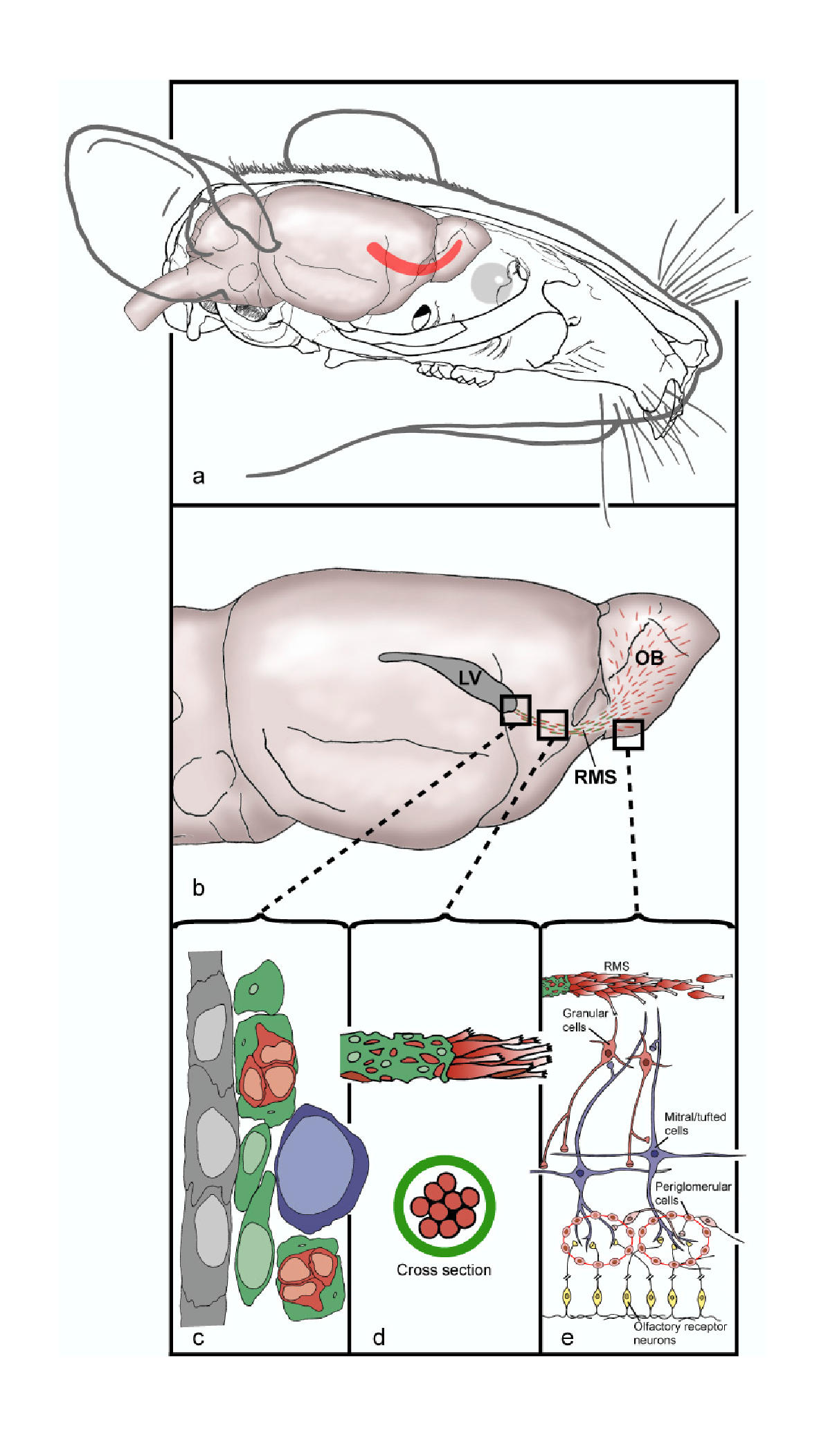
The rostral migratory stream (RMS) is a specialized migratory route found in the brain of some animals along which neuronal precursors that originated in the subventricular zone (SVZ) of the brain migrate to reach the main olfactory bulb (OB). The importance of the RMS lies in its ability to refine and even change an animal's sensitivity to smells, which explains its importance and larger size in the rodent brain as compared to the human brain, as our olfactory sense is not as developed. This pathway has been studied in the rodent, rabbit, and both the squirrel monkey and rhesus monkey. When the neurons reach the OB they differentiate into GABAergic interneurons as they are integrated into either the granule cell layer or periglomerular layer. Although it was originally believed that neurons could not regenerate in the adult brain, neurogenesis has been shown to occur in mammalian brains, including those of primates. However, neurogenesis is limited to the hippocampus and SV ... [...More Info...] [...Related Items...] OR: [Wikipedia] [Google] [Baidu] |


