|
Intertrochanteric Line
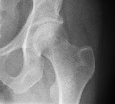
The intertrochanteric line (or ''spiral line of the femur''White (2005), p 256 ) is a line located on the anterior side of the proximal end of the femur. Structure The rough, variable ridge stretches between the lesser trochanter and the greater trochanter forming the base of the neck of the femur, roughly following the direction of the shaft of the femur. The iliofemoral ligament — the largest ligament of the human body — attaches above the line which also strengthens the capsule of the hip joint. The lower half, less prominent than the upper half, gives origin to the upper part of the Vastus medialis. Just like the intertrochanteric crest on the posterior side of the femoral head, the intertrochanteric line marks the transition between the femoral neck and shaft.Platzer (2004), p 192 The distal capsular attachment on the femur follows the shape of the irregular rim between the head and the neck. As a consequence, the capsule of the hip joint attaches in the ... [...More Info...] [...Related Items...] OR: [Wikipedia] [Google] [Baidu] |
Hip-joint
In vertebrate anatomy, hip (or "coxa"Latin ''coxa'' was used by Celsus in the sense "hip", but by Pliny the Elder in the sense "hip bone" (Diab, p 77) in medical terminology) refers to either an anatomical region or a joint. The hip region is located lateral and anterior to the gluteal region, inferior to the iliac crest, and overlying the greater trochanter of the femur, or "thigh bone". In adults, three of the bones of the pelvis have fused into the hip bone or acetabulum which forms part of the hip region. The hip joint, scientifically referred to as the acetabulofemoral joint (''art. coxae''), is the joint between the head of the femur and acetabulum of the pelvis and its primary function is to support the weight of the body in both static (e.g., standing) and dynamic (e.g., walking or running) postures. The hip joints have very important roles in retaining balance, and for maintaining the pelvic inclination angle. Pain of the hip may be the result of numerous causes, ... [...More Info...] [...Related Items...] OR: [Wikipedia] [Google] [Baidu] |
Femur
The femur (; ), or thigh bone, is the proximal bone of the hindlimb in tetrapod vertebrates. The head of the femur articulates with the acetabulum in the pelvic bone forming the hip joint, while the distal part of the femur articulates with the tibia (shinbone) and patella (kneecap), forming the knee joint. By most measures the two (left and right) femurs are the strongest bones of the body, and in humans, the largest and thickest. Structure The femur is the only bone in the upper leg. The two femurs converge medially toward the knees, where they articulate with the proximal ends of the tibiae. The angle of convergence of the femora is a major factor in determining the femoral-tibial angle. Human females have thicker pelvic bones, causing their femora to converge more than in males. In the condition ''genu valgum'' (knock knee) the femurs converge so much that the knees touch one another. The opposite extreme is ''genu varum'' (bow-leggedness). In the general popu ... [...More Info...] [...Related Items...] OR: [Wikipedia] [Google] [Baidu] |
Lesser Trochanter
The lesser trochanter is a conical posteromedial bony projection of the femoral shaft. it serves as the principal insertion site of the iliopsoas muscle. Structure The lesser trochanter is a conical posteromedial projection of the shaft of the femur, projecting from the posteroinferior aspect of its junction with the femoral neck. The summit and anterior surface of the lesser trochanter are rough, whereas its posterior surface is smooth. From its apex three well-marked borders extend: * two of these are above ** a medial continuous with the lower border of the femur neck ** a lateral with the intertrochanteric crest * the inferior border is continuous with the middle division of the linea aspera Attachments The summit of the lesser trochanter gives insertion to the tendon of the psoas major muscle and the iliacus muscle; the lesser trochanter represents the principal attachment of the iliopsoas. Anatomical relations The intertrochanteric crest (which demarcates the j ... [...More Info...] [...Related Items...] OR: [Wikipedia] [Google] [Baidu] |
Greater Trochanter
The greater trochanter of the femur is a large, irregular, quadrilateral eminence and a part of the skeletal system. It is directed lateral and medially and slightly posterior. In the adult it is about 2–4 cm lower than the femoral head.Standring, Susan, editor. ''Gray’s Anatomy: The Anatomical Basis of Clinical Practice''. Forty-First edition, Elsevier Limited, 2016, p. 1327. Because the pelvic outlet in the female is larger than in the male, there is a greater distance between the greater trochanters in the female. It has two surfaces and four borders. It is a traction epiphysis. Surfaces The ''lateral surface'', quadrilateral in form, is broad, rough, convex, and marked by a diagonal impression, which extends from the postero-superior to the antero-inferior angle, and serves for the insertion of the tendon of the gluteus medius. Above the impression is a triangular surface, sometimes rough for part of the tendon of the same muscle, sometimes smooth for the interpo ... [...More Info...] [...Related Items...] OR: [Wikipedia] [Google] [Baidu] |
Neck Of Femur
The femoral neck (femur neck or neck of the femur) is a flattened pyramidal process of bone, connecting the femoral head with the femoral shaft, and forming with the latter a wide angle opening medialward. Structure The neck is flattened from before backward, contracted in the middle, and broader laterally than medially. The vertical diameter of the lateral half is increased by the obliquity of the lower edge, which slopes downward to join the body at the level of the lesser trochanter, so that it measures one-third more than the antero-posterior diameter. The medial half is smaller and of a more circular shape. The anterior surface of the neck is perforated by numerous vascular foramina. Along the upper part of the line of junction of the anterior surface with the head is a shallow groove, best marked in elderly subjects; this groove lodges the orbicular fibers of the capsule of the hip joint. The posterior surface is smooth, and is broader and more concave than the anteri ... [...More Info...] [...Related Items...] OR: [Wikipedia] [Google] [Baidu] |
Shaft Of Femur
The body of femur (or shaft of femur) is the almost cylindrical, long part of the femur. It is a little broader above than in the center, broadest and somewhat flattened from before backward below. It is slightly arched, so as to be convex in front, and concave behind, where it is strengthened by a prominent longitudinal ridge, the linea aspera. It presents for examination three borders, separating three surfaces. Of the borders, one, the linea aspera, is posterior, one is medial, and the other, lateral. Borders The borders of the femur are the linea aspera, a medial border, and a lateral border. Linea aspera border The linea aspera is a prominent longitudinal ridge or crest, on the middle third of the bone, presenting a medial and a lateral lip, and a narrow rough, intermediate line. Above, the linea aspera is prolonged by three ridges. The lateral ridge termed the gluteal tuberosity is very rough, and runs almost vertically upward to the base of the greater trochanter. It ... [...More Info...] [...Related Items...] OR: [Wikipedia] [Google] [Baidu] |
Iliofemoral Ligament
The iliofemoral ligament is a ligament of the hip joint which extends from the ilium to the femur in front of the joint. It is also referred to as the Y-ligament (see below). the ligament of Bigelow, the ligament of Bertin and any combinations of these names. With a force strength exceeding 350 kg (772 lbs), the iliofemoral ligament is not only stronger than the two other ligaments of the hip joint, the ischiofemoral and the pubofemoral, but also the strongest ligament in the human body and as such is an important constraint to the hip joint. Structure Arising from the anterior inferior iliac spine and the rim of the acetabulum, the iliofemoral ligament spreads obliquely downwards and laterally to the intertrochanteric line on the anterior side of the femoral head. It is divided into two parts or bands which act differently: the transverse part above, is strong and runs parallel to the axis of the femoral neck. The descending part below, is weaker and runs parallel ... [...More Info...] [...Related Items...] OR: [Wikipedia] [Google] [Baidu] |
Capsule Of Hip Joint
The articular capsule (capsular ligament) is strong and dense. Anterosuperiorly, it is attached to the margin of the acetabulum 5 to 6 mm. beyond the Acetabular labrum, labrum behind; but in front, it is attached to the outer margin of the labrum, and, opposite to the notch where the margin of the cavity is deficient, it is connected to the transverse acetabular ligament, transverse ligament, and by a few fibers to the edge of the obturator foramen. It surrounds the neck of the femur, and is attached, in front, to the intertrochanteric line; above, to the base of the neck; behind, to the neck, about 1.25 cm. above the intertrochanteric crest; below, to the lower part of the neck, close to the lesser trochanter. From its femoral attachment some of the fibers are reflected upward along the neck as longitudinal bands, termed ''retinacula''. The capsule is much thicker at the upper and forepart of the joint, where the most resistance is required; behind and below, it is thi ... [...More Info...] [...Related Items...] OR: [Wikipedia] [Google] [Baidu] |
Tubercle Of The Femur
{{disambig ...
Tubercle of the femur can refer to: * Quadrate tubercle * Adductor tubercle of femur The adductor tubercle is a tubercle on the lower extremity of the femur. It is formed where the medial lips of the linea aspera end below at the summit of the medial condyle. It is the insertion point of the tendon of the vertical fibers of the a ... [...More Info...] [...Related Items...] OR: [Wikipedia] [Google] [Baidu] |
Linea Aspera
The linea aspera ( la, rough line) is a ridge of roughened surface on the posterior surface of the shaft of the femur. It is the site of attachments of muscles and the intermuscular septum. Its margins diverge above and below. The linea aspera is a prominent longitudinal ridge or crest, on the middle third of the bone, presenting a medial and a lateral lip, and a narrow rough, intermediate line. It is an important insertion point for the adductors and the lateral and medial intermuscular septa that divides the thigh into three compartments. The tension generated by muscle attached to the bones is responsible for the formation of the ridges. Structure Above Above, the linea aspera is prolonged by three ridges. * The lateral ridge is very rough, and runs almost vertically upward to the base of the greater trochanter. It is termed the gluteal tuberosity, and gives attachment to part of the gluteus maximus: its upper part is often elongated into a roughened crest, on which a more ... [...More Info...] [...Related Items...] OR: [Wikipedia] [Google] [Baidu] |
Vastus Medialis
The vastus medialis (vastus internus or teardrop muscle) is an extensor muscle located medially in the thigh that extends the knee. The vastus medialis is part of the quadriceps muscle group. Structure The vastus medialis is a muscle present in the anterior compartment of thigh, and is one of the four muscles that make up the quadriceps muscle. The others are the vastus lateralis, vastus intermedius and rectus femoris. It is the most medial of the "vastus" group of muscles. The vastus medialis arises medially along the entire length of the femur, and attaches with the other muscles of the quadriceps in the quadriceps tendon. The vastus medialis muscle originates from a continuous line of attachment on the femur, which begins on the front and middle side (anteromedially) on the intertrochanteric line of the femur. It continues down and back (posteroinferiorly) along the pectineal line and then descends along the inner (medial) lip of the linea aspera and onto the media ... [...More Info...] [...Related Items...] OR: [Wikipedia] [Google] [Baidu] |
Intertrochanteric Crest
The intertrochanteric crest is a prominent bony ridge upon the posterior surface of the femur at the junction of the neck and the shaft of the femur. It extends between the greater trochanter superiorly, and the lesser trochanter inferiorly. Anatomy The intertrochanteric crest is a prominent smooth bony ridge upon the posterior surface of the femur at the junction of the neck and the shaft of the femur; together with the intertrochanteric line on the anterior side of the head, the intertrochanteric crest marks the transition between the femoral neck and shaft. The intertrochanteric crest extends between the greater trochanter superiorly, and the lesser trochanter inferiorly; it passes obliquely inferomedially from the greater trochanter to the lesser trochanter. An elevation between the middle and proximal third of the crest is known as the quadrate tubercle. Relations The distal capsular attachment on the femur follows the shape of the irregular rim between the head and th ... [...More Info...] [...Related Items...] OR: [Wikipedia] [Google] [Baidu] |


