|
Hypoplastic
Hypoplasia (from Ancient Greek ὑπo- ''hypo-'' 'under' + πλάσις ''plasis'' 'formation'; adjective form ''hypoplastic'') is underdevelopment or incomplete development of a tissue or organ. Dictionary of Cell and Molecular Biology (11 March 2008) Although the term is not always used precisely, it properly refers to an inadequate or below-normal number of cells.Hypoplasia Stedman's Medical Dictionary. lww.com Hypoplasia is similar to |
Hypoplastic Left Heart Syndrome
Hypoplastic left heart syndrome (HLHS) is a rare congenital heart defect in which the left side of the heart is severely underdeveloped and incapable of supporting the systemic circulation. It is estimated to account for 2-3% of all congenital heart disease. Early signs and symptoms include poor feeding, cyanosis, and diminished pulse in the extremities. The etiology is believed to be multifactorial resulting from a combination of genetic mutations and defects resulting in altered blood flow in the heart. Several structures can be affected including the left ventricle, aorta, aortic valve, or mitral valve all resulting in decreased systemic blood flow. Diagnosis can occur prenatally via ultrasound or shortly after birth via echocardiography. Initial management is geared to maintaining patency of the ductus arteriosus - a connection between the pulmonary artery and the aorta that closes shortly after birth. Patient subsequently undergoes a three-stage palliative repair over the ne ... [...More Info...] [...Related Items...] OR: [Wikipedia] [Google] [Baidu] |
Hypoplastic Right Heart Syndrome
Hypoplastic right heart syndrome is a congenital heart defect in which the right atrium and right ventricle are underdeveloped. This defect causes inadequate blood flow to the lungs and thus, a blue or cyanotic infant. Symptoms and signs Common symptoms include a grayish-blue (cyanosis) coloration to the skin, lips, fingernails and other parts of the body. Other pronounced symptoms can be rapid/difficulty breathing, poor feeding, cold hands or feet, or being inactive and drowsy. "In a baby with hypoplastic right heart syndrome, if the natural connections between the heart's left and right sides (foramen oval and ductus arteriosus) are allowed to close, he or she may go into shock." Signs of shock can include cool or clammy skin, a weak or rapid pulse, and dilated pupils. Causes The Notch-signaling pathway is involved in multiple processes during heart development, along with Wnt signaling. Cardiomyocyte differentiation, patterning of the different cardiac regions, valve developmen ... [...More Info...] [...Related Items...] OR: [Wikipedia] [Google] [Baidu] |
Popliteal Pterygium Syndrome
Popliteal pterygium syndrome (PPS) is an heredity, inherited condition affecting the face, Limb (anatomy), limbs, and genitalia. The syndrome goes by a number of names including the ''popliteal web syndrome'' and, more inclusively, the ''facio-genito-popliteal syndrome''. The term PPS was coined by Gorlin ''et al.'' in 1968 on the basis of the most unusual Birth defect, anomaly, the popliteal pterygium (a web behind the knee). Symptoms and signs Medical sign, Clinical expressions of PPS are highly variable, but include the following: * Limb (anatomy), Limb findings: an extensive web running from behind the knee down to the heel (90%), malformed toenails, and webbed toes. * face, Facial findings: cleft palate with or without cleft lip (75%), pits in the lower lip (40%), fibrous bands in the mouth known as syngnathia (25%), and tissue connecting the upper and lower eyelids * genitalia, Genital findings (50%): hypoplasia of the labia majora, malformation of the scrotum, and cryptorch ... [...More Info...] [...Related Items...] OR: [Wikipedia] [Google] [Baidu] |
Heart
The heart is a muscular organ in most animals. This organ pumps blood through the blood vessels of the circulatory system. The pumped blood carries oxygen and nutrients to the body, while carrying metabolic waste such as carbon dioxide to the lungs. In humans, the heart is approximately the size of a closed fist and is located between the lungs, in the middle compartment of the chest. In humans, other mammals, and birds, the heart is divided into four chambers: upper left and right atria and lower left and right ventricles. Commonly the right atrium and ventricle are referred together as the right heart and their left counterparts as the left heart. Fish, in contrast, have two chambers, an atrium and a ventricle, while most reptiles have three chambers. In a healthy heart blood flows one way through the heart due to heart valves, which prevent backflow. The heart is enclosed in a protective sac, the pericardium, which also contains a small amount of fluid. The wall ... [...More Info...] [...Related Items...] OR: [Wikipedia] [Google] [Baidu] |
Turner's Hypoplasia
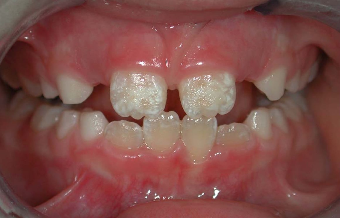
Enamel hypoplasia is a defect of the teeth in which the enamel is deficient in quantity, caused by defective enamel matrix formation during enamel development, as a result of inherited and acquired systemic condition(s). It can be identified as missing tooth structure and may manifest as pits or grooves in the crown of the affected teeth, and in extreme cases, some portions of the crown of the tooth may have no enamel, exposing the dentin. It may be generalized across the dentition or localized to a few teeth. Defects are categorized by shape or location. Common categories are pit-form, plane-form, linear-form, and localised enamel hypoplasia. Hypoplastic lesions are found in areas of the teeth where the enamel was being actively formed during a systemic or local disturbance. Since the formation of enamel extends over a long period of time, defects may be confined to one well-defined area of the affected teeth. Knowledge of chronological development of deciduous and permanen ... [...More Info...] [...Related Items...] OR: [Wikipedia] [Google] [Baidu] |
Reelin
Reelin, encoded by the ''RELN'' gene, is a large secreted extracellular matrix glycoprotein that helps regulate processes of neuronal migration and positioning in the developing brain by controlling cell–cell interactions. Besides this important role in early development, reelin continues to work in the adult brain. It modulates synaptic plasticity by enhancing the induction and maintenance of long-term potentiation. It also stimulates dendrite and dendritic spine development and regulates the continuing migration of neuroblasts generated in adult neurogenesis sites like the subventricular and subgranular zones. It is found not only in the brain but also in the liver, thyroid gland, adrenal gland, Fallopian tube, breast and in comparatively lower levels across a range of anatomical regions. Reelin has been suggested to be implicated in pathogenesis of several brain diseases. The expression of the protein has been found to be significantly lower in schizophrenia and psycho ... [...More Info...] [...Related Items...] OR: [Wikipedia] [Google] [Baidu] |
DiGeorge Syndrome
DiGeorge syndrome, also known as 22q11.2 deletion syndrome, is a syndrome caused by a microdeletion on the long arm of chromosome 22. While the symptoms can vary, they often include congenital heart problems, specific facial features, frequent infections, developmental delay, learning problems and cleft palate. Associated conditions include kidney problems, schizophrenia, hearing loss and autoimmune disorders such as rheumatoid arthritis or Graves' disease. DiGeorge syndrome is typically due to the deletion of 30 to 40 genes in the middle of chromosome 22 at a location known as ''22q11.2''. About 90% of cases occur due to a new mutation during early development, while 10% are inherited from a person's parents. It is autosomal dominant, meaning that only one affected chromosome is needed for the condition to occur. Diagnosis is suspected based on the symptoms and confirmed by genetic testing. Although there is no cure, treatment can improve symptoms. This often includes a m ... [...More Info...] [...Related Items...] OR: [Wikipedia] [Google] [Baidu] |
Corpus Callosum
The corpus callosum (Latin for "tough body"), also callosal commissure, is a wide, thick nerve tract, consisting of a flat bundle of commissural fibers, beneath the cerebral cortex in the brain. The corpus callosum is only found in placental mammals. It spans part of the longitudinal fissure, connecting the left and right cerebral hemispheres, enabling communication between them. It is the largest white matter structure in the human brain, about in length and consisting of 200–300 million axonal projections. A number of separate nerve tracts, classed as subregions of the corpus callosum, connect different parts of the hemispheres. The main ones are known as the genu, the rostrum, the trunk or body, and the splenium. Structure The corpus callosum forms the floor of the longitudinal fissure that separates the two cerebral hemispheres. Part of the corpus callosum forms the roof of the lateral ventricles. The corpus callosum has four main parts – individual nerve tracts ... [...More Info...] [...Related Items...] OR: [Wikipedia] [Google] [Baidu] |
Sacral Agenesis
Sacral may refer to: *Sacred Sacred describes something that is dedicated or set apart for the service or worship of a deity; is considered worthy of spiritual respect or devotion; or inspires awe or reverence among believers. The property is often ascribed to objects ( ..., associated with divinity and considered worthy of spiritual respect or devotion *Of the sacrum, a large, triangular bone at the base of the spine See also * {{disambiguation ... [...More Info...] [...Related Items...] OR: [Wikipedia] [Google] [Baidu] |
Sacrum
The sacrum (plural: ''sacra'' or ''sacrums''), in human anatomy, is a large, triangular bone at the base of the spine that forms by the fusing of the sacral vertebrae (S1S5) between ages 18 and 30. The sacrum situates at the upper, back part of the pelvic cavity, between the two wings of the pelvis. It forms joints with four other bones. The two projections at the sides of the sacrum are called the alae (wings), and articulate with the ilium at the L-shaped sacroiliac joints. The upper part of the sacrum connects with the last lumbar vertebra (L5), and its lower part with the coccyx (tailbone) via the sacral and coccygeal cornua. The sacrum has three different surfaces which are shaped to accommodate surrounding pelvic structures. Overall it is concave (curved upon itself). The base of the sacrum, the broadest and uppermost part, is tilted forward as the sacral promontory internally. The central part is curved outward toward the posterior, allowing greater room for the pel ... [...More Info...] [...Related Items...] OR: [Wikipedia] [Google] [Baidu] |
Optic Nerve Hypoplasia
Optic nerve hypoplasia (ONH) is a medical condition arising from the underdevelopment of the optic nerve(s). This condition is the most common congenital optic nerve anomaly. The optic disc appears abnormally small, because not all the optic nerve axons have developed properly.Sadun, Alfredo A., and Michelle Y. Wang. ''Handbook of Clinical Neurology''. p. 37. In press. It is often associated with endocrinopathies (hormone deficiencies), developmental delay, and brain malformations. The optic nerve, which is responsible for transmitting visual signals from the retina to the brain, has approximately 1.2 million nerve fibers in the average person. In those diagnosed with ONH, however, there are noticeably fewer nerves. Symptoms ONH may be found in isolation or in conjunction with myriad functional and anatomic abnormalities of the central nervous system. Nearly 80% of those affected with ONH will experience hypothalamic dysfunction and/or impaired development of the brain, regardless ... [...More Info...] [...Related Items...] OR: [Wikipedia] [Google] [Baidu] |
Optic Nerve
In neuroanatomy, the optic nerve, also known as the second cranial nerve, cranial nerve II, or simply CN II, is a paired cranial nerve that transmits visual system, visual information from the retina to the brain. In humans, the optic nerve is derived from optic stalks during the seventh week of development and is composed of retinal ganglion cell axons and glial cells; it extends from the optic disc to the optic chiasma and continues as the optic tract to the lateral geniculate nucleus, Pretectal area, pretectal nuclei, and superior colliculus. Structure The optic nerve has been classified as the second of twelve paired cranial nerves, but it is technically part of the central nervous system, rather than the peripheral nervous system because it is derived from an out-pouching of the diencephalon (optic stalks) during embryonic development. As a consequence, the fibers of the optic nerve are covered with myelin produced by oligodendrocytes, rather than Schwann cells of the per ... [...More Info...] [...Related Items...] OR: [Wikipedia] [Google] [Baidu] |


