|
Histopathologic Diagnosis Of Prostate Cancer
A histopathology, histopathologic diagnosis of prostate cancer is the discernment of whether there is a cancer in the prostate, as well as specifying any subdiagnosis of prostate cancer if possible. The histopathologic subdiagnosis of prostate cancer has implications for the possibility and methodology of any subsequent Gleason score, Gleason scoring. The most common histopathological subdiagnosis of prostate cancer is acinar adenocarcinoma, constituting 93% of prostate cancers. The most common form of acinar adenocarcinoma, in turn, is "adenocarcinoma, not otherwise specified", also termed conventional, or usual acinar adenocarcinoma. Sampling The main sources of tissue sampling are prostatectomy and prostate biopsy. Subdiagnoses - overview In uncertain cases, a diagnosis of malignancy can be excluded by immunohistochemical detection of basal cells (or confirmed by absence thereof), such as using the ''PIN-4'' cocktail of stains, which targets TP63, p63, Keratin 5, CK-5, Kerati ... [...More Info...] [...Related Items...] OR: [Wikipedia] [Google] [Baidu] |
Histopathology
Histopathology (compound of three Greek words: ''histos'' "tissue", πάθος ''pathos'' "suffering", and -λογία '' -logia'' "study of") refers to the microscopic examination of tissue in order to study the manifestations of disease. Specifically, in clinical medicine, histopathology refers to the examination of a biopsy or surgical specimen by a pathologist, after the specimen has been processed and histological sections have been placed onto glass slides. In contrast, cytopathology examines free cells or tissue micro-fragments (as "cell blocks"). Collection of tissues Histopathological examination of tissues starts with surgery, biopsy, or autopsy. The tissue is removed from the body or plant, and then, often following expert dissection in the fresh state, placed in a fixative which stabilizes the tissues to prevent decay. The most common fixative is 10% neutral buffered formalin (corresponding to 3.7% w/v formaldehyde in neutral buffered water, such as phosphate buf ... [...More Info...] [...Related Items...] OR: [Wikipedia] [Google] [Baidu] |
Stanford University School Of Medicine
Stanford University School of Medicine is the medical school of Stanford University and is located in Stanford, California. It traces its roots to the Medical Department of the University of the Pacific, founded in San Francisco in 1858. This medical institution, then called Cooper Medical College, was acquired by Stanford in 1908. The medical school moved to the Stanford campus near Palo Alto, California, in 1959. The School of Medicine, along with Stanford Health Care and Lucile Packard Children's Hospital, is part of Stanford Medicine. Stanford Health Care was ranked the fourth best hospital in California (behind UCLA Medical Center, Cedars-Sinai Medical Center, and UCSF Medical Center, respectively). History In 1855, Illinois physician Elias Samuel Cooper moved to San Francisco in the wake of the California Gold Rush. In cooperation with the University of the Pacific (also known as California Wesleyan College), Cooper established the Medical Department of the Univers ... [...More Info...] [...Related Items...] OR: [Wikipedia] [Google] [Baidu] |
Prostate Cancer
Prostate cancer is cancer of the prostate. Prostate cancer is the second most common cancerous tumor worldwide and is the fifth leading cause of cancer-related mortality among men. The prostate is a gland in the male reproductive system that surrounds the urethra just below the bladder. It is located in the hypogastric region of the abdomen. To give an idea of where it is located, the bladder is superior to the prostate gland as shown in the image The rectum is posterior in perspective to the prostate gland and the ischial tuberosity of the pelvic bone is inferior. Only those who have male reproductive organs are able to get prostate cancer. Most prostate cancers are slow growing. Cancerous cells may spread to other areas of the body, particularly the bones and lymph nodes. It may initially cause no symptoms. In later stages, symptoms include pain or difficulty urinating, blood in the urine, or pain in the pelvis or back. Benign prostatic hyperplasia may produce similar symptoms ... [...More Info...] [...Related Items...] OR: [Wikipedia] [Google] [Baidu] |
Keratin 14
Keratin 14 is a member of the type I keratin family of intermediate filament proteins. Keratin 14 was the first type I keratin sequence determined. Keratin 14 is also known as cytokeratin-14 (CK-14) or keratin-14 (KRT14). In humans it is encoded by the ''KRT14'' gene. Keratin 14 is usually found as a heterodimer with type II keratin 5 and form the cytoskeleton of epithelial cells. Pathology Mutations in the genes for these keratins are associated with epidermolysis bullosa simplex and dermatopathia pigmentosa reticularis, both of which are autosomal dominant mutations. See also *34βE12 34βE12, often written as 34betaE12 and also known as CK34βE12 and keratin 903 (CK903), is an antibody specific for high molecular weight cytokeratins 1, 5, 10 and 14. It is sometimes, less precisely, referred to as high-molecular weight kerat ... (keratin 903) References Further reading * * * * * * * * * * * * * * * * * External links GeneReviews/NCBI/UW/NIH ... [...More Info...] [...Related Items...] OR: [Wikipedia] [Google] [Baidu] |
Keratin 5
Keratin 5, also known as KRT5, K5, or CK5, is a protein that is encoded in humans by the ''KRT5'' gene. It dimerizes with keratin 14 and forms the intermediate filaments (IF) that make up the cytoskeleton of basal epithelial cells. This protein is involved in several diseases including epidermolysis bullosa simplex and breast and lung cancers. Structure Keratin 5, like other members of the keratin family, is an intermediate filament protein. These polypeptides are characterized by a 310 residue central rod domain that consists of four alpha helix segments (helix 1A, 1B, 2A, and 2B) connected by three short linker regions (L1, L1-2, and L2). The ends of the central rod domain, which are called the helix initiation motif (HIM) and the helix termination motif (HTM), are highly conserved. They are especially important for helix stabilization, heterodimer formation, and filament formation.Shinkuma, Satoru, et al. "A Novel Keratin 5 Mutation in an African Family with Epidermolysis ... [...More Info...] [...Related Items...] OR: [Wikipedia] [Google] [Baidu] |
TP63
Tumor protein p63, typically referred to as p63, also known as transformation-related protein 63 is a protein that in humans is encoded by the ''TP63'' (also known as the '' p63'') gene. The ''TP63'' gene was discovered 20 years after the discovery of the ''p53'' tumor suppressor gene and along with '' p73'' constitutes the ''p53'' gene family based on their structural similarity. Despite being discovered significantly later than ''p53'', phylogenetic analysis of ''p53'', ''p63'' and ''p73'', suggest that ''p63'' was the original member of the family from which ''p53'' and ''p73'' evolved. Function Tumor protein p63 is a member of the p53 family of transcription factors. p63 -/- mice have several developmental defects which include the lack of limbs and other tissues, such as teeth and mammary glands, which develop as a result of interactions between mesenchyme and epithelium. TP63 encodes for two main isoforms by alternative promoters (TAp63 and ΔNp63). ΔNp63 is involved i ... [...More Info...] [...Related Items...] OR: [Wikipedia] [Google] [Baidu] |
PIN-4 Staining Of Benign Prostate Gland And Adenocarcinoma, Annotated , targeting TP63, CK-5, CK-14 and P504S
{{dab ...
PIN4 or PIN-4 may refer to: *''PIN4'', the gene encoding the protein Peptidyl-prolyl cis-trans isomerase NIMA-interacting 4 (PIN4) *''PIN-4'', a cocktail for immunohistochemistry Immunohistochemistry (IHC) is the most common application of immunostaining. It involves the process of selectively identifying antigens (proteins) in cells of a tissue section by exploiting the principle of antibodies binding specifically to an ... [...More Info...] [...Related Items...] OR: [Wikipedia] [Google] [Baidu] |
Palisade (pathology)
In histopathology, a palisade is a single layer of relatively long cells, arranged loosely perpendicular to a surface and parallel to each other. A rosette is a palisade in a halo or spoke-and-wheel arrangement, surrounding a central core or hub. A pseudorosette is a perivascular radial arrangement of neoplastic cells around a small blood vessel. Rosette A ''rosette'' is a cell formation in a halo or spoke-and-wheel arrangement, surrounding a central core or hub. The central hub may consist of an empty-appearing lumen or a space filled with cytoplasmic processes. The cytoplasm of each of the cells in the rosette is often wedge-shaped with the apex directed toward the central core: the nuclei of the cells participating in the rosette are peripherally positioned and form a ring or halo around the hub. Pathogenesis Rosettes may be considered primary or secondary manifestations of tumor architecture. Primary rosettes form as a characteristic growth pattern of a given tumor type wh ... [...More Info...] [...Related Items...] OR: [Wikipedia] [Google] [Baidu] |
Euchromatin
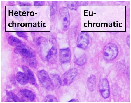
Euchromatin (also called "open chromatin") is a lightly packed form of chromatin ( DNA, RNA, and protein) that is enriched in genes, and is often (but not always) under active transcription. Euchromatin stands in contrast to heterochromatin, which is tightly packed and less accessible for transcription. 92% of the human genome is euchromatic. In eukaryotes, euchromatin comprises the most active portion of the genome within the cell nucleus. In prokaryotes, euchromatin is the ''only'' form of chromatin present; this indicates that the heterochromatin structure evolved later along with the nucleus, possibly as a mechanism to handle increasing genome size. Structure Euchromatin is composed of repeating subunits known as nucleosomes, reminiscent of an unfolded set of beads on a string, that are approximately 11 nm in diameter. At the core of these nucleosomes are a set of four histone protein pairs: H3, H4, H2A, and H2B. Each core histone protein possesses a 'tail' str ... [...More Info...] [...Related Items...] OR: [Wikipedia] [Google] [Baidu] |
Micrograph Of Signet-ring Adenocarcinoma Of The Prostate
A micrograph or photomicrograph is a photograph or digital image taken through a microscope or similar device to show a magnified image of an object. This is opposed to a macrograph or photomacrograph, an image which is also taken on a microscope but is only slightly magnified, usually less than 10 times. Micrography is the practice or art of using microscopes to make photographs. A micrograph contains extensive details of microstructure. A wealth of information can be obtained from a simple micrograph like behavior of the material under different conditions, the phases found in the system, failure analysis, grain size estimation, elemental analysis and so on. Micrographs are widely used in all fields of microscopy. Types Photomicrograph A light micrograph or photomicrograph is a micrograph prepared using an optical microscope, a process referred to as ''photomicroscopy''. At a basic level, photomicroscopy may be performed simply by connecting a camera to a microscope, the ... [...More Info...] [...Related Items...] OR: [Wikipedia] [Google] [Baidu] |
Micrograph Of Mucinous Adenocarcinoma Of The Prostate With Gleason Score 7 (3 + 4) With Individual Well-formed Glands And Minor Component Of Cribriform Glands Floating In Extracellular Mucin
A micrograph or photomicrograph is a photograph or digital image taken through a microscope or similar device to show a magnified image of an object. This is opposed to a macrograph or photomacrograph, an image which is also taken on a microscope but is only slightly magnified, usually less than 10 times. Micrography is the practice or art of using microscopes to make photographs. A micrograph contains extensive details of microstructure. A wealth of information can be obtained from a simple micrograph like behavior of the material under different conditions, the phases found in the system, failure analysis, grain size estimation, elemental analysis and so on. Micrographs are widely used in all fields of microscopy. Types Photomicrograph A light micrograph or photomicrograph is a micrograph prepared using an optical microscope, a process referred to as ''photomicroscopy''. At a basic level, photomicroscopy may be performed simply by connecting a camera to a microscope, the ... [...More Info...] [...Related Items...] OR: [Wikipedia] [Google] [Baidu] |
Micrograph Of Small-cell Carcinoma Of The Prostate
A micrograph or photomicrograph is a photograph or digital image taken through a microscope or similar device to show a magnify, magnified image of an object. This is opposed to a macrograph or photomacrograph, an image which is also taken on a microscope but is only slightly magnified, usually less than 10 times. Micrography is the practice or art of using microscopes to make photographs. A micrograph contains extensive details of microstructure. A wealth of information can be obtained from a simple micrograph like behavior of the material under different conditions, the phases found in the system, failure analysis, grain size estimation, elemental analysis and so on. Micrographs are widely used in all fields of microscopy. Types Photomicrograph A light micrograph or photomicrograph is a micrograph prepared using an optical microscope, a process referred to as ''photomicroscopy''. At a basic level, photomicroscopy may be performed simply by connecting a camera to a micros ... [...More Info...] [...Related Items...] OR: [Wikipedia] [Google] [Baidu] |


