|
Hip Spica Cast
A hip spica cast is a sort of orthopedic cast used to immobilize the hip or thigh. It is used to facilitate healing of injured hip joints or of fractured femurs. A hip spica includes the trunk of the body and one or both legs. A hip spica which covers only one leg to the ankle or foot may be referred to as a single hip spica, while one which covers both legs is called a double hip spica. A one-and-a-half hip spica encases one leg to the ankle or foot and the other to just above the knee. The extent to which the hip spica covers the trunk depends greatly on the injury and the surgeon; the spica may extend only to the navel, allowing mobility of the spine and the possibility of walking with the aid of crutches, or may extend to the rib cage or even to the armpits in some rare cases. Hip spicas were formerly common in reducing femoral fractures. Spica casts are used for treating hip dysplasia Hip dysplasia is an abnormality of the hip joint where the socket portion does not fully ... [...More Info...] [...Related Items...] OR: [Wikipedia] [Google] [Baidu] |
Hip Spica Casts
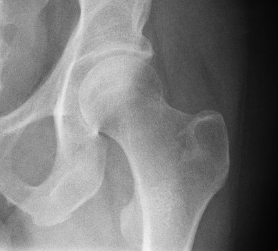
In vertebrate anatomy, hip (or "coxa"Latin ''coxa'' was used by Celsus in the sense "hip", but by Pliny the Elder in the sense "hip bone" (Diab, p 77) in medical terminology) refers to either an anatomical region or a joint. The hip region is located lateral and anterior to the gluteal region, inferior to the iliac crest, and overlying the greater trochanter of the femur, or "thigh bone". In adults, three of the bones of the pelvis have fused into the hip bone or acetabulum which forms part of the hip region. The hip joint, scientifically referred to as the acetabulofemoral joint (''art. coxae''), is the joint between the head of the femur and acetabulum of the pelvis and its primary function is to support the weight of the body in both static (e.g., standing) and dynamic (e.g., walking or running) postures. The hip joints have very important roles in retaining balance, and for maintaining the pelvic inclination angle. Pain of the hip may be the result of numerous causes, i ... [...More Info...] [...Related Items...] OR: [Wikipedia] [Google] [Baidu] |
Orthopedic Cast
An orthopedic cast, or simply cast, is a shell, frequently made from plaster or fiberglass, that encases a limb (or, in some cases, large portions of the body) to stabilize and hold anatomical structures—most often a broken bone (or bones), in place until healing is confirmed. It is similar in function to a splint. Plaster bandages consist of a cotton bandage that has been combined with plaster of paris, which hardens after it has been made wet. Plaster of Paris is calcined gypsum (roasted gypsum), ground to a fine powder by milling. When water is added, the more soluble form of calcium sulfate returns to the relatively insoluble form, and heat is produced. :2 (CaSO4·½ H2O) + 3 H2O → 2 (CaSO4.2H2O) + Heat The setting of unmodified plaster starts about 10 minutes after mixing and is complete in about 45 minutes; however, the cast is not fully dry for 72 hours. Current bandages of synthetic materials are often used, often knitted fiberglass bandages impregnated with polyu ... [...More Info...] [...Related Items...] OR: [Wikipedia] [Google] [Baidu] |
Thigh
In human anatomy, the thigh is the area between the hip (pelvis) and the knee. Anatomically, it is part of the lower limb. The single bone in the thigh is called the femur. This bone is very thick and strong (due to the high proportion of bone tissue), and forms a ball and socket joint at the hip, and a modified hinge joint at the knee. Structure Bones The femur is the only bone in the thigh and serves as an attachment site for all muscles in the thigh. The head of the femur articulates with the acetabulum in the pelvic bone forming the hip joint, while the distal part of the femur articulates with the tibia and patella forming the knee. By most measures, the femur is the strongest bone in the body. The femur is also the longest bone in the body. The femur is categorised as a long bone and comprises a diaphysis, the shaft (or body) and two epiphysis or extremities that articulate with adjacent bones in the hip and knee. Muscular compartments In cross-section, the thigh is ... [...More Info...] [...Related Items...] OR: [Wikipedia] [Google] [Baidu] |
Bone Fracture
A bone fracture (abbreviated FRX or Fx, Fx, or #) is a medical condition in which there is a partial or complete break in the continuity of any bone in the body. In more severe cases, the bone may be broken into several fragments, known as a ''comminuted fracture''. A bone fracture may be the result of high force impact or stress, or a minimal trauma injury as a result of certain medical conditions that weaken the bones, such as osteoporosis, osteopenia, bone cancer, or osteogenesis imperfecta, where the fracture is then properly termed a pathologic fracture. Signs and symptoms Although bone tissue contains no pain receptors, a bone fracture is painful for several reasons: * Breaking in the continuity of the periosteum, with or without similar discontinuity in endosteum, as both contain multiple pain receptors. * Edema and hematoma of nearby soft tissues caused by ruptured bone marrow evokes pressure pain. * Involuntary muscle spasms trying to hold bone fragments in place. D ... [...More Info...] [...Related Items...] OR: [Wikipedia] [Google] [Baidu] |
Femur
The femur (; ), or thigh bone, is the proximal bone of the hindlimb in tetrapod vertebrates. The head of the femur articulates with the acetabulum in the pelvic bone forming the hip joint, while the distal part of the femur articulates with the tibia (shinbone) and patella (kneecap), forming the knee joint. By most measures the two (left and right) femurs are the strongest bones of the body, and in humans, the largest and thickest. Structure The femur is the only bone in the upper leg. The two femurs converge medially toward the knees, where they articulate with the proximal ends of the tibiae. The angle of convergence of the femora is a major factor in determining the femoral-tibial angle. Human females have thicker pelvic bones, causing their femora to converge more than in males. In the condition ''genu valgum'' (knock knee) the femurs converge so much that the knees touch one another. The opposite extreme is ''genu varum'' (bow-leggedness). In the general populatio ... [...More Info...] [...Related Items...] OR: [Wikipedia] [Google] [Baidu] |
Hip Dysplasia
Hip dysplasia is an abnormality of the hip joint where the socket portion does not fully cover the ball portion, resulting in an increased risk for joint dislocation. Hip dysplasia may occur at birth or develop in early life. Regardless, it does not typically produce symptoms in babies less than a year old. Occasionally one leg may be shorter than the other. The left hip is more often affected than the right. Complications without treatment can include arthritis, limping, and low back pain. Females are affected more often than males. Hip dysplasia was described at least as early as the 300s BC by Hippocrates. Risk factors for hip dysplasia include female sex, family history, certain swaddling practices, and breech presentation whether an infant is delivered vaginally or by cesarean section. If one identical twin is affected, there is a 40% risk the other will also be affected. Screening all babies for the condition by physical examination is recommended. Ultrasonography may al ... [...More Info...] [...Related Items...] OR: [Wikipedia] [Google] [Baidu] |


