|
High-frequency Ventilation
High-frequency ventilation is a type of mechanical ventilation which utilizes a respiratory rate greater than four times the normal value. (>150 (Vf) breaths per minute) and very small tidal volumes. High frequency ventilation is thought to reduce ventilator-associated lung injury (VALI), especially in the context of ARDS and acute lung injury. This is commonly referred to as lung protective ventilation. There are different types of high-frequency ventilation. Each type has its own unique advantages and disadvantages. The types of HFV are characterized by the delivery system and the type of exhalation phase. High-frequency ventilation may be used alone, or in combination with conventional mechanical ventilation. In general, those devices that need conventional mechanical ventilation do not produce the same lung protective effects as those that can operate without tidal breathing. Specifications and capabilities will vary depending on the device manufacturer. Physiology With conve ... [...More Info...] [...Related Items...] OR: [Wikipedia] [Google] [Baidu] |
Mechanical Ventilation
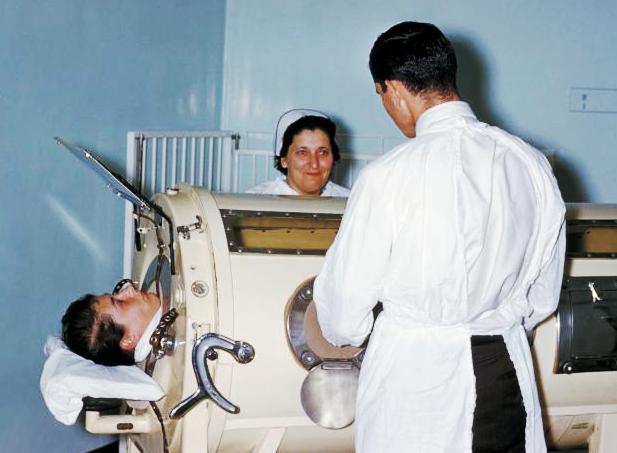
Mechanical ventilation, assisted ventilation or intermittent mandatory ventilation (IMV), is the medical term for using a machine called a ventilator to fully or partially provide artificial ventilation. Mechanical ventilation helps move air into and out of the lungs, with the main goal of helping the delivery of oxygen and removal of carbon dioxide. Mechanical ventilation is used for many reasons, including to protect the airway due to mechanical or neurologic cause, to ensure adequate oxygenation, or to remove excess carbon dioxide from the lungs. Various healthcare providers are involved with the use of mechanical ventilation and people who require ventilators are typically monitored in an intensive care unit. Mechanical ventilation is termed invasive if it involves an instrument to create an airway that is placed inside the trachea. This is done through an endotracheal tube or nasotracheal tube. For non-invasive ventilation in people who are conscious, face or nasal ... [...More Info...] [...Related Items...] OR: [Wikipedia] [Google] [Baidu] |
Pneumothorax
A pneumothorax is an abnormal collection of air in the pleural space between the lung and the chest wall. Symptoms typically include sudden onset of sharp, one-sided chest pain and shortness of breath. In a minority of cases, a one-way valve is formed by an area of damaged tissue, and the amount of air in the space between chest wall and lungs increases; this is called a tension pneumothorax. This can cause a steadily worsening oxygen shortage and low blood pressure. This leads to a type of shock called obstructive shock, which can be fatal unless reversed. Very rarely, both lungs may be affected by a pneumothorax. It is often called a "collapsed lung", although that term may also refer to atelectasis. A primary spontaneous pneumothorax is one that occurs without an apparent cause and in the absence of significant lung disease. A secondary spontaneous pneumothorax occurs in the presence of existing lung disease. Smoking increases the risk of primary spontaneous pneumothorax ... [...More Info...] [...Related Items...] OR: [Wikipedia] [Google] [Baidu] |
Hypoxemia
Hypoxemia is an abnormally low level of oxygen in the blood. More specifically, it is oxygen deficiency in arterial blood. Hypoxemia has many causes, and often causes hypoxia as the blood is not supplying enough oxygen to the tissues of the body. Definition ''Hypoxemia'' refers to the low level of oxygen in blood, and the more general term ''hypoxia'' is an abnormally low oxygen content in any tissue or organ, or the body as a whole. Hypoxemia can cause hypoxia (hypoxemic hypoxia), but hypoxia can also occur via other mechanisms, such as anemia. Hypoxemia is usually defined in terms of reduced partial pressure of oxygen (mm Hg) in arterial blood, but also in terms of reduced content of oxygen (ml oxygen per dl blood) or percentage saturation of hemoglobin (the oxygen-binding protein within red blood cells) with oxygen, which is either found singly or in combination. While there is general agreement that an arterial blood gas measurement which shows that the partial pressure ... [...More Info...] [...Related Items...] OR: [Wikipedia] [Google] [Baidu] |
Endotracheal Tube
A tracheal tube is a catheter that is inserted into the Vertebrate trachea, trachea for the primary purpose of establishing and maintaining a patent airway and to ensure the adequate Gas exchange, exchange of oxygen and carbon dioxide. Many different types of tracheal tubes are available, suited for different specific applications: * An endotracheal tube is a specific type of tracheal tube that is nearly always inserted through the mouth (orotracheal) or nose (nasotracheal). * A tracheostomy tube is another type of tracheal tube; this curved metal or plastic tube may be inserted into a tracheostomy stoma (following a tracheotomy) to maintain a patent lumen. * A tracheal button is a rigid plastic cannula about 1 inch in length that can be placed into the tracheostomy after removal of a tracheostomy tube to maintain patency of the lumen. History Portex Medical (England and France) produced the first cuff-less plastic 'Ivory' endotracheal tubes. Ivan Magill later added a cuff ( ... [...More Info...] [...Related Items...] OR: [Wikipedia] [Google] [Baidu] |
Pneumothorax
A pneumothorax is an abnormal collection of air in the pleural space between the lung and the chest wall. Symptoms typically include sudden onset of sharp, one-sided chest pain and shortness of breath. In a minority of cases, a one-way valve is formed by an area of damaged tissue, and the amount of air in the space between chest wall and lungs increases; this is called a tension pneumothorax. This can cause a steadily worsening oxygen shortage and low blood pressure. This leads to a type of shock called obstructive shock, which can be fatal unless reversed. Very rarely, both lungs may be affected by a pneumothorax. It is often called a "collapsed lung", although that term may also refer to atelectasis. A primary spontaneous pneumothorax is one that occurs without an apparent cause and in the absence of significant lung disease. A secondary spontaneous pneumothorax occurs in the presence of existing lung disease. Smoking increases the risk of primary spontaneous pneumothorax ... [...More Info...] [...Related Items...] OR: [Wikipedia] [Google] [Baidu] |
Airway Resistance
In respiratory physiology, airway resistance is the resistance of the respiratory tract to airflow during inhalation and exhalation. Airway resistance can be measured using plethysmography. Definition Analogously to Ohm's Law: :R_ = \frac Where: : = P_ - P_A So: :R_ = \frac Where: *R_ = Airway Resistance *P = Pressure Difference driving airflow *P_ = Atmospheric Pressure *P_A = Alveolar Pressure *\dot V = Volumetric Airflow (not minute ventilation which, confusingly, may be represented by the same symbol) N.B. PA and \dot V change constantly during the respiratory cycle. Determinants of airway resistance There are several important determinants of airway resistance including: *The diameter of the airways *Whether airflow is laminar or turbulent Hagen–Poiseuille equation In fluid dynamics, the Hagen–Poiseuille equation is a physical law that gives the pressure drop in a fluid flowing through a long cylindrical pipe. The assumptions of the equation are that the flow ... [...More Info...] [...Related Items...] OR: [Wikipedia] [Google] [Baidu] |
Pulmonary Compliance
The lungs are the primary organs of the respiratory system in humans and most other animals, including some snails and a small number of fish. In mammals and most other vertebrates, two lungs are located near the vertebral column, backbone on either side of the heart. Their function in the respiratory system is to extract oxygen from the air and transfer it into the bloodstream, and to release carbon dioxide from the bloodstream into the Atmosphere of Earth, atmosphere, in a process of gas exchange. Respiration (physiology), Respiration is driven by different muscle, muscular systems in different species. Mammals, reptiles and birds use their different muscles to support and foster breathing. In earlier tetrapods, air was driven into the lungs by the pharyngeal muscles via buccal pumping, a mechanism still seen in amphibians. In humans, the main muscles of respiration, muscle of respiration that drives breathing is the thoracic diaphragm, diaphragm. The lungs also provide airf ... [...More Info...] [...Related Items...] OR: [Wikipedia] [Google] [Baidu] |
Respiratory Therapist
A respiratory therapist is a specialized healthcare practitioner trained in critical care and cardio-pulmonary medicine in order to work therapeutically with people who have acute critical conditions, cardiac and pulmonary disease. Respiratory therapists graduate from a college or university with a degree in respiratory therapy and have passed a national board certifying examination. The NBRC (National Board for Respiratory Care) is responsible for credentialing as a CRT ( certified respiratory therapist), or RRT ( registered respiratory therapist), The specialty certifications of respiratory therapy include: CPFT and RPFT (Certified or Registered Pulmonary Function Technologist), ACCS (Adult Critical Care Specialist), NPS (Neonatal/Pediatric Specialist), and SDS (Sleep Disorder Specialist). Respiratory therapists work in hospitals in the intensive care units (Adult, Pediatric, and Neonatal), on hospital floors, in emergency departments, in pulmonary functioning laboratories ... [...More Info...] [...Related Items...] OR: [Wikipedia] [Google] [Baidu] |
Bronchopulmonary Dysplasia
Bronchopulmonary dysplasia (BPD; part of the spectrum of chronic lung disease of infancy) is a chronic lung disease in which premature infants, usually those who were treated with supplemental oxygen, require long-term oxygen. The alveoli that are present tend to not be mature enough to function normally. It is more common in infants with low birth weight (LBW) and those who receive prolonged mechanical ventilation to treat respiratory distress syndrome (RDS). It results in significant morbidity and mortality. The definition of BPD has continued to evolve primarily due to changes in the population, such as more survivors at earlier gestational ages, and improved neonatal management including surfactant, antenatal glucocorticoid therapy, and less aggressive mechanical ventilation. Currently the description of BPD includes the grading of its severity into mild, moderate and severe. This correlates with the infant's maturity, growth and overall severity of illness. The new system of ... [...More Info...] [...Related Items...] OR: [Wikipedia] [Google] [Baidu] |
Necrotizing Tracheobronchitis
Necrosis () is a form of cell injury which results in the premature death of cells in living tissue by autolysis. Necrosis is caused by factors external to the cell or tissue, such as infection, or trauma which result in the unregulated digestion of cell components. In contrast, apoptosis is a naturally occurring programmed and targeted cause of cellular death. While apoptosis often provides beneficial effects to the organism, necrosis is almost always detrimental and can be fatal. Cellular death due to necrosis does not follow the apoptotic signal transduction pathway, but rather various receptors are activated and result in the loss of cell membrane integrity and an uncontrolled release of products of cell death into the extracellular space. This initiates in the surrounding tissue an inflammatory response, which attracts leukocytes and nearby phagocytes which eliminate the dead cells by phagocytosis. However, microbial damaging substances released by leukocytes would crea ... [...More Info...] [...Related Items...] OR: [Wikipedia] [Google] [Baidu] |
Intraventricular Hemorrhage
Intraventricular hemorrhage (IVH), also known as intraventricular bleeding, is a bleeding into the brain's ventricular system, where the cerebrospinal fluid is produced and circulates through towards the subarachnoid space. It can result from physical trauma or from hemorrhagic stroke. 30% of intraventricular hemorrhage (IVH) are primary, confined to the ventricular system and typically caused by intraventricular trauma, aneurysm, vascular malformations, or tumors, particularly of the choroid plexus. However 70% of IVH are secondary in nature, resulting from an expansion of an existing intraparenchymal or subarachnoid hemorrhage. Intraventricular hemorrhage has been found to occur in 35% of moderate to severe traumatic brain injuries. Thus the hemorrhage usually does not occur without extensive associated damage, and so the outcome is rarely good.Dawodu S. 2007"Traumatic Brain Injury: Definition, Epidemiology, Pathophysiology"Emedicine.com. Retrieved on June 19, 2007.Vinas FC and ... [...More Info...] [...Related Items...] OR: [Wikipedia] [Google] [Baidu] |
Pulmonary Interstitial Emphysema
Pulmonary interstitial emphysema (PIE) is a collection of air outside of the normal air space of the pulmonary alveoli, found instead inside the connective tissue of the peribronchovascular sheaths, interlobular septa, and visceral pleura. (This supportive tissue is called the pulmonary interstitium.) This collection of air develops as a result of alveolar and terminal bronchiolar rupture. Pulmonary interstitial emphysema is more frequent in premature infants who require mechanical ventilation for severe lung disease. Infants with pulmonary interstitial emphysema are typically recommended for admission to a neonatal intensive care unit. Cause Pulmonary interstitial emphysema is a concern in any of the following: * Prematurity *Infant respiratory distress syndrome (IRDS) * Meconium aspiration syndrome (MAS) *Amniotic fluid aspiration *Sepsis *Infections *Mechanical ventilation Pathophysiology Pulmonary interstitial emphysema is created when air bursts or ruptures through ti ... [...More Info...] [...Related Items...] OR: [Wikipedia] [Google] [Baidu] |


