|
Hepatic Adenoma
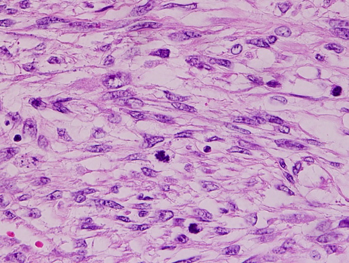
Hepatocellular adenoma (also known as hepatic adenoma or hepadenoma) is a rare, benign liver tumor. It most commonly occurs in people with elevated systemic levels of estrogen, classically in women taking estrogen-containing oral contraceptive medication. Signs and symptoms About 25–50% of hepatic adenomas cause pain in the right upper quadrant or epigastric region of the abdomen. Since hepatic adenomas can be large (8–15 cm), patients may notice a palpable mass. However, hepatic adenomas are usually asymptomatic, and may be discovered incidentally on imaging ordered for some unrelated reason. Large hepatic adenomas have a tendency to rupture and bleed massively inside the abdomen. If not treated, there is a 30% risk of bleeding. Bleeding may lead to hypotension, tachycardia, and sweating ( diaphoresis). Related Conditions Hepatic adenomas are related to glycogen storage diseases, type 1 diabetes, as well as anabolic steroid use. Diagnosis Hepatic adenoma is usually de ... [...More Info...] [...Related Items...] OR: [Wikipedia] [Google] [Baidu] |
Micrograph
A micrograph or photomicrograph is a photograph or digital image taken through a microscope or similar device to show a magnified image of an object. This is opposed to a macrograph or photomacrograph, an image which is also taken on a microscope but is only slightly magnified, usually less than 10 times. Micrography is the practice or art of using microscopes to make photographs. A micrograph contains extensive details of microstructure. A wealth of information can be obtained from a simple micrograph like behavior of the material under different conditions, the phases found in the system, failure analysis, grain size estimation, elemental analysis and so on. Micrographs are widely used in all fields of microscopy. Types Photomicrograph A light micrograph or photomicrograph is a micrograph prepared using an optical microscope, a process referred to as ''photomicroscopy''. At a basic level, photomicroscopy may be performed simply by connecting a camera to a microscope ... [...More Info...] [...Related Items...] OR: [Wikipedia] [Google] [Baidu] |
Fatty Change
Steatosis, also called fatty change, is abnormal retention of fat (lipids) within a cell or organ. Steatosis most often affects the liver – the primary organ of lipid metabolism – where the condition is commonly referred to as fatty liver disease. Steatosis can also occur in other organs, including the kidneys, heart, and muscle. When the term is not further specified (as, for example, in 'cardiac steatosis'), it is assumed to refer to the liver. Risk factors associated with steatosis are varied, and may include diabetes mellitus, protein malnutrition, hypertension, cell toxins, obesity, anoxia, and sleep apnea. Steatosis reflects an impairment of the normal processes of synthesis and elimination of triglyceride fat. Excess lipid accumulates in vesicles that displace the cytoplasm. When the vesicles are large enough to distort the nucleus, the condition is known as macrovesicular steatosis; otherwise, the condition is known as microvesicular steatosis. While not particul ... [...More Info...] [...Related Items...] OR: [Wikipedia] [Google] [Baidu] |
Reticulin
Reticular fibers, reticular fibres or reticulin is a type of fiber in connective tissue composed of type III collagen secreted by reticular cells. Reticular fibers crosslink to form a fine meshwork (reticulin). This network acts as a supporting mesh in soft tissues such as liver, bone marrow, and the tissues and organs of the lymphatic system. History The term reticulin was coined in 1892 by M. Siegfried. Today, the term reticulin or reticular fiber is restricted to referring to fibers composed of type III collagen. However, during the pre-molecular era, there was confusion in the use of the term ''reticulin'', which was used to describe two structures: *the argyrophilic (silver staining) fibrous structures present in basement membranes *histologically similar fibers present in developing connective tissue. The history of the reticulin silver stain is reviewed by Puchtler ''et al.'' (1978). The abstract of this paper says: Maresch (1905) introduced Bielschowsky's silver i ... [...More Info...] [...Related Items...] OR: [Wikipedia] [Google] [Baidu] |
Histologic
Histology, also known as microscopic anatomy or microanatomy, is the branch of biology which studies the microscopic anatomy of biological tissues. Histology is the microscopic counterpart to gross anatomy, which looks at larger structures visible without a microscope. Although one may divide microscopic anatomy into ''organology'', the study of organs, ''histology'', the study of tissues, and ''cytology'', the study of cells, modern usage places all of these topics under the field of histology. In medicine, histopathology is the branch of histology that includes the microscopic identification and study of diseased tissue. In the field of paleontology, the term paleohistology refers to the histology of fossil organisms. Biological tissues Animal tissue classification There are four basic types of animal tissues: muscle tissue, nervous tissue, connective tissue, and epithelial tissue. All animal tissues are considered to be subtypes of these four principal tissue types ... [...More Info...] [...Related Items...] OR: [Wikipedia] [Google] [Baidu] |
Cytoplasm
In cell biology, the cytoplasm is all of the material within a eukaryotic cell, enclosed by the cell membrane, except for the cell nucleus. The material inside the nucleus and contained within the nuclear membrane is termed the nucleoplasm. The main components of the cytoplasm are cytosol (a gel-like substance), the organelles (the cell's internal sub-structures), and various cytoplasmic inclusions. The cytoplasm is about 80% water and is usually colorless. The submicroscopic ground cell substance or cytoplasmic matrix which remains after exclusion of the cell organelles and particles is groundplasm. It is the hyaloplasm of light microscopy, a highly complex, polyphasic system in which all resolvable cytoplasmic elements are suspended, including the larger organelles such as the ribosomes, mitochondria, the plant plastids, lipid droplets, and vacuoles. Most cellular activities take place within the cytoplasm, such as many metabolic pathways including glycolysis, ... [...More Info...] [...Related Items...] OR: [Wikipedia] [Google] [Baidu] |
Vacuole
A vacuole () is a membrane-bound organelle which is present in plant and fungal cells and some protist, animal, and bacterial cells. Vacuoles are essentially enclosed compartments which are filled with water containing inorganic and organic molecules including enzymes in solution, though in certain cases they may contain solids which have been engulfed. Vacuoles are formed by the fusion of multiple membrane vesicles and are effectively just larger forms of these. The organelle has no basic shape or size; its structure varies according to the requirements of the cell. Discovery Contractile vacuoles ("stars") were first observed by Spallanzani (1776) in protozoa, although mistaken for respiratory organs. Dujardin (1841) named these "stars" as ''vacuoles''. In 1842, Schleiden applied the term for plant cells, to distinguish the structure with cell sap from the rest of the protoplasm. In 1885, de Vries named the vacuole membrane as tonoplast. Function The function and sig ... [...More Info...] [...Related Items...] OR: [Wikipedia] [Google] [Baidu] |
Hepatocyte
A hepatocyte is a cell of the main parenchymal tissue of the liver. Hepatocytes make up 80% of the liver's mass. These cells are involved in: * Protein synthesis * Protein storage * Transformation of carbohydrates * Synthesis of cholesterol, bile salts and phospholipids * Detoxification, modification, and excretion of exogenous and endogenous substances * Initiation of formation and secretion of bile Structure The typical hepatocyte is cubical with sides of 20-30 μm, (in comparison, a human hair has a diameter of 17 to 180 μm).The diameter of human hair ranges from 17 to 181 μm. The typical volume of a hepatocyte is 3.4 x 10⁻⁹ cm3. Smooth endoplasmic reticulum is abundant in hepatocytes, in contrast to most other cell types. Microanatomy Hepatocytes display an eosinophilic cytoplasm, reflecting numerous mitochondria, and basophilic stippling due to large amounts of smooth endoplasmic reticulum and free ribosomes. Brown lipofuscin granules are also obser ... [...More Info...] [...Related Items...] OR: [Wikipedia] [Google] [Baidu] |
Hepatic Adenoma High Mag Reticulin
The liver is a major organ only found in vertebrates which performs many essential biological functions such as detoxification of the organism, and the synthesis of proteins and biochemicals necessary for digestion and growth. In humans, it is located in the right upper quadrant of the abdomen, below the diaphragm. Its other roles in metabolism include the regulation of glycogen storage, decomposition of red blood cells, and the production of hormones. The liver is an accessory digestive organ that produces bile, an alkaline fluid containing cholesterol and bile acids, which helps the breakdown of fat. The gallbladder, a small pouch that sits just under the liver, stores bile produced by the liver which is later moved to the small intestine to complete digestion. The liver's highly specialized tissue, consisting mostly of hepatocytes, regulates a wide variety of high-volume biochemical reactions, including the synthesis and breakdown of small and complex molecules, man ... [...More Info...] [...Related Items...] OR: [Wikipedia] [Google] [Baidu] |
Nodular Regenerative Hyperplasia
Nodular regenerative hyperplasia (NRH) is a rare liver disease, characterised by the growth of nodules within the liver, resulting in liver hyperplasia. While in many cases it is asymptomatic and thus goes undetected – or is only discovered incidentally while investigating some other medical condition – in some people it results in non-cirrhotic portal hypertension (NCPH). NCPH is generally less severe than the much more common portal hypertension due to cirrhosis. Complications of NCPH can include jaundice, ascites, splenomegaly, and bleeding esophageal varices. Most people with NRH retain normal liver function – even among the subset who go on to develop NCPH – and liver failure in NRH is uncommon. Only a small proportion of NRH patients will ever require liver transplantation. The causes of NRH are poorly understood, although it is believed to be related to abnormal blood flow in the liver. Some cases are known to be caused by treatment with azathioprine Azathio ... [...More Info...] [...Related Items...] OR: [Wikipedia] [Google] [Baidu] |
Lymphoma
Lymphoma is a group of blood and lymph tumors that develop from lymphocytes (a type of white blood cell). In current usage the name usually refers to just the cancerous versions rather than all such tumours. Signs and symptoms may include enlarged lymph nodes, fever, drenching sweats, unintended weight loss, itching, and constantly feeling tired. The enlarged lymph nodes are usually painless. The sweats are most common at night. Many subtypes of lymphomas are known. The two main categories of lymphomas are the non-Hodgkin lymphoma (NHL) (90% of cases) and Hodgkin lymphoma (HL) (10%). The World Health Organization (WHO) includes two other categories as types of lymphoma – multiple myeloma and immunoproliferative diseases. Lymphomas and leukemias are a part of the broader group of tumors of the hematopoietic and lymphoid tissues. Risk factors for Hodgkin lymphoma include infection with Epstein–Barr virus and a history of the disease in the family. Risk factors for ... [...More Info...] [...Related Items...] OR: [Wikipedia] [Google] [Baidu] |
Leiomyosarcoma
Leiomyosarcoma is a malignant (cancerous) smooth muscle tumor. A benign tumor originating from the same tissue is termed leiomyoma. While leiomyosarcomas are not thought to arise from leiomyomas, some leiomyoma variants' classification is evolving. About one in 100,000 people are diagnosed with leiomyosarcoma (LMS) each year. LMS is one of the more common types of soft-tissue sarcoma, representing 10 to 20% of new cases. (Leiomyosarcoma of the bone is more rare.) Sarcoma is rare, consisting of only 1% of cancer cases in adults. Leiomyosarcomas can be very unpredictable; they can remain dormant for long periods of time and recur after years. It is a resistant cancer, meaning generally not very responsive to chemotherapy or radiation. The best outcomes occur when it can be removed surgically with wide margins early, while small and still ''in situ''. Mechanism Smooth muscle cells make up the involuntary muscles, which are found in most parts of the body, including the uterus, ... [...More Info...] [...Related Items...] OR: [Wikipedia] [Google] [Baidu] |
Inflammatory Pseudotumor
According to the WHO classification, three lesional patterns can be observed * Inflammatory myofibroblastic tumour, that can be associated with an ALK gene rearrangement * Plasmocytic pattern (" plasma cell granuloma"), that can be linked to IgG4-related disease IgG4-related disease (IgG4-RD), formerly known as IgG4-related systemic disease, is a chronic inflammatory condition characterized by tissue infiltration with lymphocytes and IgG4-secreting plasma cells, various degrees of fibrosis (scarring) and ... * Fibrous and hyalinizing pattern: Pulmonary hyalinizing granuloma References Lesion {{med-sign-stub ... [...More Info...] [...Related Items...] OR: [Wikipedia] [Google] [Baidu] |


