|
Hemangioblasts
Hemangioblasts are the multipotent precursor cells that can differentiate into both hematopoietic and endothelial cells. In the mouse embryo, the emergence of blood islands in the yolk sac at embryonic day 7 marks the onset of hematopoiesis. From these blood islands, the hematopoietic cells and vasculature are formed shortly after. Hemangioblasts are the progenitors that form the blood islands. To date, the hemangioblast has been identified in human, mouse and zebrafish embryos. Hemangioblasts have been first extracted from embryonic cultures and manipulated by cytokines to differentiate along either hematopoietic or endothelial route. It has been shown that these pre-endothelial/pre-hematopoietic cells in the embryo arise out of a phenotype CD34 population. It was then found that hemangioblasts are also present in the tissue of post-natal individuals, such as in newborn infants and adults. Adult hemangioblast There is now emerging evidence of hemangioblasts that continue to exist ... [...More Info...] [...Related Items...] OR: [Wikipedia] [Google] [Baidu] |
Angioblast
Angioblasts (or vasoformative cells) are embryonic cells from which the endothelium of blood vessels arises. They are derived from embryonic mesoderm. Blood vessels first make their appearance in several scattered vascular areas that are developed simultaneously between the endoderm and the mesoderm of the yolk-sac, i. e., outside the body of the embryo. Here a new type of cell, the angioblast, is differentiated from the mesoderm. These cells as they divide form small, dense syncytial masses, which soon join with similar masses by means of fine processes to form plexuses. They form capillaries through vasculogenesis and angiogenesis. Angioblasts are one of the two products formed from hemangioblasts (the other being multipotential hemopoietic stem cells). See also * Blood islands See also *List of human cell types derived from the germ layers This is a list of cells in humans derived from the three embryonic germ layers – ectoderm, mesoderm, and endoderm. Cells derived ... [...More Info...] [...Related Items...] OR: [Wikipedia] [Google] [Baidu] |
Mesenchyme
Mesenchyme () is a type of loosely organized animal embryonic connective tissue of undifferentiated cells that give rise to most tissues, such as skin, blood or bone. The interactions between mesenchyme and epithelium help to form nearly every organ in the developing embryo. Vertebrates Structure Mesenchyme is characterized morphologically by a prominent ground substance matrix containing a loose aggregate of reticular fibers and unspecialized mesenchymal stem cells. Mesenchymal cells can migrate easily (in contrast to epithelial cells, which lack mobility), are organized into closely adherent sheets, and are polarized in an apical-basal orientation. Development The mesenchyme originates from the mesoderm. From the mesoderm, the mesenchyme appears as an embryologically primitive "soup". This "soup" exists as a combination of the mesenchymal cells plus serous fluid plus the many different tissue proteins. Serous fluid is typically stocked with the many serous elements, such a ... [...More Info...] [...Related Items...] OR: [Wikipedia] [Google] [Baidu] |
Bone Marrow
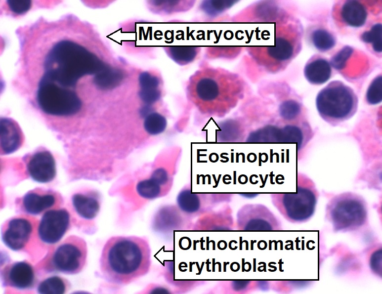
Bone marrow is a semi-solid tissue found within the spongy (also known as cancellous) portions of bones. In birds and mammals, bone marrow is the primary site of new blood cell production (or haematopoiesis). It is composed of hematopoietic cells, marrow adipose tissue, and supportive stromal cells. In adult humans, bone marrow is primarily located in the ribs, vertebrae, sternum, and bones of the pelvis. Bone marrow comprises approximately 5% of total body mass in healthy adult humans, such that a man weighing 73 kg (161 lbs) will have around 3.7 kg (8 lbs) of bone marrow. Human marrow produces approximately 500 billion blood cells per day, which join the systemic circulation via permeable vasculature sinusoids within the medullary cavity. All types of hematopoietic cells, including both myeloid and lymphoid lineages, are created in bone marrow; however, lymphoid cells must migrate to other lymphoid organs (e.g. thymus) in order to complete maturation. ... [...More Info...] [...Related Items...] OR: [Wikipedia] [Google] [Baidu] |
Hemangioblastoma
Hemangioblastomas, or haemangioblastomas, are vascular tumors of the central nervous system that originate from the vascular system, usually during middle age. Sometimes, these tumors occur in other sites such as the spinal cord and retina. They may be associated with other diseases such as polycythemia (increased blood cell count), pancreatic cysts and Von Hippel–Lindau syndrome (VHL syndrome). Hemangioblastomas are most commonly composed of stromal cells in small blood vessels and usually occur in the cerebellum, brainstem or spinal cord. They are classed as grade I tumors under the World Health Organization's classification system. Presentation Complications Hemangioblastomas can cause an abnormally high number of red blood cells in the bloodstream due to ectopic production of the hormone erythropoietin as a paraneoplastic syndrome. Pathogenesis Hemangioblastomas are composed of endothelial cells, pericytes and stromal cells. In VHL syndrome the von Hippel-Lindau protein ... [...More Info...] [...Related Items...] OR: [Wikipedia] [Google] [Baidu] |
Vasculogenesis
Vasculogenesis is the process of blood vessel formation, occurring by a '' de novo'' production of endothelial cells. It is sometimes paired with angiogenesis, as the first stage of the formation of the vascular network, closely followed by angiogenesis. Process In the sense distinguished from angiogenesis, vasculogenesis is different in one aspect: whereas angiogenesis is the formation of new blood vessels from pre-existing ones, vasculogenesis is the formation of new blood vessels, in blood islands, when there are no pre-existing ones. For example, if a monolayer of endothelial cells begins sprouting to form capillaries, angiogenesis is occurring. Vasculogenesis, in contrast, is when endothelial precursor cells (angioblasts) migrate and differentiate in response to local cues (such as growth factors and extracellular matrices) to form new blood vessels. These vascular trees are then pruned and extended through angiogenesis. Occurrences Vasculogenesis occurs during embryologic ... [...More Info...] [...Related Items...] OR: [Wikipedia] [Google] [Baidu] |
Endothelial Progenitor Cell
Endothelial progenitor cell (or EPC) is a term that has been applied to multiple different cell types that play roles in the regeneration of the endothelial lining of blood vessels. Outgrowth endothelial cells are an EPC subtype committed to endothelial cell formation. Despite the history and controversy, the EPC in all its forms remains a promising target of regenerative medicine research. History and controversy Developmentally, the endothelium arises in close contact with the hematopoietic system. This, and the existence of hemogenic endothelium, led to a belief and search for adult hemangioblast- or angioblast-like cells; cells which could give rise to functional vasculature in adults. The existence of endothelial progenitor cells has been posited since the mid-twentieth century, however their existence was not confirmed until the 1990s when Asahara et al. published the discovery of the first putative EPC. Recently, controversy has developed over the definition of true endothe ... [...More Info...] [...Related Items...] OR: [Wikipedia] [Google] [Baidu] |
Hemogenic Endothelium
Hemogenic endothelium is a special subset of endothelial cells scattered within blood vessels that can differentiate into haematopoietic cells. The development of hematopoietic cells in the embryo proceeds sequentially from mesoderm through the hemangioblast to the hemogenic endothelium and hematopoietic progenitors.C. Lancrin, P. Sroczynska, C. Stephenson, et al. The haemangioblast generates haematopoietic cells through a haemogenic endothelium stage. Nature 2009;457:892–895 See also *Hemangioblast Hemangioblasts are the multipotent precursor cells that can differentiate into both hematopoietic and endothelial cells. In the mouse embryo, the emergence of blood islands in the yolk sac at embryonic day 7 marks the onset of hematopoiesis. From ... References Cell biology {{Molecular-cell-biology-stub ... [...More Info...] [...Related Items...] OR: [Wikipedia] [Google] [Baidu] |
Mesoderm
The mesoderm is the middle layer of the three germ layers that develops during gastrulation in the very early development of the embryo of most animals. The outer layer is the ectoderm, and the inner layer is the endoderm.Langman's Medical Embryology, 11th edition. 2010. The mesoderm forms mesenchyme, mesothelium, non-epithelial blood cells and coelomocytes. Mesothelium lines coeloms. Mesoderm forms the muscles in a process known as myogenesis, septa (cross-wise partitions) and mesenteries (length-wise partitions); and forms part of the gonads (the rest being the gametes). Myogenesis is specifically a function of mesenchyme. The mesoderm differentiates from the rest of the embryo through intercellular signaling, after which the mesoderm is polarized by an organizing center. The position of the organizing center is in turn determined by the regions in which beta-catenin is protected from degradation by GSK-3. Beta-catenin acts as a co-factor that alters the activity of ... [...More Info...] [...Related Items...] OR: [Wikipedia] [Google] [Baidu] |
Gordon M
Gordon may refer to: People * Gordon (given name), a masculine given name, including list of persons and fictional characters * Gordon (surname), the surname * Gordon (slave), escaped to a Union Army camp during the U.S. Civil War * Clan Gordon, aka the House of Gordon, a Scottish clan Education * Gordon State College, a public college in Barnesville, Georgia * Gordon College (Massachusetts), a Christian college in Wenham, Massachusetts * Gordon College (Pakistan), a Christian college in Rawalpindi, Pakistan * Gordon College (Philippines), a public university in Subic, Zambales * Gordon College of Education, a public college in Haifa, Israel Places Australia *Gordon, Australian Capital Territory *Gordon, New South Wales * Gordon, South Australia *Gordon, Victoria *Gordon River, Tasmania *Gordon River (Western Australia) Canada *Gordon Parish, New Brunswick * Gordon/Barrie Island, municipality in Ontario * Gordon River (Chochocouane River), a river in Quebec Scotland *Gordo ... [...More Info...] [...Related Items...] OR: [Wikipedia] [Google] [Baidu] |
Wilhelm His Jr
Wilhelm His Jr. (29 December 1863 – 10 November 1934) was a Swiss cardiologist and anatomist, son of Wilhelm His Sr. In 1893, His discovered the bundle of His, the collection of specialized cardiac muscle cells in the heart that transmits electrical impulses and helps synchronize contraction of the cardiac muscles. Later in life, as a professor of medicine at the University of Berlin, he was one of the first to recognize that "the heartbeat has its origin in the individual cells of heart muscle." Werner–His disease (or trench fever) was also named after him. Angle of His (or incisura cardiaca) was posthumously named after him by Daniel John Cunningham in 1906. Works * ''Die Front der Ärzte'' . Velhagen & Klasing, Bielefeld .a.193Digital editionby the University and State Library Düsseldorf The University and State Library Düsseldorf (german: Universitäts- und Landesbibliothek Düsseldorf, abbreviated ULB Düsseldorf) is a central service institution of Heinri ... [...More Info...] [...Related Items...] OR: [Wikipedia] [Google] [Baidu] |
CD133
CD133 antigen, also known as prominin-1, is a glycoprotein that in humans is encoded by the ''PROM1'' gene. It is a member of pentaspan transmembrane glycoproteins, which specifically localize to cellular protrusions. When embedded in the cell membrane, the membrane topology of prominin-1 is such that the N-terminus extends into the extracellular space and the C-terminus resides in the intracellular compartment. The protein consists of five transmembrane segments, with the first and second segments and the third and fourth segments connected by intracellular loops while the second and third as well as fourth and fifth transmembrane segments are connected by extracellular loops. While the precise function of CD133 remains unknown, it has been proposed that it acts as an organizer of cell membrane topology. Tissue distribution CD133 is expressed in hematopoietic stem cells, endothelial progenitor cells, glioblastoma, neuronal and glial stem cells, various pediatric brain tum ... [...More Info...] [...Related Items...] OR: [Wikipedia] [Google] [Baidu] |
Lateral Mesoderm
The lateral plate mesoderm is the mesoderm that is found at the periphery of the embryo. It is to the side of the paraxial mesoderm, and further to the axial mesoderm. The lateral plate mesoderm is separated from the paraxial mesoderm by a narrow region of intermediate mesoderm. The mesoderm is the middle layer of the three germ layers, between the outer ectoderm and inner endoderm. During the third week of embryonic development the lateral plate mesoderm splits into two layers forming the intraembryonic coelom. The outer layer of lateral plate mesoderm adheres to the ectoderm to become the somatic or parietal layer known as the somatopleure. The inner layer adheres to the endoderm to become the splanchnic or visceral layer known as the splanchnopleure. Development The lateral plate mesoderm will split into two layers, the somatopleuric mesenchyme, and the splanchnopleuric mesenchyme. * The ''somatopleuric layer'' forms the future body wall. * The ''splanchnopleuric layer'' form ... [...More Info...] [...Related Items...] OR: [Wikipedia] [Google] [Baidu] |

