|
Heart Valve Disease
Valvular heart disease is any cardiovascular disease process involving one or more of the four valves of the heart (the aortic and mitral valves on the left side of heart and the pulmonic and tricuspid valves on the right side of heart). These conditions occur largely as a consequence of aging,Burden of valvular heart diseases: a population-based study. Nkomo VT, Gardin JM, Skelton TN, Gottdiener JS, Scott CG, Enriquez-Sarano. Lancet. 2006 Sep;368(9540):1005-11. but may also be the result of congenital (inborn) abnormalities or specific disease or physiologic processes including rheumatic heart disease and pregnancy. Anatomically, the valves are part of the dense connective tissue of the heart known as the cardiac skeleton and are responsible for the regulation of blood flow through the heart and great vessels. Valve failure or dysfunction can result in diminished heart functionality, though the particular consequences are dependent on the type and severity of valvular diseas ... [...More Info...] [...Related Items...] OR: [Wikipedia] [Google] [Baidu] |
Phonocardiogram
A phonocardiogram (or PCG) is a plot of high-fidelity recording of the sounds and murmurs made by the heart with the help of the machine called the phonocardiograph; thus, phonocardiography is the recording of all the sounds made by the heart during a cardiac cycle. Medical use Heart sounds result from vibrations created by the closure of the heart valves. There are at least two; the first (S1) is produced when the atrioventricular valves (tricuspid and mitral) close at the beginning of systole and the second (S2) when the aortic valve and pulmonary valve (semilunar valves) close at the end of systole. Phonocardiography allows the detection of subaudible sounds and murmurs and makes a permanent record of these events. In contrast, the stethoscope cannot always detect all such sounds or murmurs and provides no record of their occurrence. The ability to quantitate the sounds made by the heart provides information not readily available from more sophisticated tests and provides ... [...More Info...] [...Related Items...] OR: [Wikipedia] [Google] [Baidu] |
Mitral Valve Stenosis
Mitral stenosis is a valvular heart disease characterized by the narrowing of the opening of the mitral valve of the heart. It is almost always caused by rheumatic valvular heart disease. Normally, the mitral valve is about 5 cm2 during diastole. Any decrease in area below 2 cm2 causes mitral stenosis. Early diagnosis of mitral stenosis in pregnancy is very important as the heart cannot tolerate increased cardiac output demand as in the case of exercise and pregnancy. Atrial fibrillation is a common complication of resulting left atrial enlargement, which can lead to systemic thromboembolic complications like stroke. Signs and symptoms Signs and symptoms of mitral stenosis include the following: * Heart failure symptoms, such as dyspnea on exertion, orthopnea and paroxysmal nocturnal dyspnea (PND) * Palpitations * Chest pain * Hemoptysis * Thromboembolism in later stages when the left atrial volume is increased (i.e., dilation). The latter leads to increase risk of ... [...More Info...] [...Related Items...] OR: [Wikipedia] [Google] [Baidu] |
Aneurysm Of Sinus Of Valsalva
Aneurysm of the aortic sinus, also known as the sinus of Valsalva, is a rare abnormality of the aorta, the largest artery in the body. The aorta normally has three small pouches that sit directly above the aortic valve (the sinuses of Valsalva), and an aneurysm of one of these sinuses is a thin-walled swelling. Aneurysms may affect the right (65–85%), non-coronary (10–30%), or rarely the left (< 5%) coronary sinus. These aneurysms may not cause any symptoms but if large can cause , or blackouts. Aortic sinus aneurysms can burst or rupture into adjacent cardiac chambers, which can lead to |
Systemic Lupus Erythematosus
Lupus, technically known as systemic lupus erythematosus (SLE), is an autoimmune disease in which the body's immune system mistakenly attacks healthy tissue in many parts of the body. Symptoms vary among people and may be mild to severe. Common symptoms include painful and swollen joints, fever, chest pain, hair loss, mouth ulcers, swollen lymph nodes, feeling tired, and a red rash which is most commonly on the face. Often there are periods of illness, called flares, and periods of remission during which there are few symptoms. The cause of SLE is not clear. It is thought to involve a mixture of genetics combined with environmental factors. Among identical twins, if one is affected there is a 24% chance the other one will also develop the disease. Female sex hormones, sunlight, smoking, vitamin D deficiency, and certain infections are also believed to increase a person's risk. The mechanism involves an immune response by autoantibodies against a person's own tissues. T ... [...More Info...] [...Related Items...] OR: [Wikipedia] [Google] [Baidu] |
Marfan Syndrome
Marfan syndrome (MFS) is a multi-systemic genetic disorder that affects the connective tissue. Those with the condition tend to be tall and thin, with long arms, legs, fingers, and toes. They also typically have exceptionally flexible joints and abnormally curved spines. The most serious complications involve the heart and aorta, with an increased risk of mitral valve prolapse and aortic aneurysm. The lungs, eyes, bones, and the covering of the spinal cord are also commonly affected. The severity of the symptoms is variable. MFS is caused by a mutation in ''FBN1'', one of the genes that makes fibrillin, which results in abnormal connective tissue. It is an autosomal dominant disorder. In about 75% of cases, it is inherited from a parent with the condition, while in about 25% it is a new mutation. Diagnosis is often based on the Ghent criteria. There is no known cure for MFS. Many of those with the disorder have a normal life expectancy with proper treatment. Management of ... [...More Info...] [...Related Items...] OR: [Wikipedia] [Google] [Baidu] |
Idiopathy
An idiopathic disease is any disease with an unknown cause or mechanism of apparent spontaneous origin. From Greek ἴδιος ''idios'' "one's own" and πάθος ''pathos'' "suffering", ''idiopathy'' means approximately "a disease of its own kind". For some medical conditions, one or more causes are somewhat understood, but in a certain percentage of people with the condition, the cause may not be readily apparent or characterized. In these cases, the origin of the condition is said to be idiopathic. With some other medical conditions, the root cause for a large percentage of all cases have not been established—for example, focal segmental glomerulosclerosis or ankylosing spondylitis; the majority of these cases are deemed idiopathic. Medical advances and this term Advances in medical science improve the understanding of causes of diseases and the classification of diseases; thus, regarding any particular condition or disease, as more root causes are discovered and as events t ... [...More Info...] [...Related Items...] OR: [Wikipedia] [Google] [Baidu] |
Systole
Systole ( ) is the part of the cardiac cycle during which some chambers of the heart contract after refilling with blood. The term originates, via New Latin, from Ancient Greek (''sustolē''), from (''sustéllein'' 'to contract'; from ''sun'' 'together' + ''stéllein'' 'to send'), and is similar to the use of the English term ''to squeeze''. The mammalian heart has four chambers: the left atrium above the left ventricle (lighter pink, see graphic), which two are connected through the mitral (or bicuspid) valve; and the right atrium above the right ventricle (lighter blue), connected through the tricuspid valve. The atria are the receiving blood chambers for the circulation of blood and the ventricles are the discharging chambers. In late ventricular diastole, the atrial chambers contract and send blood to the larger, lower ventricle chambers. This flow fills the ventricles with blood, and the resulting pressure closes the valves to the atria. The ventricles now perform i ... [...More Info...] [...Related Items...] OR: [Wikipedia] [Google] [Baidu] |
Bicuspid Aortic Valve
Bicuspid aortic valve (aka BAV) is a form of heart disease in which two of the leaflets of the aortic valve fuse during development in the womb resulting in a two-leaflet (bicuspid) valve instead of the normal three-leaflet (tricuspid) valve. BAV is the most common cause of heart disease present at birth and affects approximately 1.3% of adults. Normally, the mitral valve is the only bicuspid valve and this is situated between the heart's left atrium and left ventricle. Heart valves play a crucial role in ensuring the unidirectional flow of blood from the atrium to the ventricles, or from the ventricle to the aorta or pulmonary trunk. BAV is normally inherited. Signs and symptoms In many cases, a bicuspid aortic valve will cause no problems. People with BAV may become tired more easily than those with normal valvular function and have difficulty maintaining stamina for cardio-intensive activities due to poor heart performance caused by stress on the aortic wall. Complica ... [...More Info...] [...Related Items...] OR: [Wikipedia] [Google] [Baidu] |
Right Heart
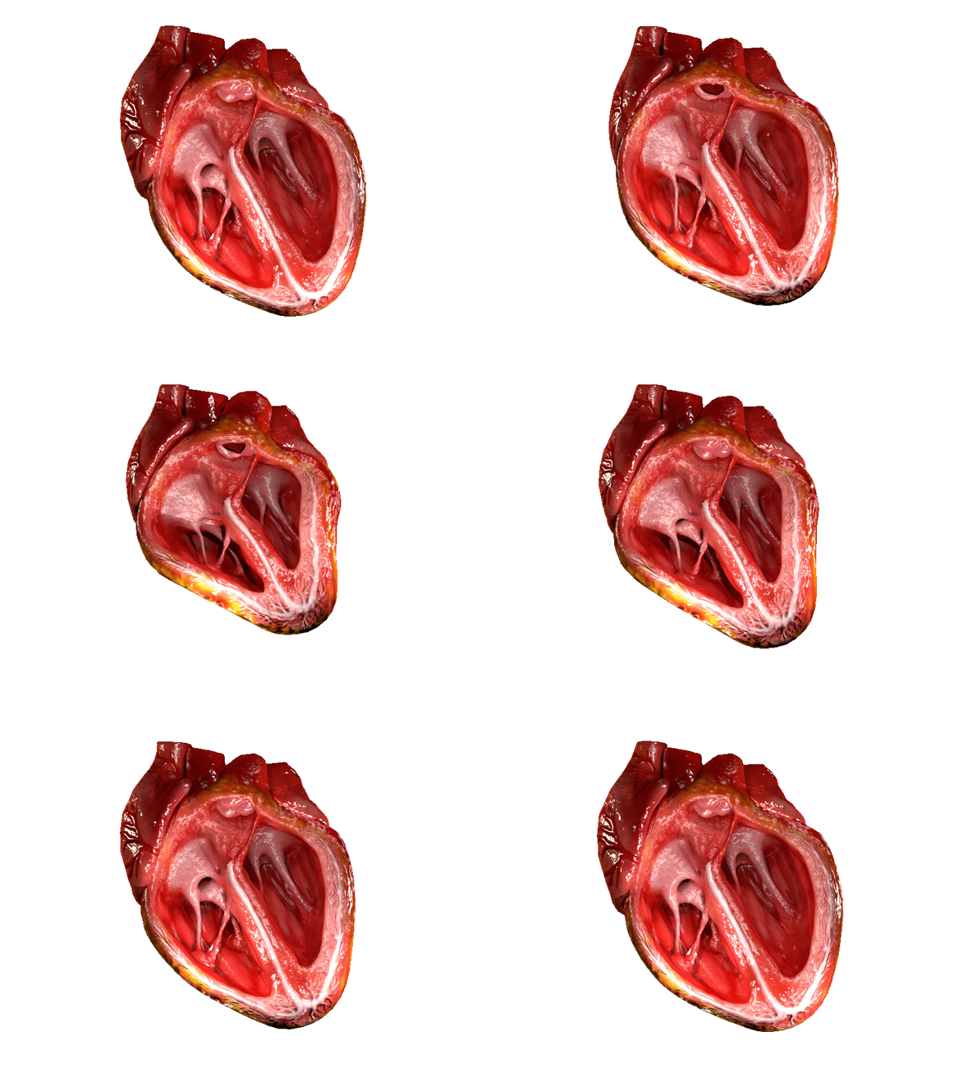
The heart is a muscular Organ (biology), organ in most animals. This organ pumps blood through the blood vessels of the circulatory system. The pumped blood carries oxygen and nutrients to the body, while carrying metabolic waste such as carbon dioxide to the lungs. In humans, the heart is approximately the size of a closed fist and is located between the lungs, in the mediastinum, middle compartment of the thorax, chest. In humans, other mammals, and birds, the heart is divided into four chambers: upper left and right Atrium (heart), atria and lower left and right Ventricle (heart), ventricles. Commonly the right atrium and ventricle are referred together as the right heart and their left counterparts as the left heart. Fish, in contrast, have two chambers, an atrium and a ventricle, while most reptiles have three chambers. In a healthy heart blood flows one way through the heart due to heart valves, which prevent cardiac regurgitation, backflow. The heart is enclosed in a ... [...More Info...] [...Related Items...] OR: [Wikipedia] [Google] [Baidu] |
Left Heart
The heart is a muscular organ in most animals. This organ pumps blood through the blood vessels of the circulatory system. The pumped blood carries oxygen and nutrients to the body, while carrying metabolic waste such as carbon dioxide to the lungs. In humans, the heart is approximately the size of a closed fist and is located between the lungs, in the middle compartment of the chest. In humans, other mammals, and birds, the heart is divided into four chambers: upper left and right atria and lower left and right ventricles. Commonly the right atrium and ventricle are referred together as the right heart and their left counterparts as the left heart. Fish, in contrast, have two chambers, an atrium and a ventricle, while most reptiles have three chambers. In a healthy heart blood flows one way through the heart due to heart valves, which prevent backflow. The heart is enclosed in a protective sac, the pericardium, which also contains a small amount of fluid. The wall of ... [...More Info...] [...Related Items...] OR: [Wikipedia] [Google] [Baidu] |
Pulmonary Insufficiency
Pulmonary (or pulmonic) insufficiency (or incompetence, or regurgitation) is a condition in which the pulmonary valve is incompetent and allows backflow from the pulmonary artery to the right ventricle of the heart during diastole. While a small amount of backflow may occur ordinarily, it is usually only shown on an echocardiogram and is harmless. More pronounced regurgitation that is noticed through a routine physical examination is a medical sign of disease and warrants further investigation. If it is secondary to pulmonary hypertension it is referred to as a Graham Steell murmur. Signs and symptoms Because pulmonic regurgitation is the result of other factors in the body, any noticeable symptoms are ultimately caused by an underlying medical condition rather than the regurgitation itself. However, more severe regurgitation may contribute to right ventricular enlargement by dilation, and in later stages, right heart failure. A diastolic decrescendo murmur can sometimes be ident ... [...More Info...] [...Related Items...] OR: [Wikipedia] [Google] [Baidu] |
Pulmonary Valve Stenosis
Pulmonary valve stenosis (PVS) is a heart valve disorder. Blood going from the heart to the lungs goes through the pulmonary valve, whose purpose is to prevent blood from flowing back to the heart. In pulmonary valve stenosis this opening is too narrow, leading to a reduction of flow of blood to the lungs. While the most common cause of pulmonary valve stenosis is congenital heart disease, it may also be due to a malignant carcinoid tumor. Both stenosis of the pulmonary artery and pulmonary valve stenosis are forms of pulmonic stenosis (nonvalvular and valvular, respectively) but pulmonary valve stenosis accounts for 80% of pulmonic stenosis. PVS was the key finding that led Jacqueline Noonan to identify the syndrome now called Noonan syndrome. Symptoms and signs Among some of the symptoms consistent with pulmonary valve stenosis are the following: * Heart murmur * Cyanosis * Dyspnea * Dizziness * Upper thorax pain * Developmental disorders Cause In regards to the cause of pul ... [...More Info...] [...Related Items...] OR: [Wikipedia] [Google] [Baidu] |


