|
Hippocampal Formation
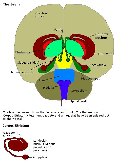
The hippocampal formation is a compound structure in the Temporal lobe#Medial temporal lobe, medial temporal lobe of the brain. It forms a c-shaped bulge on the floor of the temporal horn of the Lateral ventricles, lateral ventricle. There is no consensus concerning which brain regions are encompassed by the term, with some authors defining it as the dentate gyrus, the hippocampus proper and the subiculum; and others including also the Brodmann area 27, presubiculum, parasubiculum, and entorhinal cortex. The hippocampal formation is thought to play a role in memory, spatial navigation and control of attention. The neural layout and pathways within the hippocampal formation are very similar in all mammals. History and function During the nineteenth and early twentieth centuries, based largely on the observation that, between species, the size of the olfactory bulb varies with the size of the parahippocampal gyrus, the hippocampal formation was thought to be part of the olfactory sy ... [...More Info...] [...Related Items...] OR: [Wikipedia] [Google] [Baidu] |
Amygdala
The amygdala (; plural: amygdalae or amygdalas; also '; Latin from Greek, , ', 'almond', 'tonsil') is one of two almond-shaped clusters of nuclei located deep and medially within the temporal lobes of the brain's cerebrum in complex vertebrates, including humans. Shown to perform a primary role in the processing of memory, decision making, and emotional responses (including fear, anxiety, and aggression), the amygdalae are considered part of the limbic system. The term "amygdala" was first introduced by Karl Friedrich Burdach in 1822. Structure The regions described as amygdala nuclei encompass several structures of the cerebrum with distinct connectional and functional characteristics in humans and other animals. Among these nuclei are the basolateral complex, the cortical nucleus, the medial nucleus, the central nucleus, and the intercalated cell clusters. The basolateral complex can be further subdivided into the lateral, the basal, and the accessory basal nucle ... [...More Info...] [...Related Items...] OR: [Wikipedia] [Google] [Baidu] |
Grid Cells
A grid cell is a type of neuron within the entorhinal cortex that fires at regular intervals as an animal navigates an open area, allowing it to understand its position in space by storing and integrating information about location, distance, and direction. Grid cells have been found in many animals, including rats, mice, bats, monkeys, and humans. Grid cells were discovered in 2005 by Edvard Moser, May-Britt Moser, and their students Torkel Hafting, Marianne Fyhn, and Sturla Molden at the Centre for the Biology of Memory (CBM) in Norway. They were awarded the 2014 Nobel Prize in Physiology or Medicine together with John O'Keefe for their discoveries of cells that constitute a positioning system in the brain. The arrangement of spatial firing fields, all at equal distances from their neighbors, led to a hypothesis that these cells encode a neural representation of Euclidean space. The discovery also suggested a mechanism for dynamic computation of self-position based on continuo ... [...More Info...] [...Related Items...] OR: [Wikipedia] [Google] [Baidu] |
Head Direction Cells
Head direction (HD) cells are neurons found in a number of brain regions that increase their firing rates above baseline levels only when the animal's head points in a specific direction. They have been reported in rats, monkeys, mice, chinchillas and bats, but are thought to be common to all mammals, perhaps all vertebrates and perhaps even some invertebrates, and to underlie the "sense of direction". When the animal's head is facing in the cell's "preferred firing direction" these neurons fire at a steady rate (i.e., they do not show adaptation), but firing decreases back to baseline rates as the animal's head turns away from the preferred direction (usually about 45° away from this direction). HD cells are found in many brain areas, including the cortical regions of postsubiculum (also known as the dorsal presubiculum), retrosplenial cortex, and entorhinal cortex, and subcortical regions including the thalamus (the anterior dorsal and the lateral dorsal thalamic nuclei), late ... [...More Info...] [...Related Items...] OR: [Wikipedia] [Google] [Baidu] |
Lynn Nadel
Lynn Nadel (born November 12, 1942) is an American psychologist who is the Regents' Professor of psychology at the University of Arizona. Nadel specializes in memory, and has investigated the role of the hippocampus in memory formation. Together with John O'Keefe, he coauthored the influential 1978 book ''The Hippocampus as a Cognitive Map'', which defended the theory that the hippocampus learns and stores cognitive maps of portions of space. With Morris Moscovitch, he advanced the multiple trace theory that the hippocampus is always involved in storage and retrieval of episodic memory, but that semantic memory can be established in the neocortex. Nadel received a Ph.D. from McGill University in 1967, and joined the faculty of the University of Arizona in 1985, where he is now an Emeritus Professor of Psychology and Cognitive Science. Nadel, together with John O'Keefe, received the 2006 Grawemeyer Award for their work in identifying the brain's mapping system. He was named recip ... [...More Info...] [...Related Items...] OR: [Wikipedia] [Google] [Baidu] |
Place Cells
A place cell is a kind of pyramidal neuron in the hippocampus that becomes active when an animal enters a particular place in its environment, which is known as the place field. Place cells are thought to act collectively as a cognitive representation of a specific location in space, known as a cognitive map. Place cells work with other types of neurons in the hippocampus and surrounding regions to perform this kind of spatial processing. They have been found in a variety of animals, including rodents, bats, monkeys and humans. Place-cell firing patterns are often determined by stimuli in the environment such as visual landmarks, and olfactory and vestibular stimuli. Place cells have the ability to suddenly change their firing pattern from one pattern to another, a phenomenon known as remapping. This remapping may occur in either some of the place cells or in all place cells at once. It may be caused by a number of changes, such as in the odor of the environment. Place cells ar ... [...More Info...] [...Related Items...] OR: [Wikipedia] [Google] [Baidu] |
John O'Keefe (neuroscientist)
John O'Keefe, (born November 18, 1939) is an American-British neuroscientist, psychologist and a professor at the Sainsbury Wellcome Centre for Neural Circuits and Behaviour and the Research Department of Cell and Developmental Biology at University College London. He discovered place cells in the hippocampus, and that they show a specific kind of temporal coding in the form of theta phase precession. He shared the Nobel Prize in Physiology or Medicine in 2014, together with May-Britt Moser and Edvard Moser; he has received several other awards. He has worked at University College London for his entire career, but also held a part-time chair at the Norwegian University of Science and Technology at the behest of his Norwegian collaborators, the Mosers. Education and early life Born in New York City to Irish immigrant parents, O'Keefe attended Regis High School (Manhattan) and received a BA degree from the City College of New York. He went on to study at McGill University in M ... [...More Info...] [...Related Items...] OR: [Wikipedia] [Google] [Baidu] |
Anterior Cingulate Cortex
In the human brain, the anterior cingulate cortex (ACC) is the frontal part of the cingulate cortex that resembles a "collar" surrounding the frontal part of the corpus callosum. It consists of Brodmann areas 24, 32, and 33. It is involved in certain higher-level functions, such as attention allocation, reward anticipation, decision-making, ethics and morality, impulse control (e.g. performance monitoring and error detection), and emotion. Anatomy The anterior cingulate cortex can be divided anatomically based on cognitive (dorsal), and emotional (ventral) components. The dorsal part of the ACC is connected with the prefrontal cortex and parietal cortex, as well as the motor system and the frontal eye fields, making it a central station for processing top-down and bottom-up stimuli and assigning appropriate control to other areas in the brain. By contrast, the ventral part of the ACC is connected with the amygdala, nucleus accumbens, hypothalamus, hippocampus, and ant ... [...More Info...] [...Related Items...] OR: [Wikipedia] [Google] [Baidu] |
Vladimir Bekhterev
Vladimir Mikhailovich Bekhterev ( rus, Влади́мир Миха́йлович Бе́хтерев, p=ˈbʲextʲɪrʲɪf; January 20, 1857 – December 24, 1927) was a Russian neurologist and the father of objective psychology. He is best known for noting the role of the hippocampus in memory, his study of reflexes, and Bekhterev’s disease. Moreover, he is known for his competition with Ivan Pavlov regarding the study of conditioned reflexes. Early life Vladimir Bekhterev was born in Sorali, a village in the Vyatka Governorate of the Russian Empire between the Volga River and the Ural Mountains. V. M. Bekhterev's father – Mikhail Pavlovich – was a district police officer; his mother, Maria Mikhailovna – was a daughter of a titular councilor, was educated at a boarding school which also provided lessons of music and the French language. Beside Vladimir they had two more sons in the family: Nikolai and Aleksandr, older than he by 6 and 3 years respectively. In 1864 the ... [...More Info...] [...Related Items...] OR: [Wikipedia] [Google] [Baidu] |
Axon
An axon (from Greek ἄξων ''áxōn'', axis), or nerve fiber (or nerve fibre: see spelling differences), is a long, slender projection of a nerve cell, or neuron, in vertebrates, that typically conducts electrical impulses known as action potentials away from the nerve cell body. The function of the axon is to transmit information to different neurons, muscles, and glands. In certain sensory neurons (pseudounipolar neurons), such as those for touch and warmth, the axons are called afferent nerve fibers and the electrical impulse travels along these from the periphery to the cell body and from the cell body to the spinal cord along another branch of the same axon. Axon dysfunction can be the cause of many inherited and acquired neurological disorders that affect both the peripheral and central neurons. Nerve fibers are classed into three typesgroup A nerve fibers, group B nerve fibers, and group C nerve fibers. Groups A and B are myelinated, and group C are unmyelinated. ... [...More Info...] [...Related Items...] OR: [Wikipedia] [Google] [Baidu] |
Alf Brodal
Alf Brodal (25 January 1910 – 29 February 1988) was a Norwegian professor of anatomy. Personal life He was born in Kristiania as a son of the doctor of engineering Peter Brodal (1872–1935) and his wife Helene Kathrine Obenauer (1879–1934). He was a brother of violinist Jon Brodal (1911-1998) and psalm writer Anne Margarethe Brodal (1914-1999). Brodal was married to physiotherapist Inger Olivia Hannestad (1910–1986). He died in 1988 in Bærum. Their son Per Alf Brodal also became a professor of medicine. Career He finished his secondary education in 1929 and the cand.med. degree in 1937. He took the dr.med. degree in 1940 on the thesis ''Experimentelle Untersuchungen über die olivocerebellare Lokalisation''. He was hired at the University of Oslo in 1943, and was promoted to professor in 1950. He was a specialist in neuroanatomy, and was particularly interested in the cerebellum, the reticular substance and the vestibular nuclei. He worked closely with Jan Birger J ... [...More Info...] [...Related Items...] OR: [Wikipedia] [Google] [Baidu] |
Medial Surface Of Cerebral Cortex - Entorhinal Cortex
Medial may refer to: Mathematics * Medial magma, a mathematical identity in algebra Geometry * Medial axis, in geometry the set of all points having more than one closest point on an object's boundary * Medial graph, another graph that represents the adjacencies between edges in the faces of a plane graph * Medial triangle, the triangle whose vertices lie at the midpoints of an enclosing triangle's sides * Polyhedra: ** Medial deltoidal hexecontahedron ** Medial disdyakis triacontahedron ** Medial hexagonal hexecontahedron ** Medial icosacronic hexecontahedron ** Medial inverted pentagonal hexecontahedron ** Medial pentagonal hexecontahedron ** Medial rhombic triacontahedron Linguistics * A medial sound or letter is one that is found in the middle of a larger unit (like a word) ** Syllable medial, a segment located between the onset and the rime of a syllable * In the older literature, a term for the voiced stops (like ''b'', ''d'', ''g'') * Medial or second person demonstr ... [...More Info...] [...Related Items...] OR: [Wikipedia] [Google] [Baidu] |
.png)


