|
High Pressure Receptor Zones
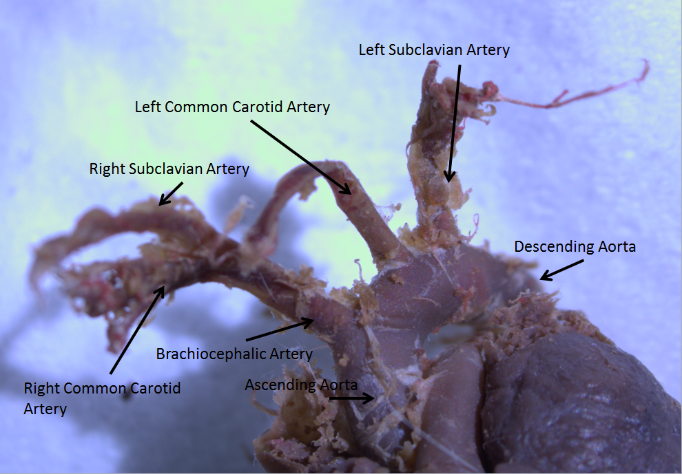
High pressure receptors are the baroreceptors found within the aortic arch and carotid sinus. They are only sensitive to blood pressures above 60 mmHg. When these receptors are activated they elicit a depressor response; which decreases the heart rate and causes a general vasodilation. An increase in arterial blood pressure reflexively elicits an increase in vagal neuronal activity to the heart (i.e. the resulting decreased heart rate). The afferent nerves from the baroreceptors are called buffer nerves. See also * Low pressure receptors * Bainbridge reflex The Bainbridge reflex or Bainbridge effect, also called the atrial reflex, is an increase in heart rate due to an increase in central venous pressure. Increased blood volume is detected by stretch receptors (Cardiac Receptors) located in both sides ... References *"Principles of medical physiology" by A Fonyo page 577 {{neuroanatomy-stub Sensory receptors ... [...More Info...] [...Related Items...] OR: [Wikipedia] [Google] [Baidu] |
Baroreceptor
Baroreceptors (or archaically, pressoreceptors) are sensors located in the carotid sinus (at the bifurcation of external and internal carotids) and in the aortic arch. They sense the blood pressure and relay the information to the brain, so that a proper blood pressure can be maintained. Baroreceptors are a type of mechanoreceptor sensory neuron that are excited by a stretch of the blood vessel. Thus, increases in the pressure of blood vessel triggers increased action potential generation rates and provides information to the central nervous system. This sensory information is used primarily in autonomic reflexes that in turn influence the heart cardiac output and vascular smooth muscle to influence vascular resistance. Baroreceptors act immediately as part of a negative feedback system called the baroreflex, as soon as there is a change from the usual mean arterial blood pressure, returning the pressure toward a normal level. These reflexes help regulate short-term blood pressur ... [...More Info...] [...Related Items...] OR: [Wikipedia] [Google] [Baidu] |
Aortic Arch
The aortic arch, arch of the aorta, or transverse aortic arch () is the part of the aorta between the ascending and descending aorta. The arch travels backward, so that it ultimately runs to the left of the trachea. Structure The aorta begins at the level of the upper border of the second/third sternocostal articulation of the right side, behind the ventricular outflow tract and pulmonary trunk. The right atrial appendage overlaps it. The first few centimeters of the ascending aorta and pulmonary trunk lies in the same pericardial sheath. and runs at first upward, arches over the pulmonary trunk, right pulmonary artery, and right main bronchus to lie behind the right second coastal cartilage. The right lung and sternum lies anterior to the aorta at this point. The aorta then passes posteriorly and to the left, anterior to the trachea, and arches over left main bronchus and left pulmonary artery, and reaches to the left side of the T4 vertebral body. Apart from T4 verte ... [...More Info...] [...Related Items...] OR: [Wikipedia] [Google] [Baidu] |
Carotid Sinus
In human anatomy, the carotid sinus is a dilated area at the base of the internal carotid artery just superior to the bifurcation of the internal carotid and external carotid at the level of the superior border of thyroid cartilage. The carotid sinus extends from the bifurcation to the "true" internal carotid artery. The carotid sinus is sensitive to pressure changes in the arterial blood at this level. It is the major baroreception site in humans and most mammals. Structure The carotid sinus is the reflex area of the carotid artery, consisting of baroreceptors which monitor blood pressure. Function The carotid sinus contains numerous baroreceptors which function as a "sampling area" for many homeostatic mechanisms for maintaining blood pressure. The carotid sinus baroreceptors are innervated by the carotid sinus nerve, which is a branch of the glossopharyngeal nerve (CN IX). The neurons which innervate the carotid sinus centrally project to the solitary nucleus in th ... [...More Info...] [...Related Items...] OR: [Wikipedia] [Google] [Baidu] |
Blood Pressure
Blood pressure (BP) is the pressure of circulating blood against the walls of blood vessels. Most of this pressure results from the heart pumping blood through the circulatory system. When used without qualification, the term "blood pressure" refers to the pressure in the large arteries. Blood pressure is usually expressed in terms of the systolic pressure (maximum pressure during one heartbeat) over diastolic pressure (minimum pressure between two heartbeats) in the cardiac cycle. It is measured in millimeters of mercury (mmHg) above the surrounding atmospheric pressure. Blood pressure is one of the vital signs—together with respiratory rate, heart rate, oxygen saturation, and body temperature—that healthcare professionals use in evaluating a patient's health. Normal resting blood pressure, in an adult is approximately systolic over diastolic, denoted as "120/80 mmHg". Globally, the average blood pressure, age standardized, has remained about the same sin ... [...More Info...] [...Related Items...] OR: [Wikipedia] [Google] [Baidu] |
MmHg
A millimetre of mercury is a manometric unit of pressure, formerly defined as the extra pressure generated by a column of mercury one millimetre high, and currently defined as exactly pascals. It is denoted mmHg or mm Hg. Although not an SI unit, the millimetre of mercury is still routinely used in medicine, meteorology, aviation, and many other scientific fields. One millimetre of mercury is approximately 1 Torr, which is of standard atmospheric pressure ( ≈ ). Although the two units are not equal, the relative difference (less than ) is negligible for most practical uses. History For much of human history, the pressure of gases like air was ignored, denied, or taken for granted, but as early as the 6th century BC, Greek philosopher Anaximenes of Miletus claimed that all things are made of air that is simply changed by varying levels of pressure. He could observe water evaporating, changing to a gas, and felt that this applied even to solid matter. Mor ... [...More Info...] [...Related Items...] OR: [Wikipedia] [Google] [Baidu] |
Heart Rate
Heart rate (or pulse rate) is the frequency of the heartbeat measured by the number of contractions (beats) of the heart per minute (bpm). The heart rate can vary according to the body's physical needs, including the need to absorb oxygen and excrete carbon dioxide, but is also modulated by numerous factors, including, but not limited to, genetics, physical fitness, stress or psychological status, diet, drugs, hormonal status, environment, and disease/illness as well as the interaction between and among these factors. It is usually equal or close to the pulse measured at any peripheral point. The American Heart Association states the normal resting adult human heart rate is 60–100 bpm. Tachycardia is a high heart rate, defined as above 100 bpm at rest. Bradycardia is a low heart rate, defined as below 60 bpm at rest. When a human sleeps, a heartbeat with rates around 40–50 bpm is common and is considered normal. When the heart is not beating in a regular pattern, this is refer ... [...More Info...] [...Related Items...] OR: [Wikipedia] [Google] [Baidu] |
Vasodilation
Vasodilation is the widening of blood vessels. It results from relaxation of smooth muscle cells within the vessel walls, in particular in the large veins, large arteries, and smaller arterioles. The process is the opposite of vasoconstriction, which is the narrowing of blood vessels. When blood vessels dilate, the flow of blood is increased due to a decrease in vascular resistance and increase in cardiac output. Therefore, dilation of arterial blood vessels (mainly the arterioles) decreases blood pressure. The response may be intrinsic (due to local processes in the surrounding tissue) or extrinsic (due to hormones or the nervous system). In addition, the response may be localized to a specific organ (depending on the metabolic needs of a particular tissue, as during strenuous exercise), or it may be systemic (seen throughout the entire systemic circulation). Endogenous substances and drugs that cause vasodilation are termed vasodilators. Such vasoactivity is necessar ... [...More Info...] [...Related Items...] OR: [Wikipedia] [Google] [Baidu] |
Vagal
The vagus nerve, also known as the tenth cranial nerve, cranial nerve X, or simply CN X, is a cranial nerve that interfaces with the parasympathetic control of the heart, lungs, and digestive tract. It comprises two nerves—the left and right vagus nerves—but they are typically referred to collectively as a single subsystem. The vagus is the longest nerve of the autonomic nervous system in the human body and comprises both sensory and motor fibers. The sensory fibers originate from neurons of the nodose ganglion, whereas the motor fibers come from neurons of the dorsal motor nucleus of the vagus and the nucleus ambiguus. The vagus was also historically called the pneumogastric nerve. Structure Upon leaving the medulla oblongata between the olive and the inferior cerebellar peduncle, the vagus nerve extends through the jugular foramen, then passes into the carotid sheath between the internal carotid artery and the internal jugular vein down to the neck, chest, and abdomen, w ... [...More Info...] [...Related Items...] OR: [Wikipedia] [Google] [Baidu] |
Heart
The heart is a muscular organ in most animals. This organ pumps blood through the blood vessels of the circulatory system. The pumped blood carries oxygen and nutrients to the body, while carrying metabolic waste such as carbon dioxide to the lungs. In humans, the heart is approximately the size of a closed fist and is located between the lungs, in the middle compartment of the chest. In humans, other mammals, and birds, the heart is divided into four chambers: upper left and right atria and lower left and right ventricles. Commonly the right atrium and ventricle are referred together as the right heart and their left counterparts as the left heart. Fish, in contrast, have two chambers, an atrium and a ventricle, while most reptiles have three chambers. In a healthy heart blood flows one way through the heart due to heart valves, which prevent backflow. The heart is enclosed in a protective sac, the pericardium, which also contains a small amount of fluid. The wall of ... [...More Info...] [...Related Items...] OR: [Wikipedia] [Google] [Baidu] |
Afferent Nerves
A sensory nerve, or afferent nerve, is a general anatomic term for a nerve which contains predominantly somatic afferent nerve fibers. Afferent nerve fibers in a sensory nerve carry sensory information toward the central nervous system (CNS) from different sensory receptors of sensory neurons in the peripheral nervous system. A motor nerve carries information from the CNS to the PNS, and both types of nerve are called peripheral nerves. Afferent nerve fibers link the sensory neurons throughout the body, in pathways to the relevant processing circuits in the central nervous system. Afferent nerve fibers are often paired with efferent nerve fibers from the motor neurons (that travel from the CNS to the PNS), in mixed nerves. Stimuli cause nerve impulses in the receptors and alter the potentials, which is known as sensory transduction. Spinal cord entry Afferent nerve fibers leave the sensory neuron from the dorsal root ganglia of the spinal cord, and motor commands carried b ... [...More Info...] [...Related Items...] OR: [Wikipedia] [Google] [Baidu] |
Low Pressure Receptors
Low pressure baroreceptors are baroreceptors that relay information derived from blood pressure within the autonomic nervous system. They are stimulated by stretching of the vessel wall. They are located in large systemic veins and in the walls of the atria of the heart, and pulmonary vasculature. Low pressure baroreceptors are also referred to as volume receptors and cardiopulmonary baroreceptors.Armstrong, Maggie, et al. ''Physiology, Baroreceptors - Statpearls - NCBI Bookshelf''. 9 Mar. 2022, https://www.ncbi.nlm.nih.gov/books/NBK538172/. Structure There are two types of cardiopulmonary baroreceptors. Type A receptors and Type B receptors are both within the atria of the heart. Type A receptors are activated by wall tension, which develops by atrial contraction during ventricular diastole. Type B receptors are activated by wall stretch, which develops by atrial filling during ventricular systole. In the right atrium, the stretch receptors occur at the junction of the ve ... [...More Info...] [...Related Items...] OR: [Wikipedia] [Google] [Baidu] |
Bainbridge Reflex
The Bainbridge reflex or Bainbridge effect, also called the atrial reflex, is an increase in heart rate due to an increase in central venous pressure. Increased blood volume is detected by stretch receptors (Cardiac Receptors) located in both sides of atria at the venoatrial junctions. History Francis Arthur Bainbridge described this as a reflex in 1918 when he was experimenting on dogs. Bainbridge found that infusing blood or saline into the animal increased heart rate. This phenomenon occurred even if arterial blood pressure did not increase. He further observed that heart rate increased when venous pressure rose high enough to distend the right atrium, but denervation of the vagi to the heart eliminated these effects. Subsequent work demonstrated that a stretch-induced increase in cardiac beating rate can also be observed in isolated hearts or even the fully separated SAN. Thus, the positive chronotropic (from Χρόνος, Greek for 'time', and τρέπειν, Greek for 'to be ... [...More Info...] [...Related Items...] OR: [Wikipedia] [Google] [Baidu] |


