|
High-power Field
A high-power field (HPF), when used in relation to microscopy, references the field of view under the maximum magnification power of the objective being used. Often, this represents a 400-fold magnification when referenced in scientific papers. Area Area per high-power field for some microscope types: *Olympus BX50, BX40 or BH2 or AO: 0.096 mm2 *AO with 10x eyepiece: 0.12 mm2 *Olympus with 10x eyepiece: 0.16 mm2 *Nikon Eclipse E400 with 10x eyepiece and 40x objective: 0.25mm2 *Leitz Ortholux: 0.27 mm2 *Leitz Diaplan: 0.31 mm2 Examples of usage The area provides a reference unit, for example in reference ranges for urine tests.Normal Reference Range Table from the |
Microscopy
Microscopy is the technical field of using microscopes to view objects and areas of objects that cannot be seen with the naked eye (objects that are not within the resolution range of the normal eye). There are three well-known branches of microscopy: optical, electron, and scanning probe microscopy, along with the emerging field of X-ray microscopy. Optical microscopy and electron microscopy involve the diffraction, reflection, or refraction of electromagnetic radiation/electron beams interacting with the specimen, and the collection of the scattered radiation or another signal in order to create an image. This process may be carried out by wide-field irradiation of the sample (for example standard light microscopy and transmission electron microscopy) or by scanning a fine beam over the sample (for example confocal laser scanning microscopy and scanning electron microscopy). Scanning probe microscopy involves the interaction of a scanning probe with the surface of the objec ... [...More Info...] [...Related Items...] OR: [Wikipedia] [Google] [Baidu] |
Field Of View
The field of view (FoV) is the extent of the observable world that is seen at any given moment. In the case of optical instruments or sensors it is a solid angle through which a detector is sensitive to electromagnetic radiation. Humans and animals In the context of human and primate vision, the term "field of view" is typically only used in the sense of a restriction to what is visible by external apparatus, like when wearing spectacles or virtual reality goggles. Note that eye movements are allowed in the definition but do not change the field of view when understood this way. If the analogy of the eye's retina working as a sensor is drawn upon, the corresponding concept in human (and much of animal vision) is the visual field. It is defined as "the number of degrees of visual angle during stable fixation of the eyes".Strasburger, Hans; Pöppel, Ernst (2002). Visual Field. In G. Adelman & B.H. Smith (Eds): ''Encyclopedia of Neuroscience''; 3rd edition, on CD-ROM. El ... [...More Info...] [...Related Items...] OR: [Wikipedia] [Google] [Baidu] |
Magnification
Magnification is the process of enlarging the apparent size, not physical size, of something. This enlargement is quantified by a calculated number also called "magnification". When this number is less than one, it refers to a reduction in size, sometimes called ''minification'' or ''de-magnification''. Typically, magnification is related to scaling up visuals or images to be able to see more detail, increasing resolution, using microscope, printing techniques, or digital processing. In all cases, the magnification of the image does not change the perspective of the image. Examples of magnification Some optical instruments provide visual aid by magnifying small or distant subjects. * A magnifying glass, which uses a positive (convex) lens to make things look bigger by allowing the user to hold them closer to their eye. * A telescope, which uses its large objective lens or primary mirror to create an image of a distant object and then allows the user to examine the image clo ... [...More Info...] [...Related Items...] OR: [Wikipedia] [Google] [Baidu] |
University Of Texas Southwestern Medical Center
The University of Texas Southwestern Medical Center (UT Southwestern or UTSW) is a public academic health science center in Dallas, Texas. With approximately 18,800 employees, more than 2,900 full-time faculty, and nearly 4 million outpatient visits per year, UT Southwestern is the largest medical school in the University of Texas System and state of Texas.https://utsystem.edu/sites/default/files/documents/publication/2018/fast-facts/fast-facts-09-2018.pdf UT Southwestern's William P. Clements Jr University Hospital is nationally ranked in nine specialties by '' U.S. News & World Report'' and ranked the best hospital in the Dallas-Fort Worth/North Texas region. Forbes ranked UT Southwestern Medical Center as the top health care employer in the state of Texas and the top health care employer to new graduates in the United States. UT Southwestern's operating budget in 2021 was more than $4.1 billion, and is the largest medical institution in the Dallas–Fort Worth Metroplex ... [...More Info...] [...Related Items...] OR: [Wikipedia] [Google] [Baidu] |
Mitoses
In cell biology, mitosis () is a part of the cell cycle in which replicated chromosomes are separated into two new nuclei. Cell division by mitosis gives rise to genetically identical cells in which the total number of chromosomes is maintained. Therefore, mitosis is also known as equational division. In general, mitosis is preceded by S phase of interphase (during which DNA replication occurs) and is often followed by telophase and cytokinesis; which divides the cytoplasm, organelles and cell membrane of one cell into two new cells containing roughly equal shares of these cellular components. The different stages of mitosis altogether define the mitotic (M) phase of an animal cell cycle—the division of the mother cell into two daughter cells genetically identical to each other. The process of mitosis is divided into stages corresponding to the completion of one set of activities and the start of the next. These stages are preprophase (specific to plant cells), prophase, p ... [...More Info...] [...Related Items...] OR: [Wikipedia] [Google] [Baidu] |
Pleomorphism (cytology)
Pleomorphism is a term used in histology and cytopathology to describe variability in the size, shape and staining of cells and/or their nuclei. Several key determinants of cell and nuclear size, like ploidy and the regulation of cellular metabolism, are commonly disrupted in tumors. Therefore, cellular and nuclear pleomorphism is one of the earliest hallmarks of cancer progression and a feature characteristic of malignant neoplasms and dysplasia. Certain benign cell types may also exhibit pleomorphism, e.g. neuroendocrine cells, Arias-Stella reaction. A rare type of rhabdomyosarcoma that is found in adults is known as pleomorphic rhabdomyosarcoma. Despite the prevalence of pleomorphism in human pathology, its role in disease progression is unclear. In epithelial tissue, pleomorphism in cellular size can induce packing defects and disperse aberrant cells. But the consequence of atypical cell and nuclear morphology in other tissues is unknown. See also *Anaplasia *Cell growt ... [...More Info...] [...Related Items...] OR: [Wikipedia] [Google] [Baidu] |
Necrosis
Necrosis () is a form of cell injury which results in the premature death of cells in living tissue by autolysis. Necrosis is caused by factors external to the cell or tissue, such as infection, or trauma which result in the unregulated digestion of cell components. In contrast, apoptosis is a naturally occurring programmed and targeted cause of cellular death. While apoptosis often provides beneficial effects to the organism, necrosis is almost always detrimental and can be fatal. Cellular death due to necrosis does not follow the apoptotic signal transduction pathway, but rather various receptors are activated and result in the loss of cell membrane integrity and an uncontrolled release of products of cell death into the extracellular space. This initiates in the surrounding tissue an inflammatory response, which attracts leukocytes and nearby phagocytes which eliminate the dead cells by phagocytosis. However, microbial damaging substances released by leukocytes would crea ... [...More Info...] [...Related Items...] OR: [Wikipedia] [Google] [Baidu] |
Classification Of Breast Cancer
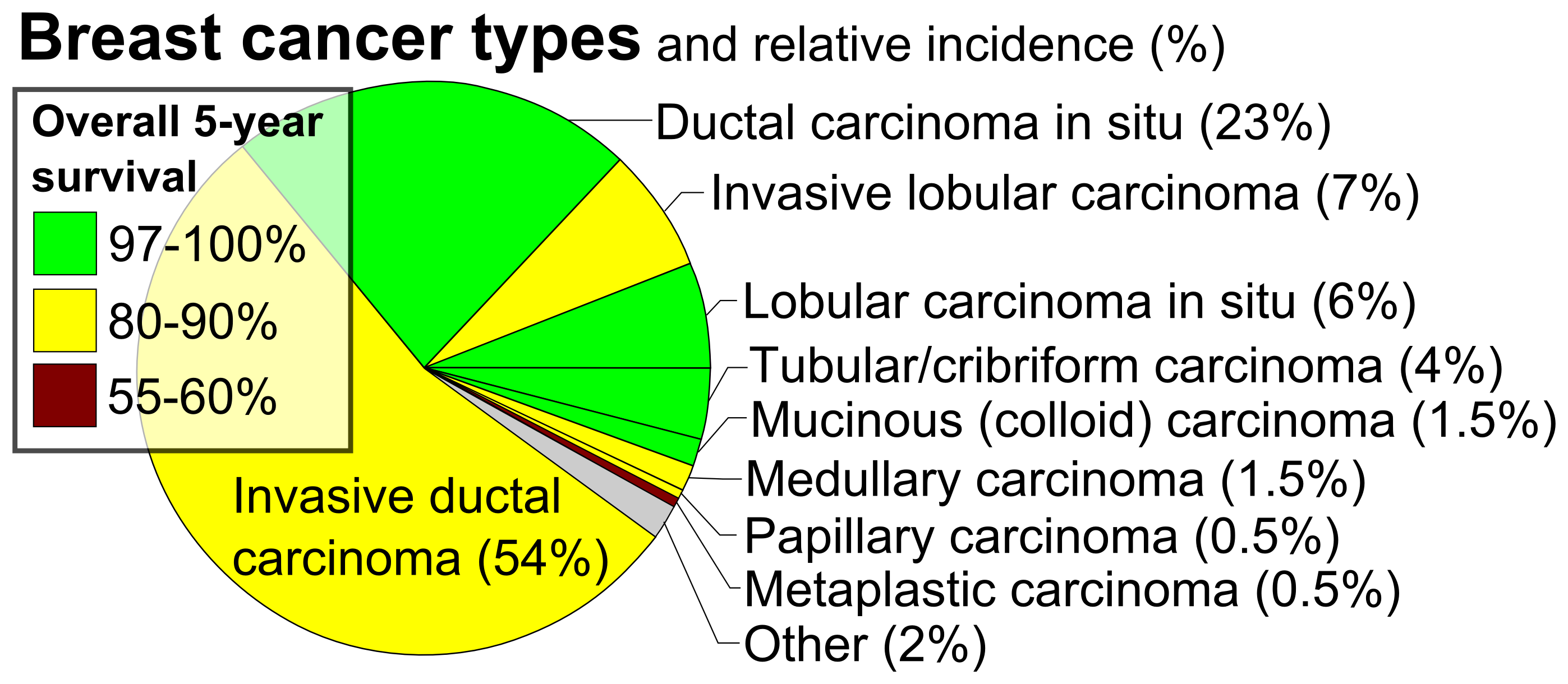
Breast cancer classification divides breast cancer into categories according to different schemes criteria and serving a different purpose. The major categories are the histopathological type, the grade of the tumor, the stage of the tumor, and the expression of proteins and genes. As knowledge of cancer cell biology develops these classifications are updated. The purpose of classification is to select the best treatment. The effectiveness of a specific treatment is demonstrated for a specific breast cancer (usually by randomized, controlled trials). That treatment may not be effective in a different breast cancer. Some breast cancers are aggressive and life-threatening, and must be treated with aggressive treatments that have major adverse effects. Other breast cancers are less aggressive and can be treated with less aggressive treatments, such as lumpectomy. Treatment algorithms rely on breast cancer classification to define specific subgroups that are each treated according t ... [...More Info...] [...Related Items...] OR: [Wikipedia] [Google] [Baidu] |
Stanford University School Of Medicine
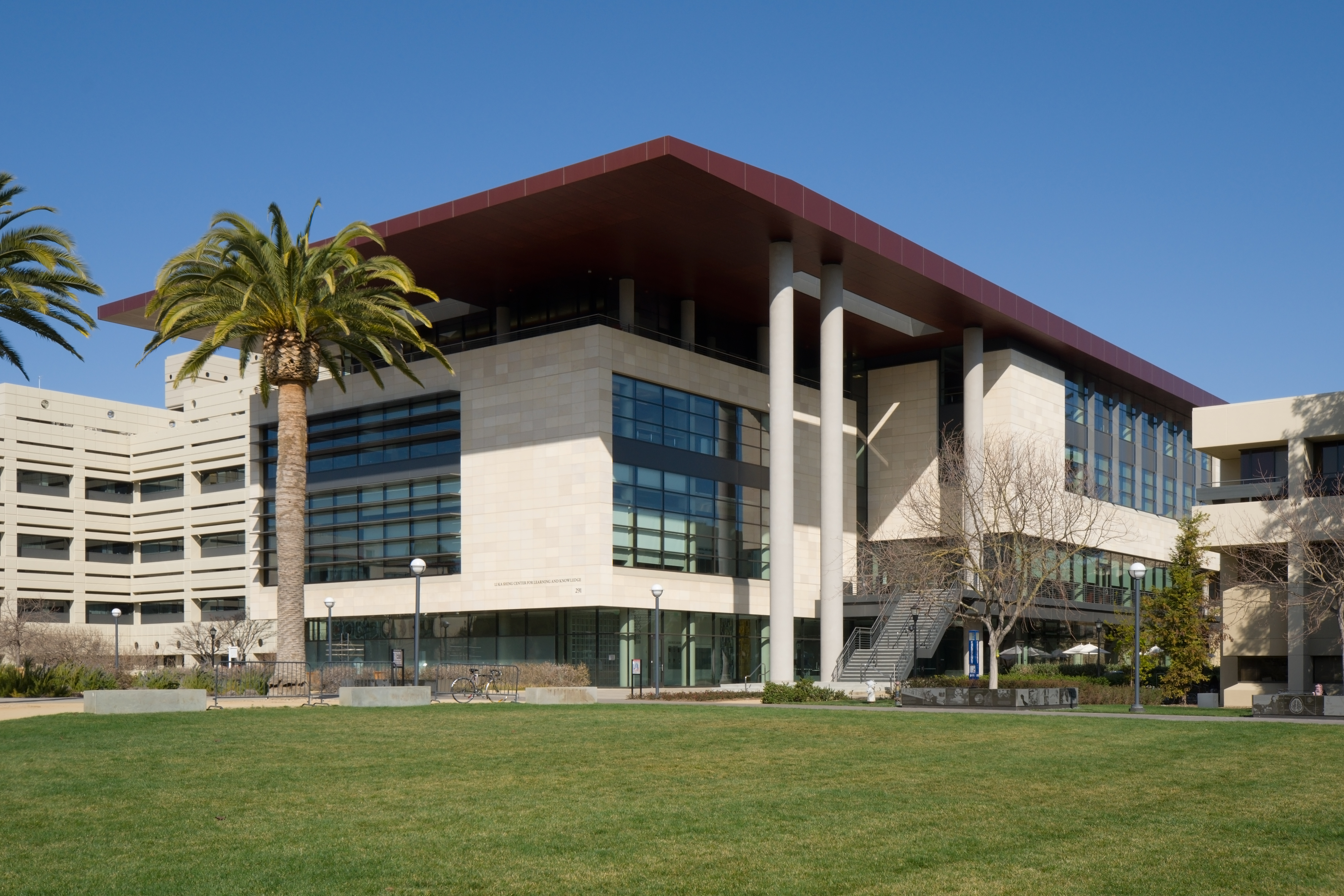
Stanford University School of Medicine is the medical school of Stanford University and is located in Stanford, California. It traces its roots to the Medical Department of the University of the Pacific, founded in San Francisco in 1858. This medical institution, then called Cooper Medical College, was acquired by Stanford in 1908. The medical school moved to the Stanford campus near Palo Alto, California, in 1959. The School of Medicine, along with Stanford Health Care and Lucile Packard Children's Hospital, is part of Stanford Medicine. Stanford Health Care was ranked the fourth best hospital in California (behind UCLA Medical Center, Cedars-Sinai Medical Center, and UCSF Medical Center, respectively). History In 1855, Illinois physician Elias Samuel Cooper moved to San Francisco in the wake of the California Gold Rush. In cooperation with the University of the Pacific (also known as California Wesleyan College), Cooper established the Medical Department of the Univers ... [...More Info...] [...Related Items...] OR: [Wikipedia] [Google] [Baidu] |


