|
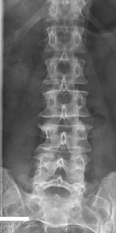
Geroderma Osteodysplasticum
Gerodermia osteodysplastica (GO) is a rare autosomal recessive connective tissue disorder included in the spectrum of cutis laxa syndromes. Usage of the name "Walt Disney dwarfism" is attributed to the first known case of the disorder, documented in a 1950 journal report, in which the authors described five affected members from a Swiss family as having the physical appearance of dwarves from a Walt Disney film. The terms "geroderma" or "gerodermia" can be used interchangeably with "osteodysplastica" or "osteodysplasticum", with the term "hereditaria" sometimes appearing at the end. Presentation Gerodermia osteodysplastica is characterized by symptoms and features which affect the connective tissues, skin and skeletal system. These are: wrinkly, loose skin over the face, abdomen, and extremities (hands, feet) on the dorsal sides usually worsened by chronic joint laxity and hyperextensibility; fragmented elastic fibers of the skin that are reduced in number, with disorientation ... [...More Info...] [...Related Items...] OR: [Wikipedia] [Google] [Baidu] |
Autosome
An autosome is any chromosome that is not a sex chromosome. The members of an autosome pair in a diploid cell have the same morphology, unlike those in allosome, allosomal (sex chromosome) pairs, which may have different structures. The DNA in autosomes is collectively known as atDNA or auDNA. For example, humans have a diploid human genome, genome that usually contains 22 pairs of autosomes and one allosome pair (46 chromosomes total). The autosome pairs are labeled with numbers (1–22 in humans) roughly in order of their sizes in base pairs, while allosomes are labelled with their letters. By contrast, the allosome pair consists of two X chromosomes in females or one X and one Y chromosome in males. Unusual combinations of XYY syndrome, XYY, Klinefelter syndrome, XXY, Triple X syndrome, XXX, XXXX syndrome, XXXX, XXXXX syndrome, XXXXX or XXYY syndrome, XXYY, among Aneuploidy, other Salome combinations, are known to occur and usually cause developmental abnormalities. Autosomes ... [...More Info...] [...Related Items...] OR: [Wikipedia] [Google] [Baidu] |
Collagen
Collagen () is the main structural protein in the extracellular matrix found in the body's various connective tissues. As the main component of connective tissue, it is the most abundant protein in mammals, making up from 25% to 35% of the whole-body protein content. Collagen consists of amino acids bound together to form a triple helix of elongated fibril known as a collagen helix. It is mostly found in connective tissue such as cartilage, bones, tendons, ligaments, and skin. Depending upon the degree of mineralization, collagen tissues may be rigid (bone) or compliant (tendon) or have a gradient from rigid to compliant (cartilage). Collagen is also abundant in corneas, blood vessels, the gut, intervertebral discs, and the dentin in teeth. In muscle tissue, it serves as a major component of the endomysium. Collagen constitutes one to two percent of muscle tissue and accounts for 6% of the weight of the skeletal muscle tissue. The fibroblast is the most common cell that crea ... [...More Info...] [...Related Items...] OR: [Wikipedia] [Google] [Baidu] |
Vertebra
The spinal column, a defining synapomorphy shared by nearly all vertebrates,Hagfish are believed to have secondarily lost their spinal column is a moderately flexible series of vertebrae (singular vertebra), each constituting a characteristic irregular bone whose complex structure is composed primarily of bone, and secondarily of hyaline cartilage. They show variation in the proportion contributed by these two tissue types; such variations correlate on one hand with the cerebral/caudal rank (i.e., location within the backbone), and on the other with phylogenetic differences among the vertebrate taxa. The basic configuration of a vertebra varies, but the bone is its ''body'', with the central part of the body constituting the ''centrum''. The upper (closer to) and lower (further from), respectively, the cranium and its central nervous system surfaces of the vertebra body support attachment to the intervertebral discs. The posterior part of a vertebra forms a vertebral arch ... [...More Info...] [...Related Items...] OR: [Wikipedia] [Google] [Baidu] |
Vertebral Column
The vertebral column, also known as the backbone or spine, is part of the axial skeleton. The vertebral column is the defining characteristic of a vertebrate in which the notochord (a flexible rod of uniform composition) found in all chordata, chordates has been replaced by a segmented series of bone: vertebrae separated by intervertebral discs. Individual vertebrae are named according to their region and position, and can be used as anatomical landmarks in order to guide procedures such as Lumbar puncture, lumbar punctures. The vertebral column houses the spinal canal, a cavity that encloses and protects the spinal cord. There are about 50,000 species of animals that have a vertebral column. The human vertebral column is one of the most-studied examples. Many different diseases in humans can affect the spine, with spina bifida and scoliosis being recognisable examples. The general structure of human vertebrae is fairly typical of that found in mammals, reptiles, and birds. Th ... [...More Info...] [...Related Items...] OR: [Wikipedia] [Google] [Baidu] |
Platyspondyly
Congenital vertebral anomalies are a collection of malformations of the spine. Most, around 85%, are not clinically significant, but they can cause compression of the spinal cord by deforming the vertebral canal or causing instability. This condition occurs in the womb. Congenital vertebral anomalies include alterations of the shape and number of vertebrae. Lumbarization and sacralization ''Lumbarization'' is an anomaly in the spine. It is defined by the nonfusion of the first and second segments of the sacrum. The lumbar spine subsequently appears to have six vertebrae or segments, not five. This sixth lumbar vertebra is known as a transitional vertebra. Conversely the sacrum appears to have only four segments instead of its designated five segments. Lumbosacral transitional vertebrae consist of the process of the last lumbar vertebra fusing with the first sacral segment. While only around 10 percent of adults have a spinal abnormality due to genetics, a sixth lumbar vertebr ... [...More Info...] [...Related Items...] OR: [Wikipedia] [Google] [Baidu] |
Long Bone
The long bones are those that are longer than they are wide. They are one of five types of bones: long, Short bone, short, Flat bone, flat, Irregular bone, irregular and Sesamoid bone, sesamoid. Long bones, especially the femur and tibia, are subjected to most of the load during daily activities and they are crucial for skeletal mobility. They grow primarily by elongation of the diaphysis, with an epiphysis at each end of the growing bone. The ends of epiphyses are covered with hyaline cartilage ("articular cartilage"). The longitudinal growth of long bones is a result of endochondral ossification at the epiphyseal plate. Bone growth in length is stimulated by the production of growth hormone (GH), a secretion of the Anterior pituitary, anterior lobe of the pituitary gland. The long bone category includes the femora, tibiae, and fibulae of the legs; the humerus, humeri, Radius (bone), radii, and ulnae of the arms; metacarpals and metatarsals of the hands and feet, the Phalanx bone ... [...More Info...] [...Related Items...] OR: [Wikipedia] [Google] [Baidu] |
Chin
The chin is the forward pointed part of the anterior mandible (List_of_human_anatomical_regions#Regions, mental region) below the lower lip. A fully developed human skull has a chin of between 0.7 cm and 1.1 cm. Evolution The presence of a well-developed chin is considered to be one of the morphological characteristics of ''Homo sapiens'' that differentiates them from other human ancestors such as the closely related Neanderthal, Neanderthals. Early human ancestors have varied Mandibular symphysis, symphysial morphology, but none of them have a well-developed chin. The origin of the chin is traditionally associated with the anterior–posterior breadth shortening of the dental arch or tooth row; however, its general mechanical or functional advantage during feeding, developmental origin, and link with human speech, physiology, and social influence are highly debated. Functional perspectives Robinson (1913) suggests that the demand to resist Mastication, masticatory stresses ... [...More Info...] [...Related Items...] OR: [Wikipedia] [Google] [Baidu] |
Prognathism
Prognathism, also called Habsburg jaw or Habsburgs' jaw primarily in the context of its prevalence amongst members of the House of Habsburg, is a positional relationship of the mandible or maxilla to the skeletal base where either of the jaws protrudes beyond a predetermined imaginary line in the coronal plane of the skull. In general dentistry, oral and maxillofacial surgery, and orthodontics, this is assessed clinically or radiographically ( cephalometrics). The word ''prognathism'' derives from Greek πρό (''pro'', meaning 'forward') and γνάθος (''gnáthos'', 'jaw'). One or more types of prognathism can result in the common condition of malocclusion, in which an individual's top teeth and lower teeth do not align properly. Presentation Prognathism in humans can occur due to normal variation among phenotypes. In human populations where prognathism is not the norm, it may be a malformation, the result of injury, a disease state or a hereditary condition. Prognathism ... [...More Info...] [...Related Items...] OR: [Wikipedia] [Google] [Baidu] |
Human Mandible
In anatomy, the mandible, lower jaw or jawbone is the largest, strongest and lowest bone in the human facial skeleton. It forms the lower jaw and holds the lower tooth, teeth in place. The mandible sits beneath the maxilla. It is the only movable bone of the skull (discounting the ossicles of the middle ear). It is connected to the temporal bones by the temporomandibular joints. The bone is formed prenatal development, in the fetus from a fusion of the left and right mandibular prominences, and the point where these sides join, the mandibular symphysis, is still visible as a faint ridge in the midline. Like other symphyses in the body, this is a midline articulation where the bones are joined by fibrocartilage, but this articulation fuses together in early childhood.Illustrated Anatomy of the Head and Neck, Fehrenbach and Herring, Elsevier, 2012, p. 59 The word "mandible" derives from the Latin word ''mandibula'', "jawbone" (literally "one used for chewing"), from ''wikt:mandere ... [...More Info...] [...Related Items...] OR: [Wikipedia] [Google] [Baidu] |
Maxilla
The maxilla (plural: ''maxillae'' ) in vertebrates is the upper fixed (not fixed in Neopterygii) bone of the jaw formed from the fusion of two maxillary bones. In humans, the upper jaw includes the hard palate in the front of the mouth. The two maxillary bones are fused at the intermaxillary suture, forming the anterior nasal spine. This is similar to the mandible (lower jaw), which is also a fusion of two mandibular bones at the mandibular symphysis. The mandible is the movable part of the jaw. Structure In humans, the maxilla consists of: * The body of the maxilla * Four processes ** the zygomatic process ** the frontal process of maxilla ** the alveolar process ** the palatine process * three surfaces – anterior, posterior, medial * the Infraorbital foramen * the maxillary sinus * the incisive foramen Articulations Each maxilla articulates with nine bones: * two of the cranium: the frontal and ethmoid * seven of the face: the nasal, zygomatic, lacrimal, inferior n ... [...More Info...] [...Related Items...] OR: [Wikipedia] [Google] [Baidu] |
Hypoplasia
Hypoplasia (from Ancient Greek ὑπo- ''hypo-'' 'under' + πλάσις ''plasis'' 'formation'; adjective form ''hypoplastic'') is underdevelopment or incomplete development of a tissue or organ. Dictionary of Cell and Molecular Biology (11 March 2008) Although the term is not always used precisely, it properly refers to an inadequate or below-normal number of cells.Hypoplasia Stedman's Medical Dictionary. lww.com Hypoplasia is similar to |
Zygomatic Bone
In the human skull, the zygomatic bone (from grc, ζῠγόν, zugón, yoke), also called cheekbone or malar bone, is a paired irregular bone which articulates with the maxilla, the temporal bone, the sphenoid bone and the frontal bone. It is situated at the upper and lateral part of the face and forms the prominence of the cheek, part of the lateral wall and floor of the orbit, and parts of the temporal fossa and the infratemporal fossa. It presents a malar and a temporal surface; four processes (the frontosphenoidal, orbital, maxillary, and temporal), and four borders. Etymology The term ''zygomatic'' derives from the Ancient Greek , ''zygoma'', meaning "yoke". The zygomatic bone is occasionally referred to as the zygoma, but this term may also refer to the zygomatic arch. Structure Surfaces The ''malar surface'' is convex and perforated near its center by a small aperture, the zygomaticofacial foramen, for the passage of the zygomaticofacial nerve and vessels; below ... [...More Info...] [...Related Items...] OR: [Wikipedia] [Google] [Baidu] |


