|
Gaze Palsy
Conjugate gaze palsies are neurological disorder (medicine), disorders affecting the ability to move both eyes in the same direction. These palsies can affect Gaze (physiology), gaze in a horizontal, upward, or downward direction. These entities overlap with ophthalmoparesis, ophthalmoparesis and ophthalmoplegia. Signs and symptoms Symptoms of conjugate gaze palsies include the impairment of gaze in various directions and different types of movement, depending on the type of gaze palsy. Signs of a person with a gaze palsy may be frequent movement of the head instead of the eyes. For example, a person with a horizontal saccadic palsy may jerk their head around while watching a movie or high action event instead of keeping their head steady and moving their eyes, which usually goes unnoticed. Someone with a nonselective horizontal gaze palsy may slowly rotate their head back and forth while reading a book instead of slowly scanning their eyes across the page. Cause A lesion, wh ... [...More Info...] [...Related Items...] OR: [Wikipedia] [Google] [Baidu] |
Neurology
Neurology (from el, wikt:νεῦρον, νεῦρον (neûron), "string, nerve" and the suffix wikt:-logia, -logia, "study of") is the branch of specialty (medicine), medicine dealing with the diagnosis and treatment of all categories of conditions and disease involving the brain, the spinal cord and the peripheral nerves. Neurological practice relies heavily on the field of neuroscience, the scientific study of the nervous system. A neurologist is a physician specializing in neurology and trained to investigate, diagnose and treat neurological disorders. Neurologists treat a myriad of neurologic conditions, including stroke, seizures, movement disorders such as Parkinson's disease, autoimmune neurologic disorders such as multiple sclerosis, headache disorders like migraine and dementias such as Alzheimer's disease. Neurologists may also be involved in clinical research, clinical trials, and basic research, basic or translational research. While neurology is a nonsurgical sp ... [...More Info...] [...Related Items...] OR: [Wikipedia] [Google] [Baidu] |
Medial Rectus Muscle
The medial rectus muscle is a muscle in the orbit near the eye. It is one of the extraocular muscles. It originates from the common tendinous ring, and inserts into the anteromedial surface of the eye. It is supplied by the inferior division of the oculomotor nerve (III). It rotates the eye medially (adduction). Structure The medial rectus muscle shares an origin with several other extrinsic eye muscles, the common tendinous ring. It inserts into the anteromedial surface of the eye. This insertion has a width of around 11 mm. Nerve supply The medial rectus muscle is supplied by the inferior division of the oculomotor nerve (III). A branch of it enters the muscle around two fifths along its length. It usually divides into 2 smaller branches, occasionally 3. These further subdivide, becoming smaller down the length of the muscle until they become imperceptible to standard staining around 17 mm from the insertion of the muscle. Relations The insertion of the medial rectus ... [...More Info...] [...Related Items...] OR: [Wikipedia] [Google] [Baidu] |
Eye Diseases
This is a partial list of human eye diseases and disorders. The World Health Organization publishes a classification of known diseases and injuries, the International Statistical Classification of Diseases and Related Health Problems, or ICD-10. This list uses that classification. H00-H06 Disorders of eyelid, lacrimal system and orbit * (H02.1) Ectropion * (H02.2) Lagophthalmos * (H02.3) Blepharochalasis * (H02.4) Ptosis * (H02.5) Stye, an acne type infection of the sebaceous glands on or near the eyelid. * (H02.6) Xanthelasma of eyelid * (H03.0*) Parasitic infestation of eyelid in diseases classified elsewhere ** Dermatitis of eyelid due to Demodex species ( B88.0+ ) ** Parasitic infestation of eyelid in: *** leishmaniasis ( B55.-+ ) *** loiasis ( B74.3+ ) *** onchocerciasis ( B73+ ) *** phthiriasis ( B85.3+ ) * (H03.1*) Involvement of eyelid in other infectious diseases classified elsewhere ** Involvement of eyelid in: *** herpesviral (herpes simplex) infection ( B00.5+ ) * ... [...More Info...] [...Related Items...] OR: [Wikipedia] [Google] [Baidu] |
Ischemic Optic Neuropathy
Ischemic optic neuropathy (ION) is the loss of structure and function of a portion of the optic nerve due to obstruction of blood flow to the nerve (i.e. ischemia). Ischemic forms of optic neuropathy are typically classified as either anterior ischemic optic neuropathy or posterior ischemic optic neuropathy according to the part of the optic nerve that is affected. People affected will often complain of a loss of visual acuity and a visual field, the latter of which is usually in the superior or inferior field. When ION occurs in patients below the age of 50 years old, other causes should be considered, such as juvenile diabetes mellitus, antiphospholipid antibody-associated clotting disorders, collagen-vascular disease, and migraines. Rarely, complications of intraocular surgery or acute blood loss may cause an ischemic event in the optic nerve. Presentation Anterior ION presents with sudden, painless visual loss, developing over hours to days. Diagnosis Examination finding ... [...More Info...] [...Related Items...] OR: [Wikipedia] [Google] [Baidu] |
Optic Neuritis
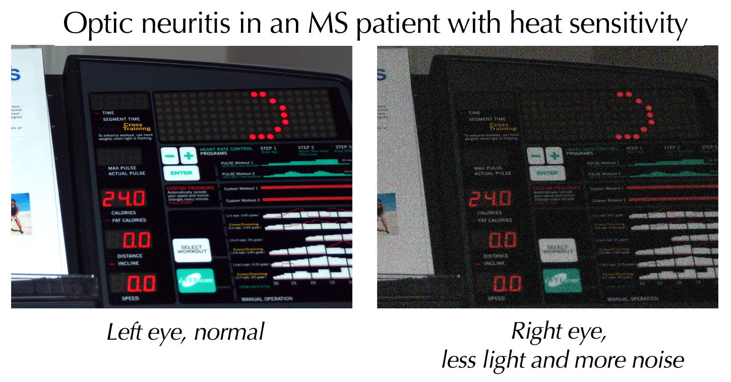
Optic neuritis describes any condition that causes inflammation of the optic nerve; it may be associated with demyelinating diseases, or infectious or inflammatory processes. It is also known as optic papillitis (when the head of the optic nerve is involved), neuroretinitis (when there is a combined involvement of the optic disc and surrounding retina in the macular area) and retrobulbar neuritis (when the posterior part of the nerve is involved). Prelaminar optic neuritis describes involvement of the non-myelinated axons in the retina. It is most often associated with multiple sclerosis, and it may lead to complete or partial loss of vision in one or both eyes. Other causes include: # Leber's hereditary optic neuropathy # Parainfectious optic neuritis (associated with viral infections such as measles, mumps, chickenpox, whooping cough and glandular fever) # Infectious optic neuritis (sinus related or associated with cat-scratch fever, tuberculosis, Lyme disease and crypt ... [...More Info...] [...Related Items...] OR: [Wikipedia] [Google] [Baidu] |
Facial Colliculus
The facial colliculus is an elevated area located in the pontine tegmentum (dorsal pons), within the floor of the fourth ventricle (i.e. the rhomboid fossa). It is formed by fibres from the facial motor nucleus looping over the abducens nucleus. The facial colliculus is an essential landmark of the rhomboid fossa. Anatomy The facial colliculus occurs within the rhomboid fossa (i.e. the floor of the fourth ventricle) where it is placed lateral to its (midline) median sulcus. Structure The facial colliculus is formed by brachial motor nerve fibres of the facial nerve (CN VII) looping over the (ipsilateral) abducens nucleus, forming a bump upon the surface. Clinical significance A facial colliculus lesion would result in ipsilateral facial paralysis (i.e. Bell's palsy Bell's palsy is a type of facial paralysis that results in a temporary inability to control the facial muscles on the affected side of the face. In most cases, the weakness is temporary and significantly imp ... [...More Info...] [...Related Items...] OR: [Wikipedia] [Google] [Baidu] |
Horizontal Gaze Palsy
A gaze palsy is the paresis of conjugate eye movements. Horizontal gaze palsy may be caused by lesions in the cerebral hemispheres, which cause paresis of gaze away from the side of the lesion, or from brain stem lesions, which, if they occur below the crossing of the fibers from the frontal eye fields in the caudal midbrain, will cause weakness of gaze In critical theory, sociology, and psychoanalysis, the gaze (French ''le regard''), in the philosophical and figurative sense, is an individual's (or a group's) awareness and perception of other individuals, other groups, or oneself. The concept ... toward the side of the lesion. These will result in horizontal gaze deviations from unopposed action of the unaffected extraocular muscles. Another way to remember this is that patients with hemisphere lesions look towards their lesion, while patients with pontine gaze palsies look away from their lesions. The human Robo gene acts as a receptor for a midline repulsive cue. When Robo ... [...More Info...] [...Related Items...] OR: [Wikipedia] [Google] [Baidu] |
Nystagmus
Nystagmus is a condition of involuntary (or voluntary, in some cases) eye movement. Infants can be born with it but more commonly acquire it in infancy or later in life. In many cases it may result in reduced or limited vision. Due to the involuntary movement of the eye, it has been called "dancing eyes". In normal eyesight, while the head rotates about an axis, distant visual images are sustained by rotating eyes in the opposite direction of the respective axis. The semicircular canals in the vestibule of the ear sense angular acceleration, and send signals to the nuclei for eye movement in the brain. From here, a signal is relayed to the extraocular muscles to allow one's gaze to fix on an object as the head moves. Nystagmus occurs when the semicircular canals are stimulated (e.g., by means of the caloric test, or by disease) while the head is stationary. The direction of ocular movement is related to the semicircular canal that is being stimulated. There are two key form ... [...More Info...] [...Related Items...] OR: [Wikipedia] [Google] [Baidu] |
Medial Longitudinal Fasciculus
The medial longitudinal fasciculus (MLF) is an area of crossed over tracts, on each side of the brainstem. These bundles of axons are situated near the midline of the brainstem. They are made up of both ascending and descending fibers that arise from a number of sources and terminate in different areas, including the superior colliculus, the vestibular nuclei, and the cerebellum. It contains the interstitial nucleus of Cajal, responsible for oculomotor control, head posture, and vertical eye movement. The medial longitudinal fasciculus is the main central connection for the oculomotor nerve, trochlear nerve, and abducens nerve. It carries information about the direction that the eyes should move. Lesions of the medial longitudinal fasciculus can cause nystagmus and diplopia, which may be associated with multiple sclerosis, a neoplasm, or a stroke. Structure The medial longitudinal fasciculus is an area of crossed over tracts, on each side of the brainstem. It is medial, ... [...More Info...] [...Related Items...] OR: [Wikipedia] [Google] [Baidu] |
One And A Half Syndrome
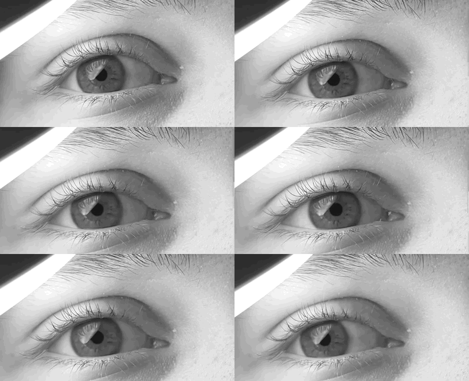
The one and a half syndrome is a rare weakness in eye movement affecting both eyes, in which one cannot move laterally at all, and the other can move only in outward direction. More formally, it is characterized by "''a conjugate horizontal gaze palsy in one direction and an internuclear ophthalmoplegia in the other''". Nystagmus is also present when the eye on the opposite side of the lesion is abducted. Convergence is classically spared as cranial nerve III (oculomotor nerve) and its nucleus is spared bilaterally. Causes Causes of the one and a half syndrome include pontine haemorrhage, ischemia, tumors, infective mass lesions such as tuberculomas, demyelinating conditions like multiple sclerosis, Arteriovenous malformation, Basilar artery aneurysms and Trauma. Anatomy The syndrome usually results from single unilateral lesion of the paramedian pontine reticular formation and the ipsilateral medial longitudinal fasciculus. An alternative anatomical cause is a lesion of th ... [...More Info...] [...Related Items...] OR: [Wikipedia] [Google] [Baidu] |
Oculomotor Nucleus
The fibers of the oculomotor nerve arise from a nucleus in the midbrain, which lies in the gray substance of the floor of the cerebral aqueduct and extends in front of the aqueduct for a short distance into the floor of the third ventricle. From this nucleus the fibers pass forward through the tegmentum, the red nucleus, and the medial part of the substantia nigra, forming a series of curves with a lateral convexity, and emerge from the oculomotor sulcus on the medial side of the cerebral peduncle. The nucleus of the oculomotor nerve does not consist of a continuous column of cells, but is broken up into a number of smaller nuclei, which are arranged in two groups, anterior and posterior. Those of the posterior group are six in number, five of which are symmetrical on the two sides of the middle line, while the sixth is centrally placed and is common to the nerves of both sides. The anterior group consists of two nuclei, an antero-medial and an antero-lateral. The nucleus of the ... [...More Info...] [...Related Items...] OR: [Wikipedia] [Google] [Baidu] |
Efferent Nerve Fiber
Efferent nerve fibers refer to axonal projections that ''exit'' a particular region; as opposed to afferent projections that ''arrive'' at the region. These terms have a slightly different meaning in the context of the peripheral nervous system (PNS) and central nervous system (CNS). The efferent fiber is a long process projecting far from the neuron's body that carries nerve impulses away from the central nervous system toward the peripheral effector organs (mainly muscles and glands). A bundle of these fibers is called an efferent nerve (if it connects to muscles, then it is a motor nerve). The opposite direction of neural activity is afferent conduction, which carries impulses by way of the afferent nerve fibers of sensory neurons. In the nervous system there is a "closed loop" system of sensation, decision, and reactions. This process is carried out through the activity of sensory neurons, interneurons, and motor neurons. In the CNS, afferent and efferent projections ... [...More Info...] [...Related Items...] OR: [Wikipedia] [Google] [Baidu] |


