|
Genitofemoral Nerve
The genitofemoral nerve refers to a nerve that is found in the abdomen. Its branches, the genital branch and femoral branch supply sensation to the upper anterior thigh, as well as the skin of the anterior scrotum in males and mons pubis in females. The femoral branch is different from the femoral nerve, which also arises from the lumbar plexus. Anatomy The genitofemoral nerve originates from the upper L1-2 segments of the lumbar plexus. It passes downwards, pierces the psoas major and emerges from its anterior surface. The nerve divides into two branches, the genital branch and the lumboinguinal nerve also known as the femoral branch, both of which then continue downwards and medially to the inguinal and femoral canal respectively. Genital Branch The genital branch continues downward on the surface of the psoas major muscle and then enters the inguinal canal through the deep inguinal ring. In men, the genital branch supplies the cremaster and scrotal skin. In women, the genit ... [...More Info...] [...Related Items...] OR: [Wikipedia] [Google] [Baidu] |
Lumbar Plexus
The lumbar plexus is a web of nerves (a nervous plexus) in the lumbar region of the body which forms part of the larger lumbosacral plexus. It is formed by the Ventral ramus of spinal nerve, divisions of the first four lumbar nerves (L1-L4) and from contributions of the subcostal nerve (T12), which is the last Thoracic nerves, thoracic nerve. Additionally, the ventral rami of the fourth lumbar nerve pass communicating branches, the lumbosacral trunk, to the sacral plexus. The nerves of the lumbar plexus pass in front of the hip joint and mainly support the anterior part of the thigh.''Thieme Atlas of anatomy'' (2006), pp 470-471 The plexus is formed lateral to the intervertebral foramina and passes through Psoas major muscle, psoas major. Its smaller motor branches are distributed directly to psoas major, while the larger branches leave the muscle at various sites to run obliquely down through the pelvis to leave under the inguinal ligament with the exception of the obturator n ... [...More Info...] [...Related Items...] OR: [Wikipedia] [Google] [Baidu] |
Inguinal Canal
The inguinal canals are the two passages in the anterior abdominal wall of humans and animals which in males convey the spermatic cords and in females the round ligament of the uterus. The inguinal canals are larger and more prominent in males. There is one inguinal canal on each side of the midline. Structure The inguinal canals are situated just above the medial half of the inguinal ligament. In both sexes the canals transmit the ilioinguinal nerves. The canals are approximately 3.75 to 4 cm long. , angled anteroinferiorly and medially. In males, its diameter is normally 2 cm (±1 cm in standard deviation) at the deep inguinal ring.The diameter has been estimated to be ±2.2cm ±1.08cm in Africans, and 2.1 cm ±0.41cm in Europeans. A first-order approximation is to visualize each canal as a cylinder. Walls To help define the boundaries, these canals are often further approximated as boxes with six sides. Not including the two rings, the remaining four sides are usually ca ... [...More Info...] [...Related Items...] OR: [Wikipedia] [Google] [Baidu] |
Cremasteric Muscle
The cremaster muscle is a paired structure made of thin layers of striated and smooth muscle that covers the testis and the spermatic cord in human males. It consists of the lateral and medial parts. Cremaster is an involuntary muscle, responsible for the cremasteric reflex; a protective and physiologic superficial reflex of the testicles. The reflex raises and lowers the testicles in order to keep them protected. Along with the dartos muscle of the scrotum, it regulates testicular temperature, thus aiding the process of spermatogenesis. Structure In human males, the cremaster muscle is a thin layer of striated muscle found in the inguinal canal and scrotum between the external and internal layers of spermatic fascia, surrounding the testis and spermatic cord. The cremaster muscle is a paired structure, there being one on each side of the body. Anatomically, the lateral cremaster muscle originates from the internal oblique muscle, just superior to the inguinal canal, and the mid ... [...More Info...] [...Related Items...] OR: [Wikipedia] [Google] [Baidu] |
Cremasteric Reflex
The cremasteric reflex is a superficial (i.e., close to the skin's surface) reflex observed in human males. This reflex is elicited by lightly stroking or poking the superior and medial (inner) part of the thigh—regardless of the direction of stroke. The normal response is an immediate contraction of the cremaster muscle that pulls up the testis ipsilaterally (on the same side of the body). The reflex utilizes sensory and motor fibers from two different nerves. When the inner thigh is stroked, sensory fibers of the ilioinguinal nerve are stimulated. These activate the motor fibers of the genital branch of the genitofemoral nerve which causes the cremaster muscle to contract and elevate the testis. Clinical conditions In boys, this reflex may be exaggerated which can occasionally lead to a misdiagnosis of cryptorchidism. The cremasteric reflex may be absent with testicular torsion, upper and lower motor neuron disorders, as well as a spine injury of L1-L2. It can also occur if ... [...More Info...] [...Related Items...] OR: [Wikipedia] [Google] [Baidu] |
Saphenous Opening
In anatomy, the saphenous opening (saphenous hiatus, also fossa ovalis) is an oval opening in the upper mid part of the fascia lata of the thigh. It lies 3–4 cm below and lateral to the pubic tubercle and is about 3 cm long and 1.5 cm wide. Structure Just inferolateral to the pubic tubercle the fascia extends downwards forming an arched (falciform) margin of the lateral boundary of the opening. It is covered by a thin perforated part of the superficial fascia called the fascia cribrosa which is pierced by the great saphenous vein, the 3 superficial branches of the femoral artery(except superficial circumflex iliac artery, which pierces fascia lata lateral to the saphenous opening), and lymphatics. It transmits the great saphenous vein and other smaller vessels including the superficial epigastric artery and superficial external pudendal artery, as well as the femoral branch of the genitofemoral nerve. The fascia cribrosa, which is pierced by the structures pass ... [...More Info...] [...Related Items...] OR: [Wikipedia] [Google] [Baidu] |
Cribriform Fascia
The cribriform fascia, fascia cribrosa also Hesselbach's fascia is the portion of fascia covering the saphenous opening in the thigh. It is perforated by the great saphenous vein and by numerous blood and lymphatic vessels. (A structure in anatomy that is pierced by several small holes is referred to as ''cribriform'' from Latin ''cribrum'' meaning sieve). Clinical significance The cribriform fascia has been proposed for use in preventing new vascularization when surgery is performed at the join between the great saphenous vein and the femoral vein. Eponym When the eponym is used, it is named for Franz Kaspar Hesselbach Franz Kaspar Hesselbach (27 January 1759 – 24 July 1816) was a German surgeon and anatomist who was a native of Hammelburg. He was a pupil, and later Prosector under Carl Caspar von Siebold (1736–1807) at Würzburg. Later Hesselbach was a lec ....F. K. Hesselbach. Anatomisch-chirurgische Abhandlung über den Urspurng der Leistenbrüche. Würzburg, Baumgärt ... [...More Info...] [...Related Items...] OR: [Wikipedia] [Google] [Baidu] |
Femoral Sheath
The femoral sheath (also called the crural sheath) is a funnel-shaped downward extension of abdominal fascia within which the femoral artery and femoral vein pass between the abdomen and the thigh. The femoral sheath is subdivided by two vertical partitions to form three compartments (medial, intermediate, and lateral); the medial compartment is known as the femoral canal and contains lymphatic vessels and a lymph node, whereas the intermediate canal and the lateral canal accommodate the femoral vein and the femoral artery (respectively). Some neurovascular structures perforate the femoral sheath. Topographically, the femoral sheath is contained within in the femoral triangle. Structure The femoral sheath is funnel-shaped fascial structure, with the wide end directed superior-ward. The femoral sheath is formed by an inferior-ward prolongation - posterior to the inguinal ligament - of abdominal fascia, with transverse fascia being continued down anterior to the femoral vessels, ... [...More Info...] [...Related Items...] OR: [Wikipedia] [Google] [Baidu] |
Inguinal Ligament
The inguinal ligament (), also known as Poupart's ligament or groin ligament, is a band running from the pubic tubercle to the anterior superior iliac spine. It forms the base of the inguinal canal through which an indirect inguinal hernia may develop. Structure The inguinal ligament runs from the anterior superior iliac crest of the ilium to the pubic tubercle of the pubic bone. It is formed by the external abdominal oblique aponeurosis and is continuous with the fascia lata of the thigh. There is some dispute over the attachments. Structures that pass deep to the inguinal ligament include: *Psoas major, iliacus, pectineus *Femoral nerve, artery, and vein *Lateral cutaneous nerve of thigh *Lymphatics Function The ligament serves to contain soft tissues as they course anteriorly from the trunk to the lower extremity. This structure demarcates the superior border of the femoral triangle. It demarcates the inferior border of the inguinal triangle. The midpoint of the ingui ... [...More Info...] [...Related Items...] OR: [Wikipedia] [Google] [Baidu] |
Labia Majora
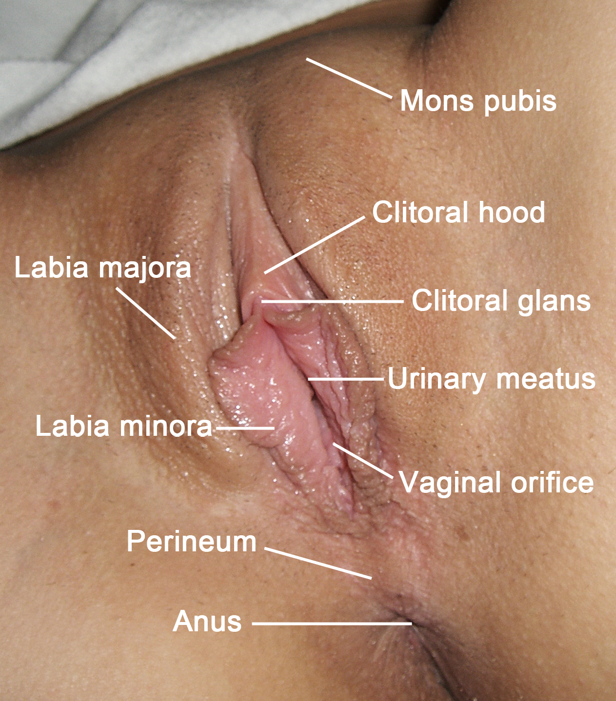
The labia majora (singular: ''labium majus'') are two prominent longitudinal cutaneous folds that extend downward and backward from the mons pubis to the perineum. Together with the labia minora they form the labia of the vulva. The labia majora are homologous to the male scrotum. Etymology ''Labia majora'' is the Latin plural for big ("major") lips; the singular is ''labium majus.'' The Latin term ''labium/labia'' is used in anatomy for a number of usually paired parallel structures, but in English it is mostly applied to two pairs of parts of female external genitals (vulva)—labia majora and labia minora. Labia majora are commonly known as the outer lips, while labia minora (Latin for ''small lips''), which run alongside between them, are referred to as the inner lips. Traditionally, to avoid confusion with other lip-like structures of the body, the labia of female genitals were termed by anatomists in Latin as ''labia majora (''or ''minora) pudendi.'' Embryology Embryolo ... [...More Info...] [...Related Items...] OR: [Wikipedia] [Google] [Baidu] |
Round Ligament Of Uterus
The round ligament of the uterus is a ligament that connects the uterus to the labia majora. Structure The round ligament of the uterus originates at the uterine horns, in the parametrium. The round ligament exits the pelvis via the deep inguinal ring. It passes through the inguinal canal, and continues on to the labia majora. At the labia majora, its fibers spread and mix with the tissue of the mons pubis. Development The round ligament develops from the gubernaculum which attaches the gonad to the labioscrotal swellings in the embryo. Blood supply The round ligament is supplied by the artery of the round ligament of uterus, also known as ''Sampson's artery''. Function The function of the round ligament is maintenance of the anteversion of the uterus (a position where the fundus of the uterus is turned forward at the junction of cervix and vagina) during pregnancy. Normally, the cardinal ligament is what supports the uterine angle (angle of anteversion). When the uterus gro ... [...More Info...] [...Related Items...] OR: [Wikipedia] [Google] [Baidu] |
Deep Inguinal Ring
The inguinal canals are the two passages in the anterior abdominal wall of humans and animals which in males convey the spermatic cords and in females the round ligament of the uterus. The inguinal canals are larger and more prominent in males. There is one inguinal canal on each side of the midline. Structure The inguinal canals are situated just above the medial half of the inguinal ligament. In both sexes the canals transmit the ilioinguinal nerves. The canals are approximately 3.75 to 4 cm long. , angled anteroinferiorly and medially. In males, its diameter is normally 2 cm (±1 cm in standard deviation) at the deep inguinal ring.The diameter has been estimated to be ±2.2cm ±1.08cm in Africans, and 2.1 cm ±0.41cm in Europeans. A first-order approximation is to visualize each canal as a cylinder. Walls To help define the boundaries, these canals are often further approximated as boxes with six sides. Not including the two rings, the remaining four sides are usually cal ... [...More Info...] [...Related Items...] OR: [Wikipedia] [Google] [Baidu] |
Psoas Major
The psoas major ( or ; from grc, ψόᾱ, psóā, muscles of the loins) is a long fusiform muscle located in the lateral lumbar region between the vertebral column and the brim of the lesser pelvis. It joins the iliacus muscle to form the iliopsoas. In animals, this muscle is equivalent to the tenderloin. Structure The psoas major is divided into a superficial and a deep part. The deep part originates from the transverse processes of lumbar vertebrae L1–L5. The superficial part originates from the lateral surfaces of the last thoracic vertebra, lumbar vertebrae L1–L4, and the neighboring intervertebral discs. The lumbar plexus lies between the two layers. Together, the iliacus muscle and the psoas major form the iliopsoas, which is surrounded by the iliac fascia. The iliopsoas runs across the iliopubic eminence through the muscular lacuna to its insertion on the lesser trochanter of the femur. The iliopectineal bursa separates the tendon of the iliopsoas muscle from th ... [...More Info...] [...Related Items...] OR: [Wikipedia] [Google] [Baidu] |


