|
Fundus (eye)
The fundus of the eye is the interior surface of the eye opposite the lens and includes the retina, optic disc, macula, fovea, and posterior pole.Cassin, B. and Solomon, S. ''Dictionary of Eye Terminology''. Gainesville, Florida: Triad Publishing Company, 1990. The fundus can be examined by ophthalmoscopy and/or fundus photography. Variation The color of the fundus varies both between and within species. In one study of primates the retina is blue, green, yellow, orange, and red; only the human fundus (from a lightly pigmented blond person) is red. The major differences noted among the "higher" primate species were size and regularity of the border of macular area, size and shape of the optic disc, apparent 'texturing' of retina, and pigmentation of retina. Clinical significance Medical signs that can be detected from observation of eye fundus (generally by funduscopy) include hemorrhages, exudates, cotton wool spots, blood vessel abnormalities (tortuosity, pulsation and n ... [...More Info...] [...Related Items...] OR: [Wikipedia] [Google] [Baidu] |
Fundus Photographs
Fundus photography involves photographing the rear of an eye, also known as the fundus. Specialized fundus cameras consisting of an intricate microscope attached to a flash enabled camera are used in fundus photography. The main structures that can be visualized on a fundus photo are the central and peripheral retina, optic disc and macula. Fundus photography can be performed with colored filters, or with specialized dyes including fluorescein and indocyanine green. The models and technology of fundus photography have advanced and evolved rapidly over the last century. Since the equipment is sophisticated and challenging to manufacture to clinical standards, only a few manufacturers/brands are available in the market: Welch Allyn, Digisight, Volk, Topcon, Zeiss, Canon, Nidek, Kowa, CSO, CenterVue, Ezer and Optos are some example of fundus camera manufacturers. History The concept of fundus photography was first introduced in the mid 19th century, after the introduction of ... [...More Info...] [...Related Items...] OR: [Wikipedia] [Google] [Baidu] |
Hemorrhage
Bleeding, hemorrhage, haemorrhage or blood loss, is blood escaping from the circulatory system from damaged blood vessels. Bleeding can occur internally, or externally either through a natural opening such as the mouth, nose, ear, urethra, vagina or anus, or through a puncture in the skin. Hypovolemia is a massive decrease in blood volume, and death by excessive loss of blood is referred to as exsanguination. Typically, a healthy person can endure a loss of 10–15% of the total blood volume without serious medical difficulties (by comparison, blood donation typically takes 8–10% of the donor's blood volume). The stopping or controlling of bleeding is called hemostasis and is an important part of both first aid and surgery. Types * Upper head ** Intracranial hemorrhage – bleeding in the skull. ** Cerebral hemorrhage – a type of intracranial hemorrhage, bleeding within the brain tissue itself. ** Intracerebral hemorrhage – bleeding in the brain caused by the ruptu ... [...More Info...] [...Related Items...] OR: [Wikipedia] [Google] [Baidu] |
Red-eye Effect
The red-eye effect in photography is the common appearance of red pupils in color photographs of the eyes of humans and several other animals. It occurs when using a photographic flash that is very close to the camera lens (as with most compact cameras) in ambient low light. Causes In flash photography the light of the flash occurs too fast for the pupil to close, so much of the very bright light from the flash passes into the eye through the pupil, reflects off the fundus at the back of the eyeball and out through the pupil. The camera records this reflected light. The main cause of the red color is the ample amount of blood in the choroid which nourishes the back of the eye and is behind the retina. The blood in the retinal circulation is far less than in the choroid, and plays virtually no role. The eye contains several photostable pigments that all absorb in the short wavelength region, and hence contribute somewhat to the red eye effect. The lens cuts off deep blue and ... [...More Info...] [...Related Items...] OR: [Wikipedia] [Google] [Baidu] |
Leukocoria
Leukocoria (also white pupillary reflex) is an abnormal white reflection from the retina of the eye. Leukocoria resembles eyeshine, but leukocoria can also occur in animals that lack eyeshine because their retina lacks a ''tapetum lucidum''. Leukocoria is a medical sign for a number of conditions, including Coats disease, congenital cataract, corneal scarring, melanoma of the ciliary body, Norrie disease, ocular toxocariasis, persistence of the tunica vasculosa lentis (PFV/PHPV), retinoblastoma, and retrolental fibroplasia. Because of the potentially life-threatening nature of retinoblastoma, a cancer, that condition is usually considered in the evaluation of leukocoria. In some rare cases (1%) the leukocoria is caused by Coats' disease (leaking retinal vessels). Diagnosis On photographs taken using a flash, instead of the familiar red-eye effect, leukocoria can cause a bright white reflection in an affected eye. Leukocoria may appear also in low indirect light, similar to eyes ... [...More Info...] [...Related Items...] OR: [Wikipedia] [Google] [Baidu] |
Fundus Camera
Fundus photography involves photographing the rear of an eye, also known as the fundus (eye), fundus. Specialized fundus cameras consisting of an intricate microscope attached to a flash (photography), flash enabled camera are used in fundus photography. The main structures that can be visualized on a fundus photo are the central and peripheral retina, optic disc and Macula of retina, macula. Fundus photography can be performed with colored filters, or with specialized dyes including Fluorescein angiography, fluorescein and indocyanine green. The models and technology of fundus photography have advanced and evolved rapidly over the last century. Since the equipment is sophisticated and challenging to manufacture to clinical standards, only a few manufacturers/brands are available in the market: Welch Allyn, Digisight, Volk, Topcon, Carl Zeiss AG, Zeiss, Canon Inc., Canon, Nidek, Kowa (company), Kowa, CSO, CenterVue, Ezer and Optos are some example of fundus camera manufacturers. ... [...More Info...] [...Related Items...] OR: [Wikipedia] [Google] [Baidu] |
Dilated Fundus Examination
Dilated fundus examination or dilated-pupil fundus examination (DFE) is a diagnostic procedure that employs the use of mydriatic eye drops (such as tropicamide) to dilate or enlarge the pupil in order to obtain a better view of the fundus of the eye. Once the pupil is dilated, examiners use ophthalmoscopy (funduscopy) to view the eye's interior, allowing assessment of the retina, optic nerve head, blood vessels, and other features. They also often use specialized equipment such as a fundus camera. DFE has been found to be a more effective method for evaluation of internal ocular health than non-dilated examination. It is frequently performed by ophthalmologists and optometrists as part of an eye examination An eye examination is a series of tests performed to assess vision and ability to focus on and discern objects. It also includes other tests and examinations pertaining to the eyes. Eye examinations are primarily performed by an optometrist, op .... References Diagno ... [...More Info...] [...Related Items...] OR: [Wikipedia] [Google] [Baidu] |
Microcirculation
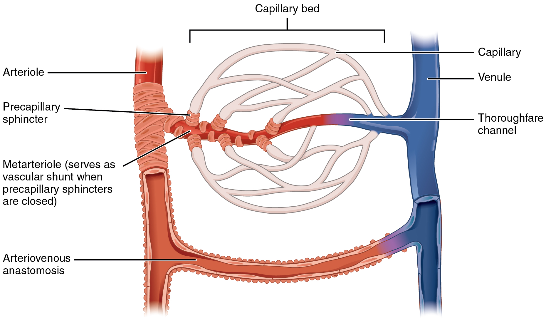
The microcirculation is the circulation of the blood in the smallest blood vessels, the microvessels of the microvasculature present within organ tissues. The microvessels include terminal arterioles, metarterioles, capillaries, and venules. Arterioles carry oxygenated blood to the capillaries, and blood flows out of the capillaries through venules into veins. In addition to these blood vessels, the microcirculation also includes lymphatic capillaries and collecting ducts. The main functions of the microcirculation are the delivery of oxygen and nutrients and the removal of carbon dioxide (CO2). It also serves to regulate blood flow and tissue perfusion thereby affecting blood pressure and responses to inflammation which can include edema (swelling). Most vessels of the microcirculation are lined by flattened cells of the endothelium and many of them are surrounded by contractile cells called pericytes. The endothelium provides a smooth surface for the flow of blood and regul ... [...More Info...] [...Related Items...] OR: [Wikipedia] [Google] [Baidu] |
Hypertensive Retinopathy
Hypertensive retinopathy is damage to the retina and retinal circulation due to high blood pressure (i.e. hypertension). Signs and symptoms Most patients with hypertensive retinopathy have no symptoms. However, some may report decreased or blurred vision, and headaches. Signs Signs of damage to the retina caused by hypertension include: * Arteriolar changes, such as generalized arteriolar narrowing, focal arteriolar narrowing, arteriovenous nicking, changes in the arteriolar wall (arteriosclerosis) and abnormalities at points where arterioles and venules cross. Manifestations of these changes include ''Copper wire arterioles'' where the central light reflex occupies most of the width of the arteriole and ''Silver wire arterioles'' where the central light reflex occupies all of the width of the arteriole, and "arterio-venular (AV) nicking" or "AV nipping", due to venous constriction and banking. * advanced retinopathy lesions, such as microaneurysms, blot hemorrhages and/or flam ... [...More Info...] [...Related Items...] OR: [Wikipedia] [Google] [Baidu] |
Pigmentation
A pigment is a colored material that is completely or nearly insoluble in water. In contrast, dyes are typically soluble, at least at some stage in their use. Generally dyes are often organic compounds whereas pigments are often inorganic compounds. Pigments of prehistoric and historic value include ochre, charcoal, and lapis lazuli. Economic impact In 2006, around 7.4 million tons of inorganic, organic, and special pigments were marketed worldwide. Estimated at around US$14.86 billion in 2018 and will rise at over 4.9% CAGR from 2019 to 2026. The global demand for pigments was roughly US$20.5 billion in 2009. According to an April 2018 report by ''Bloomberg Businessweek'', the estimated value of the pigment industry globally is $30 billion. The value of titanium dioxide – used to enhance the white brightness of many products – was placed at $13.2 billion per year, while the color Ferrari red is valued at $300 million each year. Physical principles ... [...More Info...] [...Related Items...] OR: [Wikipedia] [Google] [Baidu] |
Pulse
In medicine, a pulse represents the tactile arterial palpation of the cardiac cycle (heartbeat) by trained fingertips. The pulse may be palpated in any place that allows an artery to be compressed near the surface of the body, such as at the neck ( carotid artery), wrist (radial artery), at the groin (femoral artery), behind the knee (popliteal artery), near the ankle joint (posterior tibial artery), and on foot (dorsalis pedis artery). Pulse (or the count of arterial pulse per minute) is equivalent to measuring the heart rate. The heart rate can also be measured by listening to the heart beat by auscultation, traditionally using a stethoscope and counting it for a minute. The radial pulse is commonly measured using three fingers. This has a reason: the finger closest to the heart is used to occlude the pulse pressure, the middle finger is used get a crude estimate of the blood pressure, and the finger most distal to the heart (usually the ring finger) is used to nullify the eff ... [...More Info...] [...Related Items...] OR: [Wikipedia] [Google] [Baidu] |
Tortuosity
Tortuosity is widely used as a critical parameter to predict transport properties of porous media, such as rocks and soils. But unlike other standard microstructural properties, the concept of tortuosity is vague with multiple definitions and various evaluation methods introduced in different contexts. Hydraulic, electrical, diffusional, and thermal tortuosities are defined to describe different transport processes in porous media, while geometrical tortuosity is introduced to characterize the morphological property of porous microstructures. Tortuosity in 2-D Subjective estimation (sometimes aided by optometric grading scales) is often used. The simplest mathematical method to estimate tortuosity is the arc-chord ratio: the ratio of the length of the curve (''C'') to the distance between its ends (''L''): :\tau = \frac Arc-chord ratio equals 1 for a straight line and is infinite for a circle. Another method, proposed in 1999, is to estimate the tortuosity as the integral of ... [...More Info...] [...Related Items...] OR: [Wikipedia] [Google] [Baidu] |
Blood Vessel
The blood vessels are the components of the circulatory system that transport blood throughout the human body. These vessels transport blood cells, nutrients, and oxygen to the tissues of the body. They also take waste and carbon dioxide away from the tissues. Blood vessels are needed to sustain life, because all of the body's tissues rely on their functionality. There are five types of blood vessels: the arteries, which carry the blood away from the heart; the arterioles; the capillaries, where the exchange of water and chemicals between the blood and the tissues occurs; the venules; and the veins, which carry blood from the capillaries back towards the heart. The word ''vascular'', meaning relating to the blood vessels, is derived from the Latin ''vas'', meaning vessel. Some structures – such as cartilage, the epithelium, and the lens and cornea of the eye – do not contain blood vessels and are labeled ''avascular''. Etymology * artery: late Middle English; from Latin ... [...More Info...] [...Related Items...] OR: [Wikipedia] [Google] [Baidu] |


