|
Frontal Suture
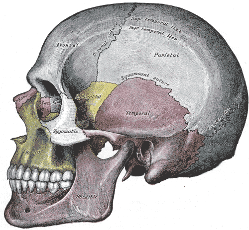
The frontal suture is a fibrous joint that divides the two halves of the frontal bone of the skull in infants and children. Typically, it completely fuses between three and nine months of age, with the two halves of the frontal bone being fused together. It is also called the metopic suture, although this term may also refer specifically to a ''persistent frontal suture''. If the suture is not present at birth because both frontal bones have fused (craniosynostosis), it will cause a keel-shaped deformity of the skull called trigonocephaly. Its presence in a fetal skull, along with other cranial sutures and fontanelles, provides a malleability to the skull that can facilitate movement of the head through the cervical canal and vagina during delivery. The dense connective tissue found between the frontal bones is replaced with bone tissue as the child grows older. Persistent frontal suture In some individuals, the suture can persist (totally or partly) into adulthood, and is ... [...More Info...] [...Related Items...] OR: [Wikipedia] [Google] [Baidu] [Amazon] |
Fibrous Joint
In anatomy, fibrous joints are joints connected by Fibrous connective tissue, fibrous tissue, consisting mainly of collagen. These are fixed joints where bones are united by a layer of white fibrous tissue of varying thickness. In the skull, the joints between the bones are called Suture (anatomy), sutures. Such immovable joints are also referred to as synarthrosis, synarthroses. Types Most fibrous joints are also called "fixed" or "immovable". These joints have no joint cavity and are connected via fibrous connective tissue. * Suture (anatomy), Sutures: The skull bones are connected by fibrous joints called ''#Sutures, sutures''. In fetus, fetal skulls, the sutures are wide to allow slight movement during birth. They later become rigid (synarthrosis, synarthrodial). * Syndesmosis: Some of the long bones in the body such as the radius (bone), radius and ulna in the forearm are joined by a ''#Syndesmosis, syndesmosis'' (along the interosseous membrane of forearm, interosseous mem ... [...More Info...] [...Related Items...] OR: [Wikipedia] [Google] [Baidu] [Amazon] |
Radiographs
Radiography is an imaging technique using X-rays, gamma rays, or similar ionizing radiation and non-ionizing radiation to view the internal form of an object. Applications of radiography include medical ("diagnostic" radiography and "therapeutic radiography") and industrial radiography. Similar techniques are used in airport security, (where "body scanners" generally use backscatter X-ray). To create an image in conventional radiography, a beam of X-rays is produced by an X-ray generator and it is projected towards the object. A certain amount of the X-rays or other radiation are absorbed by the object, dependent on the object's density and structural composition. The X-rays that pass through the object are captured behind the object by a detector (either photographic film or a digital detector). The generation of flat two-dimensional images by this technique is called projectional radiography. In computed tomography (CT scanning), an X-ray source and its associated de ... [...More Info...] [...Related Items...] OR: [Wikipedia] [Google] [Baidu] [Amazon] |
Joints Of The Head And Neck
A joint or articulation (or articular surface) is the connection made between bones, ossicles, or other hard structures in the body which link an animal's skeletal system into a functional whole.Saladin, Ken. Anatomy & Physiology. 7th ed. McGraw-Hill Connect. Webp.274/ref> They are constructed to allow for different degrees and types of movement. Some joints, such as the knee, elbow, and shoulder, are self-lubricating, almost frictionless, and are able to withstand compression and maintain heavy loads while still executing smooth and precise movements. Other joints such as suture (joint), sutures between the bones of the skull permit very little movement (only during birth) in order to protect the brain and the sense organs. The connection between a tooth and the jawbone is also called a joint, and is described as a fibrous joint known as a gomphosis. Joints are classified both structurally and functionally. Joints play a vital role in the human body, contributing to movement, sta ... [...More Info...] [...Related Items...] OR: [Wikipedia] [Google] [Baidu] [Amazon] |
Human Head And Neck
Humans (''Homo sapiens'') or modern humans are the most common and widespread species of primate, and the last surviving species of the genus ''Homo''. They are great apes characterized by their hairlessness, bipedalism, and high intelligence. Humans have large brains, enabling more advanced cognitive skills that facilitate successful adaptation to varied environments, development of sophisticated tools, and formation of complex social structures and civilizations. Humans are highly social, with individual humans tending to belong to a multi-layered network of distinct social groups — from families and peer groups to corporations and political states. As such, social interactions between humans have established a wide variety of values, social norms, languages, and traditions (collectively termed institutions), each of which bolsters human society. Humans are also highly curious: the desire to understand and influence phenomena has motivated humanity's deve ... [...More Info...] [...Related Items...] OR: [Wikipedia] [Google] [Baidu] [Amazon] |
Cranial Sutures
In anatomy, fibrous joints are joints connected by Fibrous connective tissue, fibrous tissue, consisting mainly of collagen. These are fixed joints where bones are united by a layer of white fibrous tissue of varying thickness. In the skull, the joints between the bones are called Suture (anatomy), sutures. Such immovable joints are also referred to as synarthrosis, synarthroses. Types Most fibrous joints are also called "fixed" or "immovable". These joints have no joint cavity and are connected via fibrous connective tissue. * Suture (anatomy), Sutures: The skull bones are connected by fibrous joints called ''#Sutures, sutures''. In fetus, fetal skulls, the sutures are wide to allow slight movement during birth. They later become rigid (synarthrosis, synarthrodial). * Syndesmosis: Some of the long bones in the body such as the radius (bone), radius and ulna in the forearm are joined by a ''#Syndesmosis, syndesmosis'' (along the interosseous membrane of forearm, interosseous mem ... [...More Info...] [...Related Items...] OR: [Wikipedia] [Google] [Baidu] [Amazon] |
Bones Of The Head And Neck
A bone is a rigid organ that constitutes part of the skeleton in most vertebrate animals. Bones protect the various other organs of the body, produce red and white blood cells, store minerals, provide structure and support for the body, and enable mobility. Bones come in a variety of shapes and sizes and have complex internal and external structures. They are lightweight yet strong and hard and serve multiple functions. Bone tissue (osseous tissue), which is also called bone in the uncountable sense of that word, is hard tissue, a type of specialised connective tissue. It has a honeycomb-like matrix internally, which helps to give the bone rigidity. Bone tissue is made up of different types of bone cells. Osteoblasts and osteocytes are involved in the formation and mineralisation of bone; osteoclasts are involved in the resorption of bone tissue. Modified (flattened) osteoblasts become the lining cells that form a protective layer on the bone surface. The minerali ... [...More Info...] [...Related Items...] OR: [Wikipedia] [Google] [Baidu] [Amazon] |
Ossification Of Frontal Bone
In the human skull, the frontal bone or sincipital bone is an unpaired bone which consists of two portions.''Gray's Anatomy'' (1918) These are the vertically oriented squamous part, and the horizontally oriented orbital part, making up the bony part of the forehead, part of the bony orbital cavity holding the eye, and part of the bony part of the nose respectively. The name comes from the Latin word ''frons'' (meaning "forehead"). Structure The frontal bone is made up of two main parts. These are the squamous part, and the orbital part. The squamous part marks the vertical, flat, and also the biggest part, and the main region of the forehead. The orbital part is the horizontal and second biggest region of the frontal bone. It enters into the formation of the roofs of the orbital and nasal cavities. Sometimes a third part is included as the nasal part of the frontal bone, and sometimes this is included with the squamous part. The nasal part is between the brow ridges, and ... [...More Info...] [...Related Items...] OR: [Wikipedia] [Google] [Baidu] [Amazon] |
Cranial Suture
In anatomy, fibrous joints are joints connected by fibrous tissue, consisting mainly of collagen. These are fixed joints where bones are united by a layer of white fibrous tissue of varying thickness. In the skull, the joints between the bones are called sutures. Such immovable joints are also referred to as synarthroses. Types Most fibrous joints are also called "fixed" or "immovable". These joints have no joint cavity and are connected via fibrous connective tissue. * Sutures: The skull bones are connected by fibrous joints called '' sutures''. In fetal skulls, the sutures are wide to allow slight movement during birth. They later become rigid ( synarthrodial). * Syndesmosis: Some of the long bones in the body such as the radius and ulna in the forearm are joined by a ''syndesmosis'' (along the interosseous membrane). Syndemoses are slightly moveable ( amphiarthrodial). The distal tibiofibular joint is another example. * A ''gomphosis'' is a joint between the root of a ... [...More Info...] [...Related Items...] OR: [Wikipedia] [Google] [Baidu] [Amazon] |
Glabella
The glabella, in humans, is the area of skin between the eyebrows and above the nose. The term also refers to the underlying bone that is slightly depressed, and joins the two brow ridges. It is a cephalometric landmark that is just superior to the nasion. Etymology The term for the area is derived from the Latin Latin ( or ) is a classical language belonging to the Italic languages, Italic branch of the Indo-European languages. Latin was originally spoken by the Latins (Italic tribe), Latins in Latium (now known as Lazio), the lower Tiber area aroun ... , "smooth", feminine of ''glabellus'', "hairless". Function The glabella is a key anatomical landmark used in craniofacial measurements, including interglabellar distance, which helps assess facial proportions in aesthetics and surgery. It also contributes to facial expressions through the action of muscles like the frontalis and orbicularis oculi. In medical science The skin of the glabella may be used to measure ... [...More Info...] [...Related Items...] OR: [Wikipedia] [Google] [Baidu] [Amazon] |
Bregma
The bregma is the anatomical point on the skull at which the coronal suture is intersected perpendicularly by the sagittal suture. Structure The bregma is located at the intersection of the coronal suture and the sagittal suture on the superior middle portion of the calvaria. It is the point where the frontal bone and the two parietal bones meet. Development The bregma is known as the anterior fontanelle during infancy. The anterior fontanelle is membranous and closes in the first 18-36 months of life. Clinical significance Cleidocranial dysostosis In the birth defect cleidocranial dysostosis, the anterior fontanelle never closes to form the bregma. Surgical landmark The bregma is often used as a reference point for stereotactic surgery of the brain. It may be identified by blunt scraping of the surface of the skull and washing to make the meeting point of the sutures clearer. Neonatal examination Examination of an infant includes palpating the anterior fontanelle ... [...More Info...] [...Related Items...] OR: [Wikipedia] [Google] [Baidu] [Amazon] |
Frontal Bone
In the human skull, the frontal bone or sincipital bone is an unpaired bone which consists of two portions.'' Gray's Anatomy'' (1918) These are the vertically oriented squamous part, and the horizontally oriented orbital part, making up the bony part of the forehead, part of the bony orbital cavity holding the eye, and part of the bony part of the nose respectively. The name comes from the Latin word ''frons'' (meaning "forehead"). Structure The frontal bone is made up of two main parts. These are the squamous part, and the orbital part. The squamous part marks the vertical, flat, and also the biggest part, and the main region of the forehead. The orbital part is the horizontal and second biggest region of the frontal bone. It enters into the formation of the roofs of the orbital and nasal cavities. Sometimes a third part is included as the nasal part of the frontal bone, and sometimes this is included with the squamous part. The nasal part is between the brow ridges, ... [...More Info...] [...Related Items...] OR: [Wikipedia] [Google] [Baidu] [Amazon] |


