|
Foramina Of Luschka
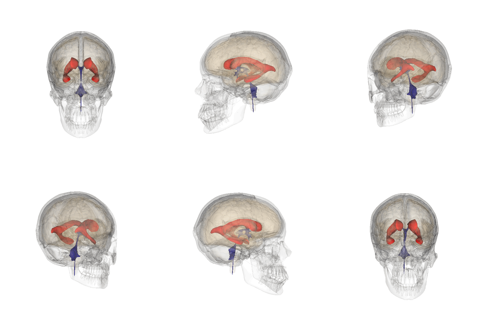
The lateral aperture is a paired structure in human anatomy. It is an opening in each lateral extremity of the lateral recess of the fourth ventricle of the human brain, which also has a single median aperture. The two lateral apertures provide a conduit for cerebrospinal fluid to flow from the brain's ventricular system into the subarachnoid space; specifically into the pontocerebellar cistern at the cerebellopontine angle. The structure is also called the lateral aperture of the fourth ventricle or the foramen of Luschka after anatomist Hubert von Luschka. Gross total resection of tumours that extend through foramen of Lushka is sometimes not possible due to bradycardia Bradycardia (also sinus bradycardia) is a slow resting heart rate, commonly under 60 beats per minute (BPM) as determined by an electrocardiogram. It is considered to be a normal heart rate during sleep, in young and healthy or elderly adults, a .... References Ventricular system {{Neuroanato ... [...More Info...] [...Related Items...] OR: [Wikipedia] [Google] [Baidu] |
Lateral Recess
The lateral recess is a projection of the fourth ventricle which extends into, or rather below, the inferior cerebellar peduncle of the brainstem. The lateral aperture ( Foramen of Luschka), an opening in each extremity of the lateral recess, provides a pathway for cerebrospinal fluid to flow from the brain's ventricular system into the subarachnoid space. The lateral aperture is the end point (and opening or foramen) of the lateral recess. Consider the lateral recess as a tunnel whose opening at one end is the lateral aperture. In the area of the lateral recess, the vestibular area (containing the vestibular nuclei) and the cochlear nuclei The cochlear nuclear (CN) complex comprises two cranial nerve nuclei in the human brainstem, the ventral cochlear nucleus (VCN) and the dorsal cochlear nucleus (DCN). The ventral cochlear nucleus is unlayered whereas the dorsal cochlear nucleus ... may be found. In the vicinity, the medullary striae may also be seen. References Ve ... [...More Info...] [...Related Items...] OR: [Wikipedia] [Google] [Baidu] |
Fourth Ventricle
The fourth ventricle is one of the four connected fluid-filled cavities within the human brain. These cavities, known collectively as the ventricular system, consist of the left and right lateral ventricles, the third ventricle, and the fourth ventricle. The fourth ventricle extends from the cerebral aqueduct (''aqueduct of Sylvius'') to the obex, and is filled with cerebrospinal fluid (CSF). The fourth ventricle has a characteristic diamond shape in cross-sections of the human brain. It is located within the pons or in the upper part of the medulla oblongata. CSF entering the fourth ventricle through the cerebral aqueduct can exit to the subarachnoid space of the spinal cord through two lateral apertures and a single, midline median aperture. Boundaries The fourth ventricle has a roof at its ''upper'' (posterior) surface and a floor at its ''lower'' (anterior) surface, and side walls formed by the cerebellar peduncles (nerve bundles joining the structure on the posterior sid ... [...More Info...] [...Related Items...] OR: [Wikipedia] [Google] [Baidu] |
Human Brain
The human brain is the central organ of the human nervous system, and with the spinal cord makes up the central nervous system. The brain consists of the cerebrum, the brainstem and the cerebellum. It controls most of the activities of the body, processing, integrating, and coordinating the information it receives from the sense organs, and making decisions as to the instructions sent to the rest of the body. The brain is contained in, and protected by, the skull bones of the head. The cerebrum, the largest part of the human brain, consists of two cerebral hemispheres. Each hemisphere has an inner core composed of white matter, and an outer surface – the cerebral cortex – composed of grey matter. The cortex has an outer layer, the neocortex, and an inner allocortex. The neocortex is made up of six neuronal layers, while the allocortex has three or four. Each hemisphere is conventionally divided into four lobes – the frontal, temporal, parietal, and occipital lo ... [...More Info...] [...Related Items...] OR: [Wikipedia] [Google] [Baidu] |
Median Aperture
In statistics and probability theory, the median is the value separating the higher half from the lower half of a data sample, a population, or a probability distribution. For a data set, it may be thought of as "the middle" value. The basic feature of the median in describing data compared to the mean (often simply described as the "average") is that it is not skewed by a small proportion of extremely large or small values, and therefore provides a better representation of a "typical" value. Median income, for example, may be a better way to suggest what a "typical" income is, because income distribution can be very skewed. The median is of central importance in robust statistics, as it is the most resistant statistic, having a breakdown point of 50%: so long as no more than half the data are contaminated, the median is not an arbitrarily large or small result. Finite data set of numbers The median of a finite list of numbers is the "middle" number, when those numbers are liste ... [...More Info...] [...Related Items...] OR: [Wikipedia] [Google] [Baidu] |
Cerebrospinal Fluid
Cerebrospinal fluid (CSF) is a clear, colorless body fluid found within the tissue that surrounds the brain and spinal cord of all vertebrates. CSF is produced by specialised ependymal cells in the choroid plexus of the ventricles of the brain, and absorbed in the arachnoid granulations. There is about 125 mL of CSF at any one time, and about 500 mL is generated every day. CSF acts as a shock absorber, cushion or buffer, providing basic mechanical and immunological protection to the brain inside the skull. CSF also serves a vital function in the cerebral autoregulation of cerebral blood flow. CSF occupies the subarachnoid space (between the arachnoid mater and the pia mater) and the ventricular system around and inside the brain and spinal cord. It fills the ventricles of the brain, cisterns, and sulci, as well as the central canal of the spinal cord. There is also a connection from the subarachnoid space to the bony labyrinth of the inner ear via the perilymphat ... [...More Info...] [...Related Items...] OR: [Wikipedia] [Google] [Baidu] |
Ventricular System
The ventricular system is a set of four interconnected cavities known as cerebral ventricles in the brain. Within each ventricle is a region of choroid plexus which produces the circulating cerebrospinal fluid (CSF). The ventricular system is continuous with the central canal of the spinal cord from the fourth ventricle, allowing for the flow of CSF to circulate. All of the ventricular system and the central canal of the spinal cord are lined with ependyma, a specialised form of epithelium connected by tight junctions that make up the blood–cerebrospinal fluid barrier. Structure The system comprises four ventricles: * lateral ventricles right and left (one for each hemisphere) * third ventricle * fourth ventricle There are several foramina, openings acting as channels, that connect the ventricles. The interventricular foramina (also called the foramina of Monro) connect the lateral ventricles to the third ventricle through which the cerebrospinal fluid can flow. Ventric ... [...More Info...] [...Related Items...] OR: [Wikipedia] [Google] [Baidu] |
Subarachnoid Space
In anatomy, the meninges (, ''singular:'' meninx ( or ), ) are the three membranes that envelop the brain and spinal cord. In mammals, the meninges are the dura mater, the arachnoid mater, and the pia mater. Cerebrospinal fluid is located in the subarachnoid space between the arachnoid mater and the pia mater. The primary function of the meninges is to protect the central nervous system. Structure Dura mater The dura mater ( la, tough mother) (also rarely called ''meninx fibrosa'' or ''pachymeninx'') is a thick, durable membrane, closest to the skull and vertebrae. The dura mater, the outermost part, is a loosely arranged, fibroelastic layer of cells, characterized by multiple interdigitating cell processes, no extracellular collagen, and significant extracellular spaces. The middle region is a mostly fibrous portion. It consists of two layers: the endosteal layer, which lies closest to the skull, and the inner meningeal layer, which lies closer to the brain. It contains large ... [...More Info...] [...Related Items...] OR: [Wikipedia] [Google] [Baidu] |
Pontine Cistern
The pontine cistern, also cisterna pontis and cisterna pontocerebellaris is a notable subarachnoid cistern on the ventral aspect of the pons. It contains the basilar artery, and is continuous behind with the subarachnoid space of the spinal cord, and with the cisterna magna, and in front of the pons with the interpeduncular cistern The interpeduncular cistern of Sweeney is the subarachnoid cistern that encloses the cerebral peduncles and the structures contained in the interpeduncular fossa and contains the arterial circle of Willis The circle of Willis (also called Wil .... References Meninges {{Portal bar, Anatomy ... [...More Info...] [...Related Items...] OR: [Wikipedia] [Google] [Baidu] |
Cerebellopontine Angle
The cerebellopontine angle (CPA) ( la, angulus cerebellopontinus) is located between the cerebellum and the pons. The cerebellopontine angle is the site of the cerebellopontine angle cistern one of the subarachnoid cisterns that contains cerebrospinal fluid, arachnoid tissue, cranial nerves, and associated vessels. The cerebellopontine angle is also the site of a set of neurological disorders known as the cerebellopontine angle syndrome. Structure The cerebellopontine angle is formed by the cerebellopontine fissure. This fissure is made when the cerebellum folds over to the pons, creating a sharply defined angle between them. The angle formed in turn creates a subarachnoid cistern, the cerebellopontine angle cistern. The pia mater follows the outline of the fissure and the arachnoid mater continues across the divide so that the subarachnoid space is dilated at this area, forming the cerebellopontine angle cistern. The anterior inferior cerebellar artery (AICA) is the principa ... [...More Info...] [...Related Items...] OR: [Wikipedia] [Google] [Baidu] |
Hubert Von Luschka
Hubert is a Germanic masculine given name, from ''hug'' "mind" and ''beraht'' "bright". It also occurs as a surname. Saint Hubertus or Hubert (c. 656 – 30 May 727) is the patron saint of hunters, mathematicians, opticians, and metalworkers. People with the given name Hubert This is a small selection of articles on people named Hubert; for a comprehensive list see instead . *Hubert Aaronson (1924–2005), F. Mehl University Professor at Carnegie Mellon University * Hubert Adair (1917–1940), World War II Royal Air Force pilot *Hubert Boulard, a French comics creator who is unusually credited as "Hubert" * Hubert Brasier (1917–1981), a Church of England clergyman, more famously the father of UK Prime Minister Theresa May * Hubert Buchanan (born 1941), a United States Air Force captain and fighter pilot *Hubert Chevis (1902–1931), a lieutenant in the Royal Artillery of the British Army who died of strychnine poisoning in June 1931 * Hubert Davies, British playwright and ... [...More Info...] [...Related Items...] OR: [Wikipedia] [Google] [Baidu] |
Bradycardia
Bradycardia (also sinus bradycardia) is a slow resting heart rate, commonly under 60 beats per minute (BPM) as determined by an electrocardiogram. It is considered to be a normal heart rate during sleep, in young and healthy or elderly adults, and in athletes. In some people, bradycardia below 60 BPM may be associated with fatigue, weakness, dizziness, sweating, and fainting. The term "relative bradycardia" is used to refer to a heart rate slower than an individual's typical resting heart rate. Athletes may have athletic heart syndrome, which includes bradycardia as part of the cardiovascular adaptations to training and participation. The word "bradycardia" is from the Greek βραδύς ''bradys'' "slow", and καρδία ''kardia'' "heart". Classification Sinus Atrial bradycardias are divided into three types. The first, respiratory sinus arrhythmia, is usually found in young and healthy adults. Heart rate increases during inhalation and decreases during exhalation. Thi ... [...More Info...] [...Related Items...] OR: [Wikipedia] [Google] [Baidu] |


