|
Fetal Rhabdomyoma
Rhabdomyoma is a benign mesenchymal tumor of skeletal muscle, separated into two major categories based on site: Cardiac and extracardiac. They are further separated by histology: fetal (myxoid and cellular), juvenile (intermediate), and adult types. Genital types are recognized, but are often part of either the fetal or juvenile types. The fetal type is thought to recapitulate immature skeletal muscle at about week six to ten of gestational development. Signs and symptoms Most fetal rhabdomyomas are tumors that develop in the head and neck or in the genital region. There are a number of cases which have been seen in association with Gorlin syndrome. However, cardiac myxomas are known to be associated with tuberous sclerosis. Pathophysiology Gross pathology The tumor may be seen within the subcutaneous tissues (below the skin), mucosal surfaces or in soft tissue. Within the head and neck, the posterior ear region, skin of the face, and the tongue are the most commonly aff ... [...More Info...] [...Related Items...] OR: [Wikipedia] [Google] [Baidu] |
Skeletal Muscle
Skeletal muscles (commonly referred to as muscles) are organs of the vertebrate muscular system and typically are attached by tendons to bones of a skeleton. The muscle cells of skeletal muscles are much longer than in the other types of muscle tissue, and are often known as muscle fibers. The muscle tissue of a skeletal muscle is striated – having a striped appearance due to the arrangement of the sarcomeres. Skeletal muscles are voluntary muscles under the control of the somatic nervous system. The other types of muscle are cardiac muscle which is also striated and smooth muscle which is non-striated; both of these types of muscle tissue are classified as involuntary, or, under the control of the autonomic nervous system. A skeletal muscle contains multiple muscle fascicle, fascicles – bundles of muscle fibers. Each individual fiber, and each muscle is surrounded by a type of connective tissue layer of fascia. Muscle fibers are formed from the cell fusion, fusion of ... [...More Info...] [...Related Items...] OR: [Wikipedia] [Google] [Baidu] |
Gorlin Syndrome
Gorlin may refer to: People *Dan Gorlin, computer game programmer, designer and founder of Dan Gorlin Productions *Eitan Gorlin, filmmaker, author and actor *Mikhail Gorlin, Russian emigre poet *Richard Gorlin, American cardiologist, co-developed the Gorlin equation * Robert J. Gorlin, a professor and researcher at the University of Minnesota In medicine *Gorlin sign, the ability to touch the tip of the nose with the tongue and touch the elbow with the tongue *Gorlin syndrome Gorlin may refer to: People *Dan Gorlin, computer game programmer, designer and founder of Dan Gorlin Productions *Eitan Gorlin, filmmaker, author and actor *Mikhail Gorlin, Russian emigre poet *Richard Gorlin, American cardiologist, co-developed ..., also known as basal cell nevus syndrome *The Gorlin equation, a method to calculate the effective area of a heart valve during cardiac catheterization {{disambig ... [...More Info...] [...Related Items...] OR: [Wikipedia] [Google] [Baidu] |
Tuberous Sclerosis
Tuberous sclerosis complex (TSC) is a rare multisystem autosomal dominant genetic disease that causes non-cancerous tumours to grow in the brain and on other vital organs such as the kidneys, heart, liver, eyes, lungs and skin. A combination of symptoms may include seizures, intellectual disability, developmental delay, behavioral problems, skin abnormalities, lung disease, and kidney disease. TSC is caused by a mutation of either of two genes, ''TSC1'' and ''TSC2'', which code for the proteins hamartin and tuberin, respectively, with ''TSC2'' mutations accounting for the majority and tending to cause more severe symptoms. These proteins act as tumor growth suppressors, agents that regulate cell proliferation and differentiation. Prognosis is highly variable and depends on the symptoms, but life expectancy is normal for many. The prevalence of the disease is estimated to be 7 to 12 in 100,000. The disease is often abbreviated to tuberous sclerosis, which refers to the har ... [...More Info...] [...Related Items...] OR: [Wikipedia] [Google] [Baidu] |
Fetal Rhabdomyoma Ear HE LDRT
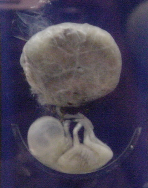
A fetus or foetus (; plural fetuses, feti, foetuses, or foeti) is the unborn offspring that develops from an animal embryo. Following embryonic development the fetal stage of development takes place. In human prenatal development, fetal development begins from the ninth week after fertilization (or eleventh week gestational age) and continues until birth. Prenatal development is a continuum, with no clear defining feature distinguishing an embryo from a fetus. However, a fetus is characterized by the presence of all the major body organs, though they will not yet be fully developed and functional and some not yet situated in their final anatomical location. Etymology The word ''fetus'' (plural '' fetuses'' or '' feti'') is related to the Latin '' fētus'' ("offspring", "bringing forth", "hatching of young") and the Greek "φυτώ" to plant. The word "fetus" was used by Ovid in Metamorphoses, book 1, line 104. The predominant British, Irish, and Commonwealth spelling is ' ... [...More Info...] [...Related Items...] OR: [Wikipedia] [Google] [Baidu] |
Periosteum
The periosteum is a membrane that covers the outer surface of all bones, except at the articular surfaces (i.e. the parts within a joint space) of long bones. Endosteum lines the inner surface of the medullary cavity of all long bones. Structure The periosteum consists of an outer fibrous layer, and an inner cambium layer (or osteogenic layer). The fibrous layer is of dense irregular connective tissue, containing fibroblasts, while the cambium layer is highly cellular containing progenitor cells that develop into osteoblasts. These osteoblasts are responsible for increasing the width of a long bone and the overall size of the other bone types. After a bone fracture, the progenitor cells develop into osteoblasts and chondroblasts, which are essential to the healing process. The outer fibrous layer and the inner cambium layer is differentiated under electron micrography. As opposed to osseous tissue, the periosteum has nociceptors, sensory neurons that make it very sensitive to ... [...More Info...] [...Related Items...] OR: [Wikipedia] [Google] [Baidu] |
Rhabdomyosarcoma
Rhabdomyosarcoma (RMS) is a highly aggressive form of cancer that develops from mesenchymal cells that have failed to fully differentiate into myocytes of skeletal muscle. Cells of the tumor are identified as rhabdomyoblasts. There are four subtypes – embryonal rhabdomyosarcoma, alveolar rhabdomyosarcoma, pleomorphic rhabdomyosarcoma, and spindle cell/sclerosing rhabdomyosarcoma. Embryonal, and alveolar are the main groups, and these types are the most common soft tissue sarcomas of childhood and adolescence. The pleomorphic type is usually found in adults. It is generally considered to be a disease of childhood, as the vast majority of cases occur in those below the age of 18. It is commonly described as one of the small-blue-round-cell tumors of childhood due to its appearance on an H&E stain. Despite being relatively rare, it accounts for approximately 40% of all recorded soft tissue sarcomas. RMS can occur in any soft tissue site in the body, but is primarily found in t ... [...More Info...] [...Related Items...] OR: [Wikipedia] [Google] [Baidu] |
Granular Cell Tumor
Granular cell tumor is a tumor that can develop on any skin or mucosal surface, but occurs on the tongue 40% of the time. It is also known as Abrikossoff's tumor, granular cell myoblastoma, granular cell nerve sheath tumor, and granular cell schwannoma. Granular cell tumors (GCTs) affect females more often than males. Pathology Granular cell tumors are derived from neural tissue, as can be demonstrated by immunohistochemistry and ultrastructural evidence using electron microscopy. These lesions characteristically consist of polygonal cells with bland nuclei, abundant cytoplasm and fine eosinophilic cytoplasmic granules. The tumor cells stain positively for S-100 as they are of Schwann cell origin. Both malignant and benign versions of the tumor exist, where malignant tumors are characterized histologically by features such as spindling, high nuclear to cytoplasmic ratios, pleomorphism, and necrosis. Multiple granular cell tumors may seen in the context of ''LEOPARD syndrome'', du ... [...More Info...] [...Related Items...] OR: [Wikipedia] [Google] [Baidu] |
Alveolar Soft Part Sarcoma
Alveolar soft part sarcoma, abbreviated ASPS, is a very rare type of soft-tissue sarcoma, that grows slowly and whose cell of origin is unknown. ASPS arises mainly in children and young adults and can migrate (metastasize) into other parts of the body, typically the lungs and the brain. Typically, ASPS arises in muscles and deep soft tissue of the thigh or the leg (lower extremities), but can also appear in the upper extremities (hands, neck, and head). While ASPS is a soft tissue sarcoma, it can also spread and grow inside the bones. Etymology * The term alveolar comes from the microscopic pattern, visible during the analysis of slides of ASPS under the microscope in histopathology. The tumor cells seem to be arranged in the same pattern as the cells of the small air sacks (alveoli) in the lungs. However, this is just a structural similarity. ASPS was first described and characterized in 1952. * ASPS is a sarcoma, and that indicates that this cancer initially arises from tissu ... [...More Info...] [...Related Items...] OR: [Wikipedia] [Google] [Baidu] |
Hibernoma
A hibernoma is a benign neoplasm of vestigial brown fat. The term was originally used by the French anatomist Louis Gery in 1914. Signs and symptoms Patients present with a slow-growing, painless, solitary mass, usually of the subcutaneous tissues. It is much less frequently noted in the intramuscular tissue. It is not uncommon for symptoms to be present for years. Benign neoplasm with brown fat is noted. Diagnosis Imaging findings In general, imaging studies show a well-defined, heterogeneous mass, usually showing a mass which is hypointense to subcutaneous fat on magnetic resonance T1-weight images. Serpentine, thin, low signal bands (septations or vessels) are often seen throughout the tumor. Pathology findings From a macroscopic perspective, there is a well-defined, encapsulated or circumscribed mass, showing a soft, yellow tan to deep brown mass. The size ranges from 1 to 27 cm, although the mean is about 10 cm. The tumors histologically resemble brown fat. ... [...More Info...] [...Related Items...] OR: [Wikipedia] [Google] [Baidu] |
Oncocytoma
An oncocytoma is a tumor made up of oncocytes, epithelial cells characterized by an excessive amount of mitochondria, resulting in an abundant acidophilic, granular cytoplasm. The cells and the tumor that they compose are often benign but sometimes may be premalignant or malignant. Presentation An oncocytoma is an epithelial tumor composed of oncocytes, large eosinophilic cells having small, round, benign-appearing nuclei with large nucleoli. Oncocytoma can arise in a number of organs. Renal oncocytoma Renal oncocytoma is thought to arise from the intercalated cells of collecting ducts of the kidney. It represents 5% to 15% of surgically resected renal neoplasms. Salivary gland oncocytoma The salivary gland oncocytoma is a well-circumscribed, benign neoplastic growth also called an oxyphilic adenoma. It comprises about 1% of all salivary gland tumors. The histopathology is marked by sheets of large swollen polyhedral epithelial oncocytes, which are granular acidophilic parot ... [...More Info...] [...Related Items...] OR: [Wikipedia] [Google] [Baidu] |


_skin.jpg)