|
Funiculus Posterior
Funiculus (''Latin'' for "slender rope") is any cord-like structure in anatomy or biology, and may refer to: * in the peripheral nervous system a bundle of axons that may be bundled into a nerve fascicle * in the central nervous system one of the paired white matter regions of the spinal cord: the anterior funiculus, the lateral funiculus, and the posterior funiculus; and in the fourth ventricle the funiculus separans a strip of ependyma. * the umbilical cord attaching a fetus to the placenta during pregnancy * the spermatic cord formed by the vas deferens and surrounding tissue * in antenna (biology), insect antennae, the funicle is the segment connecting the club with the base * in flowering plants, the funiculus is the stalk that attaches an ovule to the placenta * in mycology, the funicular cord is a sticky trailing thread that attaches the peridioles (the "eggs") to the peridium (the "nest") in some species of bird's nest fungi {{disambiguation ... [...More Info...] [...Related Items...] OR: [Wikipedia] [Google] [Baidu] |
Peripheral Nervous System
The peripheral nervous system (PNS) is one of two components that make up the nervous system of bilateral animals, with the other part being the central nervous system (CNS). The PNS consists of nerves and ganglia, which lie outside the brain and the spinal cord. The main function of the PNS is to connect the CNS to the limbs and organs, essentially serving as a relay between the brain and spinal cord and the rest of the body. Unlike the CNS, the PNS is not protected by the vertebral column and skull, or by the blood–brain barrier, which leaves it exposed to toxins. The peripheral nervous system can be divided into the somatic nervous system and the autonomic nervous system. In the somatic nervous system, the cranial nerves are part of the PNS with the exception of the optic nerve (cranial nerve II), along with the retina. The second cranial nerve is not a true peripheral nerve but a tract of the diencephalon. Cranial nerve ganglia, as with all ganglia, are part of the P ... [...More Info...] [...Related Items...] OR: [Wikipedia] [Google] [Baidu] |
Funiculus Separans
The cells of the dorsal nucleus of vagus nerve are spindle-shaped, like those of the posterior column of the spinal cord, and the nucleus is usually considered as representing the base of the posterior column. It measures about 2 cm. in length, and in the lower, closed part of the medulla oblongata is situated behind the dorsal nucleus of the vagus; whereas in the upper, open part it lies lateral to that nucleus, and corresponds to an eminence, named the vagal trigone (ala cinerea), in the rhomboid fossa. The vagal trigone is separated from the area postrema by a narrow strip of thickened ependyma The ependyma is the thin Neuroepithelial cell, neuroepithelial (Simple columnar epithelium, simple columnar ciliated epithelium) lining of the ventricular system of the brain and the central canal of the spinal cord. The ependyma is one of the fou ... – the funiculus separans. References External links * https://web.archive.org/web/20070927162218/http://www.ib.amwaw.edu.pl/an ... [...More Info...] [...Related Items...] OR: [Wikipedia] [Google] [Baidu] |
Ovule
In seed plants, the ovule is the structure that gives rise to and contains the female reproductive cells. It consists of three parts: the ''integument'', forming its outer layer, the ''nucellus'' (or remnant of the megasporangium), and the female gametophyte (formed from a haploid megaspore) in its center. The female gametophyte — specifically termed a ''megagametophyte''— is also called the ''embryo sac'' in angiosperms. The megagametophyte produces an egg cell for the purpose of fertilization. The ovule is a small structure present in the ovary. It is attached to the placenta by a stalk called a funicle. The funicle provides nourishment to the ovule. Location within the plant In flowering plants, the ovule is located inside the portion of the flower called the gynoecium. The ovary of the gynoecium produces one or more ovules and ultimately becomes the fruit wall. Ovules are attached to the placenta in the ovary through a stalk-like structure known as a ''funiculus'' ... [...More Info...] [...Related Items...] OR: [Wikipedia] [Google] [Baidu] |
Flowering Plant
Flowering plants are plants that bear flowers and fruits, and form the clade Angiospermae (), commonly called angiosperms. The term "angiosperm" is derived from the Greek words ('container, vessel') and ('seed'), and refers to those plants that produce their seeds enclosed within a fruit. They are by far the most diverse group of land plants with 64 orders, 416 families, approximately 13,000 known genera and 300,000 known species. Angiosperms were formerly called Magnoliophyta (). Like gymnosperms, angiosperms are seed-producing plants. They are distinguished from gymnosperms by characteristics including flowers, endosperm within their seeds, and the production of fruits that contain the seeds. The ancestors of flowering plants diverged from the common ancestor of all living gymnosperms before the end of the Carboniferous, over 300 million years ago. The closest fossil relatives of flowering plants are uncertain and contentious. The earliest angiosperm fossils ar ... [...More Info...] [...Related Items...] OR: [Wikipedia] [Google] [Baidu] |
Antenna (biology)
Antennae ( antenna), sometimes referred to as "feelers", are paired appendages used for sensing in arthropods. Antennae are connected to the first one or two segments of the arthropod head. They vary widely in form but are always made of one or more jointed segments. While they are typically sensory organs, the exact nature of what they sense and how they sense it is not the same in all groups. Functions may variously include sensing touch, air motion, heat, vibration (sound), and especially smell or taste. Antennae are sometimes modified for other purposes, such as mating, brooding, swimming, and even anchoring the arthropod to a substrate. Larval arthropods have antennae that differ from those of the adult. Many crustaceans, for example, have free-swimming larvae that use their antennae for swimming. Antennae can also locate other group members if the insect lives in a group, like the ant. The common ancestor of all arthropods likely had one pair of uniramous (unbranched ... [...More Info...] [...Related Items...] OR: [Wikipedia] [Google] [Baidu] |
Vas Deferens
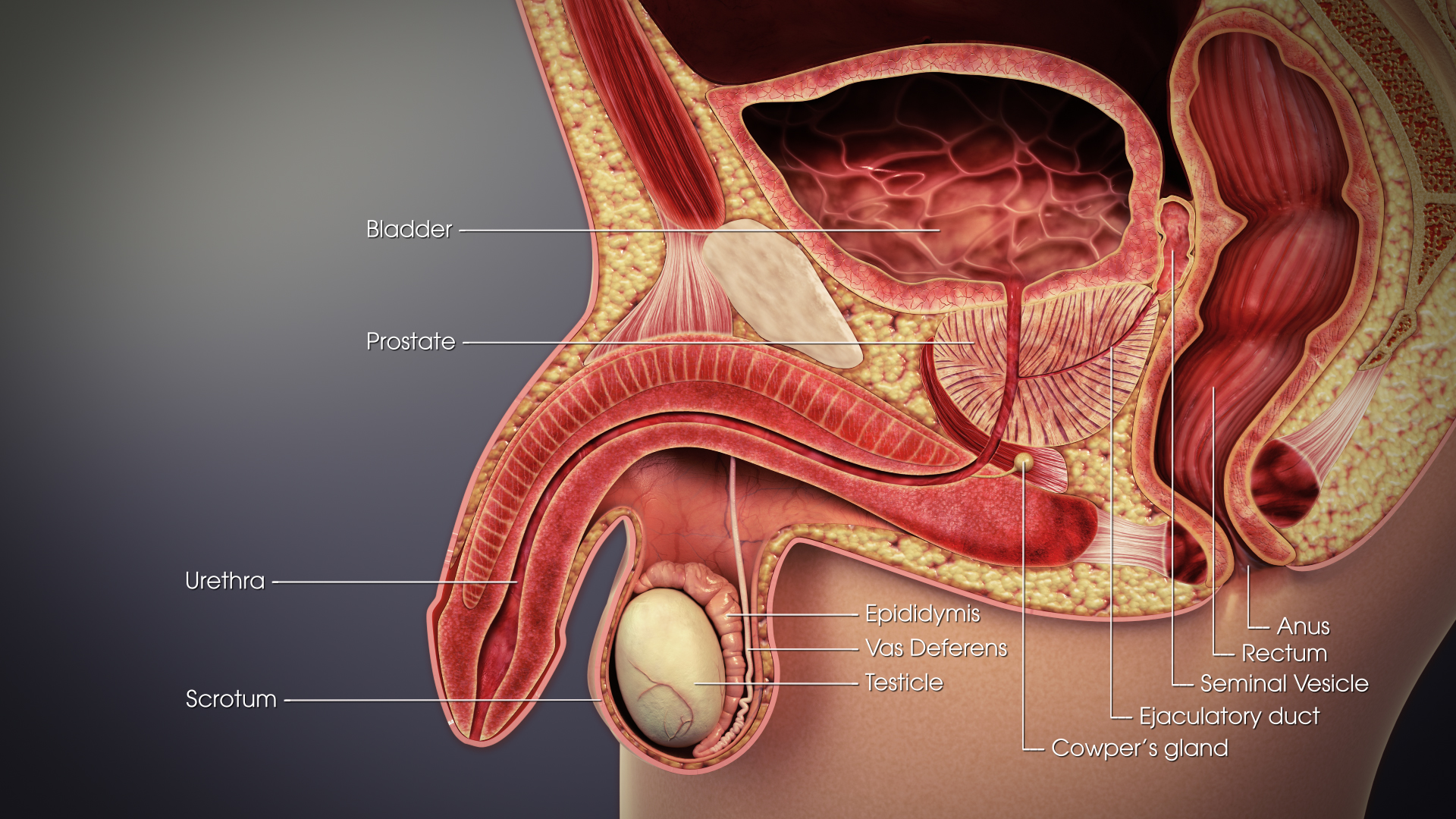
The vas deferens or ductus deferens is part of the male reproductive system of many vertebrates. The ducts transport sperm from the epididymis to the ejaculatory ducts in anticipation of ejaculation. The vas deferens is a partially coiled tube which exits the abdominal cavity through the inguinal canal. Etymology ''Vas deferens'' is Latin, meaning "carrying-away vessel"; the plural version is ''vasa deferentia''. ''Ductus deferens'' is also Latin, meaning "carrying-away duct"; the plural version is ''ducti deferentes''. Structure There are two vasa deferentia, connecting the left and right epididymis with the seminal vesicles to form the ejaculatory duct in order to move sperm. The (human) vas deferens measures 30–35 cm in length, and 2–3 mm in diameter. The vas deferens is continuous proximally with the tail of the epididymis. The vas deferens exhibits a tortuous, convoluted initial/proximal section (which measures 2–3 cm in length). Distally, it forms ... [...More Info...] [...Related Items...] OR: [Wikipedia] [Google] [Baidu] |
Spermatic Cord
The spermatic cord is the cord-like structure in males formed by the vas deferens (''ductus deferens'') and surrounding tissue that runs from the deep inguinal ring down to each testicle. Its serosal covering, the tunica vaginalis, is an extension of the peritoneum that passes through the transversalis fascia. Each testicle develops in the lower thoracic and upper lumbar region and migrates into the scrotum. During its descent it carries along with it the vas deferens, its vessels, nerves etc. There is one on each side. Structure The spermatic cord is ensheathed in three layers of tissue: * ''external spermatic fascia'', an extension of the innominate fascia that overlies the aponeurosis of the external oblique muscle. * ''cremasteric muscle and fascia'', formed from a continuation of the internal oblique muscle and its fascia. * ''internal spermatic fascia'', continuous with the transversalis fascia. The normal diameter of the spermatic cord is about 16 mm (range 11 to 22 mm). It ... [...More Info...] [...Related Items...] OR: [Wikipedia] [Google] [Baidu] |
Umbilical Cord
In placental mammals, the umbilical cord (also called the navel string, birth cord or ''funiculus umbilicalis'') is a conduit between the developing embryo or fetus and the placenta. During prenatal development, the umbilical cord is physiologically and genetically part of the fetus and (in humans) normally contains two arteries (the umbilical arteries) and one vein (the umbilical vein), buried within Wharton's jelly. The umbilical vein supplies the fetus with oxygenated, nutrient-rich blood from the placenta. Conversely, the fetal heart pumps low-oxygen, nutrient-depleted blood through the umbilical arteries back to the placenta. Structure and development The umbilical cord develops from and contains remnants of the yolk sac and allantois. It forms by the fifth week of development, replacing the yolk sac as the source of nutrients for the embryo. The cord is not directly connected to the mother's circulatory system, but instead joins the placenta, which transfers materials t ... [...More Info...] [...Related Items...] OR: [Wikipedia] [Google] [Baidu] |
Ependyma
The ependyma is the thin Neuroepithelial cell, neuroepithelial (Simple columnar epithelium, simple columnar ciliated epithelium) lining of the ventricular system of the brain and the central canal of the spinal cord. The ependyma is one of the four types of neuroglia in the central nervous system (CNS). It is involved in the production of cerebrospinal fluid (CSF), and is shown to serve as a reservoir for neuroregeneration. Structure The ependyma is made up of ependymal Cell (biology), cells called ependymocytes, a type of glial cell. These cells line the Ventricular system, ventricles in the brain and the central canal of the spinal cord, which become filled with cerebrospinal fluid. These are nervous tissue cells with simple columnar shape, much like that of some mucosal epithelial cells. Early monociliated ependymal cells are differentiated to multiciliated ependymal cells for their function in circulating cerebrospinal fluid. The epithelial polarity#Basolateral membranes, bas ... [...More Info...] [...Related Items...] OR: [Wikipedia] [Google] [Baidu] |
Fourth Ventricle
The fourth ventricle is one of the four connected fluid-filled cavities within the human brain. These cavities, known collectively as the ventricular system, consist of the left and right lateral ventricles, the third ventricle, and the fourth ventricle. The fourth ventricle extends from the cerebral aqueduct (''aqueduct of Sylvius'') to the obex, and is filled with cerebrospinal fluid (CSF). The fourth ventricle has a characteristic diamond shape in cross-sections of the human brain. It is located within the pons or in the upper part of the medulla oblongata. CSF entering the fourth ventricle through the cerebral aqueduct can exit to the subarachnoid space of the spinal cord through two lateral apertures and a single, midline median aperture. Boundaries The fourth ventricle has a roof at its ''upper'' (posterior) surface and a floor at its ''lower'' (anterior) surface, and side walls formed by the cerebellar peduncles (nerve bundles joining the structure on the posterior sid ... [...More Info...] [...Related Items...] OR: [Wikipedia] [Google] [Baidu] |
Axon
An axon (from Greek ἄξων ''áxōn'', axis), or nerve fiber (or nerve fibre: see spelling differences), is a long, slender projection of a nerve cell, or neuron, in vertebrates, that typically conducts electrical impulses known as action potentials away from the nerve cell body. The function of the axon is to transmit information to different neurons, muscles, and glands. In certain sensory neurons (pseudounipolar neurons), such as those for touch and warmth, the axons are called afferent nerve fibers and the electrical impulse travels along these from the periphery to the cell body and from the cell body to the spinal cord along another branch of the same axon. Axon dysfunction can be the cause of many inherited and acquired neurological disorders that affect both the peripheral and central neurons. Nerve fibers are classed into three typesgroup A nerve fibers, group B nerve fibers, and group C nerve fibers. Groups A and B are myelinated, and group C are unmyelinated. ... [...More Info...] [...Related Items...] OR: [Wikipedia] [Google] [Baidu] |
Posterior Funiculus
Posterior may refer to: * Posterior (anatomy), the end of an organism opposite to its head ** Buttocks, as a euphemism * Posterior horn (other) * Posterior probability The posterior probability is a type of conditional probability that results from updating the prior probability with information summarized by the likelihood via an application of Bayes' rule. From an epistemological perspective, the posterior ..., the conditional probability that is assigned when the relevant evidence is taken into account * Posterior tense, a relative future tense {{disambiguation ... [...More Info...] [...Related Items...] OR: [Wikipedia] [Google] [Baidu] |


